Effect of PLIF and TLIF on sagittal spinopelvic balance of patients with
degenerative spondylolisthesis
Mustafa Uysal
a, Metin Ozalay
b, Alihan Derincek
c, Alauddin Kochai
a,*, Mehmet Turker
aaDepartment of Orthopedics and Traumatology, Sakarya University Faculty of Medicine, Sakarya, Turkey
bDepartment of Orthopedics and Traumatology, Adana Medical Center, Baskent University School of Medicine, Adana, Turkey cDepartment of Orthopedics and Traumatology, Medline Adana Hospital, Adana, Turkey
a r t i c l e i n f o
Article history:
Received 13 February 2017 Received in revised form 20 February 2018 Accepted 19 March 2018 Available online 26 April 2018 Keywords:
Degenerative spondylolisthesis Sagittal spinopelvic balance PLIF/TLIF
Angles
Lumbar interbody fusion
a b s t r a c t
Objective: The aim of this study was to evaluate the effects of PLIF and TLIF on sagittal spinopelvic balance and to compare radiological results of two surgical procedures with regard to spinopelvic parameters.
Methods: Thirty-five patients (34 female and 1 male; mean age: 52.29 ± 13.08 (range: 35e75)) with degenerative spondylolisthesis cases were included in the study. Patients were divided into two groups according to surgical technique: PLIF and TLIF. The level and the severity of listhesis according to Meyerding classification were assessed and spinopelvic parameters including sacral slope, pelvic tilt, pelvic incidence (PI), lumbar lordosis, and segmental lumbar lordosis were measured on digital X-rays. All preoperative and postoperative parameters and the results were compared between two groups. Results: The age distribution was similar in both groups (p¼ 0.825) and there was no difference between the mean PI of the groups (p¼ 0.616). In 15 patients, spondylolisthesis level were at the L5-S1 level (PLIF: 8, TLIF: 7), in 16 patients at the L4-L5 level (PLIF: 6, TLIF: 10) and in 4 patients at the L3-L4 level (PLIF: 2, TLIF: 2). According to Meyerding classification, before the operation, the sliding grades were 0 in 4 pa-tients, 1 in 21 papa-tients, 2 in 7 papa-tients, and 3 in 3 patients. The grades changed into 0 in 28 papa-tients, 1 in 5 patients, and 2 in 2 patients after surgery. There were no differences in the grade of listhesis between PLIF and TLIF groups preoperatively (p¼ 0.190) and postoperatively (p ¼ 0.208). In both groups, the spondylolisthesis-related deformities of patients were significantly corrected after surgery (p < 0.001). Conclusion: PLIF and TLIF techniques have similar radiological results in restoring the sagittal spinopelvic balance in patients with degenerative spondylolisthesis. Both techniques are good options to achieve reduction and fusion in patients with degenerative spondylolisthesis, but have no advantage over each other for restoring spinopelvic balance.
Level of evidence: Level III, Therapeutic study.
© 2018 Turkish Association of Orthopaedics and Traumatology. Publishing services by Elsevier B.V. This is an open access article under the CC BY-NC-ND license (http://creativecommons.org/licenses/by-nc-nd/ 4.0/).
Introduction
Degenerative spondylolisthesis is defined as slipping of one lumbar vertebral body onto a subjacent vertebral body due to degenerative deformation of articular and ligamentous
structures in the elderly population.1 Compensatory mechanisms such as facet and ligament hypertrophy and displacement may lead to compression neural elements, which further potentiate pain and disability.
Spondylolisthesis changes sagittal spinal alignment, which is one of the reasons for back pain.2,3Sagittal spinal balance refers to optimal configuration between the pelvis and spinal column in standing position.4
Sagittal spinal alignment is greatly influenced by spinopelvic parameters such as sacral slope (SS), lumbar lordosis (LL), pelvic tilt (PT), and pelvic incidence (PI).2,5e7 In standing position, the morphology and position of the pelvis influence lumbar lordosis, * Corresponding author. Department of Orthopedics and Traumatology, Sakarya
University Faculty of Medicine, Adnan Menderes Blvd, Saglik Str. 193, 54100, Ada-pazari, Sakarya, Turkey.
E-mail address:[email protected](A. Kochai).
Peer review under responsibility of Turkish Association of Orthopaedics and Traumatology.
Contents lists available atScienceDirect
Acta Orthopaedica et Traumatologica Turcica
j o u r n a l h o m e p a g e : h t t p s : / / w w w . e l s e v ie r . c o m / l o c a t e / a o t thttps://doi.org/10.1016/j.aott.2018.03.001
1017-995X/© 2018 Turkish Association of Orthopaedics and Traumatology. Publishing services by Elsevier B.V. This is an open access article under the CC BY-NC-ND license (http://creativecommons.org/licenses/by-nc-nd/4.0/).
which is important for sagittal spinal alignment.6In most studies PI for pelvic morphology and PT and SS for pelvic position over the femoral heads are used as pelvic parameters. Changes in LL resulted in compensation with pelvic retroversion.8
Posterior lumbar interbody fusion (PLIF) and transforaminal lumbar interbody fusion (TLIF) are two different interbody fusion techniques, which promise better fusion rate than standard posterolateral fusion.9 The PLIF technique wasfirst described by Cloward in 1940.10The TLIF technique was a modification of PLIF and described by Harms in 1998.11The main difference is that TLIF is performed with unilateral approach, preserving contralateral facet and laminar surface. PLIF and TLIF provide good outcomes in patients with degenerative spondylolisthesis, especially when the slip is accompanied by severe stenosis and major segmental instability (generally classified Meyerding grade II or higher).12
Both techniques simultaneously offer the option of disc height restoration, which is crucial for LL.13
Biomechanical loads on intervertebral discs increase parallel to the decrease in the normal sagittal inclination of the lumbosacral vertebral column; it also shows that, in addition to other pa-rameters analyzed in sagittal morphology, the sacral table and sacral kyphosis angles are important predisposing anatomical factors for the development of intervertebral disc degeneration and herniation.14
Sagittal imbalance has negative effect on patient's clinic. One of the main interest of these surgical techniques is to restore the balance and normalize patient daily life. As we know in literature correction of spinopelvic parameters improves patient clinic7,8. Both TLIF and PLIF have different mechanism on correction of lumbar lordosis. The purpose of this study was to evaluate the effects of PLIF and TLIF on sagittal spinopelvic balance and to compare radiological results of two surgical procedures with regard to spinopelvic parameters.
Materials and methods
Ninety-eight patients with spondylolisthesis were retrospec-tively evaluated from January 2008 to December 2014. Only adult degenerative spondylolisthesis cases operated with either PLIF or TLIF were included in the study. Exclusion criteria were spondylo-listhesis caused by pathologic conditions such as infection, tumor, iatrogenic and congenital reasons. Five patients were lost to follow-up. We enrolled 35 patients with spondylolisthesis who were operated on with either PLIF or TLIF. All patients were female except one. The mean age of patients was 52.29± 13.08 (range: 35e75). Patients were evaluated in two groups, PLIF and TLIF. There were 16 patients (female: 15 and male: 1) in the PLIF group and 19 patients (female: 19) in the TLIF group. The mean of the patients was 52.87± 13.64 in the PLIF group and 51.84 ± 12.98 in the TLIF group. Surgical technique
The same surgical team performed all procedures. Surgeons randomly selected PLIF or TLIF. Both procedures were performed in similar fashion as in the literature.11,13 Patients were placed in prone position on the surgical table. Two vertebras in the spon-dylolisthesis level were exposed. Pedicular screws were implanted in the upper and lower levels of spondylolisthesis (Xia spinal sys-tem, Stryker). Posterior elements were removed, but facet joints were left intact bilaterally in PLIF. Unilateral laminectomy and partial facetectomy was performed in TLIF (Fig. 1). Dura and nerve root were exposed bilaterally in PLIF and unilaterally in TLIF. Segmental distraction was performed over the rod between two pedicular screws to facilitate decompression and reduction. The thecal sac and nerve root were protected by retracting to the
midline. After resection of disc material and denuding the carti-laginous endplates, disc space was prepared for the interbody fusion device. Double cylindrical titanium mesh (Pyramesh surgical titanium mesh, Medtronic) or rectangular peek cages (Capstone PTC spinal system, Medtronic) for PLIF (the average hight of cages was 10.12 mm) and a single banana-shaped peek cage (AVS TL peek, Styker) for TLIF were used for interbody fusion (the average hight of cages was 9.84 mm). Autographs harvested from lamina and spinous process werefilled into cages and the impacted anterior disc space. Cages were inserted into the disc space close to the midline anteriorly as far as possible. Compression was applied be-tween pedicular screws after C-arm control.
Main differences between PLIF and TLIF are the approach to access disc and the interbody devices used for fusion. Nerve root retraction is less because the disc approach is more lateral in TLIF compared to PLIF (Fig. 1).
Radiological evaluation
Lateral radiographs of the whole spine were taken for all pa-tients before and 6 months after surgery. Papa-tients were standing in lateral position, elbows fullyflexed with fingers on clavicle, knees and hips fully extended.15
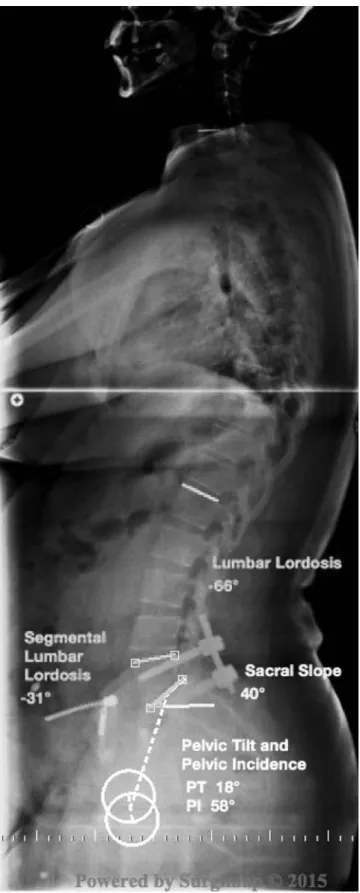
All measurements were made in preoperative and post-operative digital X-rays by using Surgimap program (Surgimap Spine, Newyork, Nemaris Inc.) (Fig. 2andFig. 3). Spondylolisthesis levels were recorded. The severity of lysthesis was assessed by using Meyerding classification.16Spinopelvic parameters including
sacral slope (SS), pelvic tilt (PT), and pelvic incidence (PI) were measured as reported by Duval et al7 Lumbar lordosis (LL) was measured to define the whole lumber curve between L1 and L5 Fig. 1. Illustration demonstrating the approach of bony removal in lamina. The two-dashed circle above represents the PLIF approach and the one-dotted circle below represents the TLIF approach.
vertebra. Segmental lumbar lordosis (SLL) was described lordosis at the lysthesis level. It was determined by measuring angle be-tween superior endplate of upper vertebra and inferior endplate of lower vertebra of lysthesis (Fig. 4).
Statistical analyses
The KolmogoroveSmirnov test was used to evaluate whether the distribution of variables were normal. Accordingly, it was seen that all variables displayed a normal distribution. Therefore, two independent sample t tests were used to compare the continuous
data between PLIF and TLIF groups. A paired sample t test was used to compare the continuous data between pre- and postoperative terms. A repeated measures two-way ANOVA test was used to analyze the alteration of continuous data between PLIF and Fig. 2. The output of the Surgimap Spine program with preoperative measurements of
spinopelvic parameters. Femoral heads were marked with circles and reference lines were drawn in end plates of vertebras. Lumbar lordosis (LL), segmental lumbar lordosis (SLL), sacral slope (SS), pelvic tilt (PT) and pelvic incidence (PI) are automatically calculated by the program.
Fig. 3. The postoperative measurements of spinopelvic parameters were made and recorded in X-ray.
TLIF groups. The continuous variables were presented as the mean± standard deviation. A p-value <0.05 was considered sig-nificant. Analyses were performed using commercial software (IBM SPSS Statistics, Version 22.0. Armonk, NY: IBM Corp.)
Results
Lumbar back pain was the main reason for the surgery in all patients. Nine patients had radiculopathy complaints. All patients had relieved pain, but two patients had sustained radiculopathy complaints up to 3 months after surgery. The patients in both groups were similar age distribution (p¼ 0.825). There was no difference between PI of groups (p ¼ 0.616) (Table 1).
Spondylolisthesis levels were at the L5-S1 level (PLIF: 8, TLIF: 7) in 15 patients, at the L4-L5 level (PLIF: 6, TLIF: 10) in 16 patients and at the L3-L4 level (PLIF: 2, TLIF: 2) in 4 patients. According to Meyerding classification, spondylolisthesis grades were distributed from 1 to 3 for all patients. Before the operations, the sliding grades were 0 in 4 patients, 1 in 21 patients, 2 in 7 patients, and 3 in 3 patients. The grades changed into 0 in 28 patients, 1 in 5 patients, and 2 in 2 patients after surgery. There were no differences in the grade of lysthesis between PLIF and TLIF groups preoperatively (p¼ 0.190) and postoperatively (p ¼ 0.208). The spondylolisthesis-related deformities of patients in both groups were significantly corrected after surgery according to the Meyerding classification (p< 0.001).
The radiographic parameters related with spinal sagittal balance are detailed and compared in Table 1. The analyses showed no significant difference between PLIF and TLIF groups according to all radiological parameters. Both surgical techniques showed the same radiological characteristics before and after surgery. There was no difference between groups according to the PI (p¼ 0.616). Discussion
The direct comparison of radiological results in a group of degenerative spondylolisthesis patients yielded equivocal results in terms of sagittal spinopelvic balance. No significant difference was found between patients treated with either PLIF or TLIF. Our results revealed that TLIF and PLIF were both successful in getting reduc-tion of sliding vertebra and fusion in all patients with degenerative spondylolisthesis. Both surgical techniques have similar effect on spinopelvic balance.
Degenerative spondylolisthesis is a contributing factor for impairment of sagittal spinopelvic balance. Unbalanced loading and compression of neural structures cause mechanical and neurological complaints. Reduction and restoration of disc height for sagittal spinal balance, stabilization of motion segments for instability, and decompression of neural structures for neurological involvement are major components of the treatment. PLIF and TLIF are surgical techniques that are able to achieve these goals. The main advantage of PLIF is the ability to make good neural decom-pression, but PLIF was associated with a higher complication rate.17 The main advantages of TLIF are limiting the risk of dural injury and shorter operative time.13,17,18 Both techniques provide enhanced segmental fusion rates.9 There was no difference in functional
outcome between PLIF and TLIF.19,20As our study revealed no dif-ference in radiological results, TLIF seems a better option for avoiding complications.
Most studies that compare PLIF and TLIF are associated with clinical results and TLIF was found to be the safer approach.17,18We aimed to observe PLIF and TLIF effect on sagittal spinopelvic balance by comparing radiological results. Sagittal spinal alignment was closely related with spinopelvic parameters including PI, LL, PT, and SS. PI is an individual constant morphological parameter.7LL, PT, and SS have significant correlation with each other.21This
radio-logical comparison indicated the mechanical effects of surgeries on lumbar vertebra and pelvis. PLIF and TLIF obtained similar radio-logical results in our study.
PI identifies the morphology of the pelvis.22The adverse effect
of high PI has been shown on occurrence of spondylolis-thesis.21,23,24Patients with a high PI are able to have a great SS leading excessive stress on posterior articular joints.25Both groups in our study have the same anatomical risk of spondylolisthesis as patients had a similar degree of PI and preoperative SS. The amount of changes in SS was the same after surgery in both techniques.
Loss of disc height due to degeneration contributes to decrease in LL.26Both PLIF and TLIF techniques aimed at restoring disc height Fig. 4. Illustration showing the reference line for measuring pelvic incidence (PI),
pelvic tilt (PT), lumbar lordosis (LL), and segmental lumbar lordosis (SLL).
Table 1
Comparisons of the patients' characteristics and spinopelvic parameters in both groups.
PLIF TLIF p-valuea
Age (52.29± 13.08) 52.87± 13.64 51.84± 12.98 0.825 Preop.Grade 1.47± 0.92 1.11± 0.66 0.190 Postop.Grade 0.33± 0.62 0.11± 0.32 0.208 p-valueb <0.001 <0.001 p-valuec 0.586 Preop.SL 19.13± 5.78 16.79± 8.3 0.360 Postop.SL 18.87± 7.54 14.89± 6.85 0.118 p-valueb 0.917 0.311 p-valuec 0.594 Preop.LL 45.47± 14.89 44.05± 10.62 0.749 Postop.LL 43.87± 15.73 47.68± 10.55 0.404 p-valueb 0.615 0.138 p-valuec 0.180 Preop.PT 16.13± 9,74 16.32± 7.35 0.951 Postop.PT 18.53± 10.47 15.58± 6.94 0.331 p-valueb 0.445 0.737 p-valuec 0.395 Preop.SS 29.33± 11.17 31.05± 10.21 0.643 Postop.SS 27.27± 10.82 31.79± 9.64 0.207 p-valueb 0.507 0.737 p-valuec 0.446 PI 45.8± 10.75 47.37± 7.3 0.616
Data were shown as mean±standard deviations.
aThe results of the comparisons between PLIF and TLIF groups.
b The results of the comparisons between pre- and postoperative terms
sepa-rately for groups.
and LL. We measured the SLL and LL to evaluate the changes in disc height after surgery. There was no difference in LL and SLL between our groups after surgery. A single interbody cage in TLIF showed the same degree in success for restoring LL and SLL as two interbody cages in PLIF.
PT indicates the pelvic rotation over the femoral heads. The limit of increasing PT depends on PI value. The PI¼SS þ PT equation is an anatomical consideration meaning that maximum PT can be as high as PI because the minimum SS can be zero.27PT changes were the same in both groups. PLIF and TLIF caused the same amount of pelvic rotation after surgery.
The present study has several limitations including small group size and lack of comparison with the normal population. We reveal that PLIF and TLIF had the same results according to radiological parameters, but we are not aware of either being successful in restoration of global spinal balance including the cervical and thoracal spine. This is a retrospective cross-sectional study. Further studies are necessary to address effects of PLIF and TLIF on global spine balance.
In conclusion, PLIF and TLIF techniques have similar radiological results in restoring the sagittal spinal balance in patients with degenerative spondylolisthesis. Our results showed that there was no significant difference in spinopelvic parameters. Both tech-niques are good options to achieve fusion and have no mechanical advantage over each other for restoring spinopelvic balance. Conflicts of interest
The authors declare no conflict of interest. Acknowledgement
We thank Mr. Unal Erkorkmaz for his editorial assistance in preparing this paper.
References
1. Grobler LJ, Robertson PA, Novotny JE, Pope MH. Etiology of spondylolisthesis. Assessment of the role played by lumbar facet joint morphology. Spine. 1993;18(1):80e91.
2. Mac-Thiong JM, Wang Z, de Guise JA, Labelle H. Postural model of sagittal spino-pelvic alignment and its relevance for lumbosacral developmental spondylolisthesis. Spine. 2008;33(21):2316e2325.
3. Le Huec JC, Charosky S, Barrey C, Rigal J, Aunoble S. Sagittal imbalance cascade for simple degenerative spine and consequences: algorithm of decision for appropriate treatment. Eur Spine J. 2011;20(Suppl 5):699e703.
4. Barrey C, Roussouly P, Perrin G, Le Huec JC. Sagittal balance disorders in severe degenerative spine. Can we identify the compensatory mechanisms? Eur Spine J. 2011;20(Suppl 5):626e633.
5. Gelb DE, Lenke LG, Bridwell KH, Blanke K, McEnery KW. An analysis of sagittal spinal alignment in 100 asymptomatic middle and older aged volunteers. Spine. 1995;20(12):1351e1358.
6. Stagnara P, De Mauroy JC, Dran G, et al. Reciprocal angulation of vertebral bodies in a sagittal plane: approach to references for the evaluation of kyphosis and lordosis. Spine. 1982;7(4):335e342.
7. Duval-Beaupere G, Schmidt C, Cosson P. A Barycentremetric study of the sagittal shape of spine and pelvis: the conditions required for an economic standing position. Ann Biomed Eng. 1992;20(4):451e462.
8. Le Huec JC, Roussouly P. Sagittal spino-pelvic balance is a crucial analysis for normal and degenerative spine. Eur Spine J. 2011;20(Suppl 5):556e557. 9. Dehoux E, Fourati E, Madi K, Reddy B, Segal P. Posterolateral versus interbody
fusion in isthmic spondylolisthesis: functional results in 52 cases with a min-imum follow-up of 6 years. Acta Orthop Belg. 2004;70(6):578e582.
10.Cloward RB. The treatment of ruptured lumbar intervertebral discs; criteria for spinal fusion. Am J Surg. 1953;86(2):145e151.
11.Harms JG, Jeszenszky D. Operat Orthop Traumatol. 1998;10(2):90e102. 12.DiPaola CP, Molinari RW. Posterior lumbar interbody fusion. J Am Acad Orthop
Surg. 2008;16(3):130e139.
13.Cole CD, McCall TD, Schmidt MH, Dailey AT. Comparison of low back fusion techniques: transforaminal lumbar interbody fusion (TLIF) or posterior lumbar interbody fusion (PLIF) approaches. Curr Rev Musculoskelet Med. 2009;2(2): 118e126.
14.Ergun T, Lakadamyalı H, S¸ükrü S¸M. The relation between sagittal morphology of the lumbosacral spine and the degree of lumbar intervertebral disc degen-eration. Acta Orthop Traumatol Turc. 2010;44(4):293e299.
15.Faro FD, Marks MC, Pawelek J, Newton PO. Evaluation of a functional position for lateral radiograph acquisition in adolescent idiopathic scoliosis. Spine. 2004;29(20):2284e2289.
16.Meyerding HW. Spondylolisthesis. Surg Gynecol Obstet. 1932;54:371e377. 17.Liu J, Deng H, Long X, Chen X, Xu R, Liu Z. A comparative study of perioperative
complications between transforaminal versus posterior lumbar interbody fusion in degenerative lumbar spondylolisthesis. Eur Spine J. 2016 May;25(5): 1575e1580.
18.Zhang Q, Yuan Z, Zhou M, Liu H, Xu Y, Ren Y. A comparison of posterior lumbar interbody fusion and transforaminal lumbar interbody fusion: a literature re-view and meta-analysis. BMC Musculoskelet Disord. 2014;15:367.
19.Fritzell P, Hagg O, Nordwall A, Swedish Lumbar Spine Study G. Complications in lumbar fusion surgery for chronic low back pain: comparison of three sur-gical techniques used in a prospective randomized study. A report from the Swedish Lumbar Spine Study Group. Eur Spine J. 2003;12(2):178e189. 20.Fritzell P, Hagg O, Wessberg P, Nordwall A, Swedish Lumbar Spine Study G.
Chronic low back pain and fusion: a comparison of three surgical techniques: a prospective multicenter randomized study from the Swedish lumbar spine study group. Spine. 2002;27(11):1131e1141.
21.Funao H, Tsuji T, Hosogane N, et al. Comparative study of spinopelvic sagittal alignment between patients with and without degenerative spondylolisthesis. Eur Spine J. 2012;21(11):2181e2187.
22.Roussouly P, Pinheiro-Franco JL. Sagittal parameters of the spine: biome-chanical approach. Eur Spine J. 2011;20(Suppl 5):578e585.
23.Barrey C, Jund J, Perrin G, Roussouly P. Spinopelvic alignment of patients with degenerative spondylolisthesis. Neurosurgery. 2007;61(5):981e986. 24.Labelle H, Roussouly P, Berthonnaud E, Dimnet J, O'Brien M. The importance of
spino-pelvic balance in L5-s1 developmental spondylolisthesis: a review of pertinent radiologic measurements. Spine. 2005;30(Suppl 6):S27eS34. 25.Roussouly P, Gollogly S, Berthonnaud E, Dimnet J. Classification of the normal
variation in the sagittal alignment of the human lumbar spine and pelvis in the standing position. Spine. 2005;30(3):346e353.
26.Schwab F, Patel A, Ungar B, Farcy JP, Lafage V. Adult spinal deformity-postoperative standing imbalance: how much can you tolerate? An overview of key parameters in assessing alignment and planning corrective surgery. Spine. 2010;35(25):2224e2231.
27.Lamartina C, Berjano P, Petruzzi M, et al. Criteria to restore the sagittal balance in deformity and degenerative spondylolisthesis. Eur Spine J. 2012;21(Suppl 1): 27e31.