Faculty £ıf Veterinaı)' Medicine, Department of Pathological Anatomy
SALIVARY GL AND VIRUS DISEASE IN
GUINEA PIGS.
Hüseyin K. Urnıan*
A virus disease of man and animals characterized by intranuc-lear and intracytoplasmic inelusion bodies in greatly hypertrophied ce lls primarily in the salivary glands and several organs was desc-ribed many years ago.
Strains of the vırus are species-specific, although the similarity of the morphologie changes they produce suggests that theyare bio-logically elosely relatcd.
it is now well recognized that there exist in man, monkey and seveal rodents elosdy related viruses called cytomegaloviruse~, also
salivary gland, submaxillary gland, and eytomegalic inelusion
disease viruses which may lie dormant in the salivary glands, but are capable of causing fatal generalized infection in guinea pigs. A generalized blood dyscrasia, congenital defects such as
micro-cephaly, cerebral cakification and hepatosplenomcgaly has been
noted in infants by a number of investigators 3, ıı, 14• Salivary
gland disease İn a 4-week-old dog with degeneration of the ductal epithdium of the submaxyllary gland and eosinophilic intranuelear inelusion bodies was reported by Haberman at all 4. This is
prob-bably the first recorded case of domesticated animals.
Avoiding confusion it has been proposed that the so-called "Sallivary gland virus (SeV)" or "Cytomegalic inelusion disease virus (CID)" viruses of man and anilmas be referred to as the "Cytomegalovirus" group \
In 1954 Margaret Smith 9 reported the suceesful culture of the
virus of salivary gland disease of micc. The culture of the virus
of human eytomegalic inelusion disease was then reported by
*
Dr. Med. Vet., Fueulty of Veterinary Medicine, Department of Pathologieal Ana-tomy. Ankara - Turkey,ıl. K. Vrınan
Row and his assoeitates 7, by Margaret Smith 10, by Weller and his assoeiates 14, and by Gear and le Roux 3.
The guniea pig salivary gland disease virus have been well charaeterized by Andrewes ı, and later by Hartley S, More reeen-tly, strains of eytomegaloviruses were reeoverd and eultivated from monkeys by Black in 1963 2.
Studies have indicated that the behavior of the guinea pıg cytomegalovirus in tissue eulture is strikingly similar to that of the human eytomegalovirus 5, 7, 14. The eytopathie effects of the agents are very similar, and are produced only in fibroblasts 5, 7. The cytopathic changes in tissue culture are initially foeal and are ehar-aeterized by eytomegaly and intranudear inclusions 5, 7, ID, 14.
Repeated attempts to infeet tissue eultures of human eelles with the guinea pig agent, and viee versa, were negative 5. Similarly, non-human tissues inoculated with human eytomegalie virus gaye also negative results 14. However, Black and his assoeiates 2 report on the abilityol' some monkey eytomegalovirus strains to multiply in both human and monkey tissue eulture eells is the first excep-tion to this speeifity stated above.
The purpose of the present paper is to atraet attention to lesions of guinea pigs when used for rcseareh on other viruses might be confused with those of eytomegalovirus.
Materials and methods
The present investigation is based on i5 animals whieh were
randomly eolleeted from the guinea pig eollonies of the Veterinary-Bacteriology Department.
Immediately after the animals wc re killed the sallivary glands, liver, and kidneyswere removed and immersed in buffered neutral formalin solution, Zenker's, and Susa's fluids. Paraffin seetions were eut and stained with hematoxylin and eosin, Mallory's modi-fied trippel stain, and periodie aeid-Sehiff reeation (PAS).
Results
Eight of 15 guinea pigs revealed foeal mononuclear eell infilt-rations of the su bmaxillary glands with typical intranuclear and intraeytoplasmie inclusions in the epithelial cells of the ducts. Altough the lesions were eonfined to the serous part of the glands, only in one case both serous and mucous glands were similary affected.
Salivary Cıand Virus Diseas
The disease was always accompanied by inflammatory chan-ges such as a patchy interacinar and periductal accumulatİon of Iymphocytes and plasma cells. Dilated and denuded ducts within such inflammatory foci were observed .. Intranuclear and eytop-lasmic inclusions were prominent in epithelial eclis of the ducts within or adjacent to the infiltrates described. In the much eiı.lar-ged celles in which the inclusions were well develaped, the nuclear chromatin was marginated, leaving a c1ear zane between the nuclear membrane. and the inclusion. This inclusions were acidophilic and were quite granular in apperance. In the zone and on the inclusi-ons body itself one or several dense basophilic granules suggesting chromatin could usually be found.
The cytoplasm of the affected cells projects into the lumen of the ducts. Many of these eclis containing intranuclear inclusions, cytoplasmic inclusions were found as welL. They appeared as raund basophilic bodies and were located on the projecting pole of the eclis, and same of them were surrounded by ahalo. Occasionaly, large numbers of these cytoplasmic inclusions lay free in the lumen of the ducts. Exfoliated inclusion bodies harboring duct cells were observed rarely. The cytoplasmic inclusions gaye a strong PAS positive reaction. Minute PAS positive raund granules in normal ductus cells were always present.
Di s c u ss i o n
Eight out of 15 guinea pigs exhibit pathognomonic changes. of the "Salivary Gland Virus Disease."
These preliminary findings suggestthat the disease is wides-pread and in an active stage in the salivary glands. No lesions were observed in any other part of the body but submaxillary glands. Only in one case mucous and serous salivary glands wcre both invol-ved in the disease.
Intranuclear inclusion bodies were observed in the epithelium of several ductules; no cytomegaly was found in other parts of the gland tissue. it seems that the virus has a predilection for the duct epithelium of the serous glands, but little affinity for the mu cous glands. Sabai 8 claimes that inclusion bodies did alsa occur in the salivary acinar epithclium of the guinea pigs, which were never observed in our cases.
The intracytoplasmic inclusİons of the duct cells were basop-hilic and gaye a strong reaction for mucopolysaccharides, they were
H. K. Unn"n
always located on the projecting pole of the enlarged ceııs. It is bclieved that these inciusions are metabolic proceses and are the result of a hyperactivity of the hypertrophying cytoplasm. This suggestion is based on the observation that normaııy numerous PAS positive minute globules does always occur in the cytoplasm of these ductules. Only the differances are that in normal ceııs the globules are distributed in the whole cytoplasm, whereas the so-caııed inciusions are much more larger and located in the projecting pole of the cytoplasm. Symmers 11 however, stated that these inciu-sions appear to be mainly mucopolysaccharide secretory products which have accumulated because the presence of virus interferes with nuciear function. The üccurence of cytüplasmic inciusions in tissue cultures has not becn repürted so far.
Exfülieted ceııs having both type of inciusiori bodies were occasionaııy übserved in the duct lumina. This may indicate that the virus is discharged by the saliva. In infants, ceııs characteristic of the disease could bc found in the urine and can be confirmed by cultivation the respectiye specimens of the urine in tissue cultu-res of human fibroblasts. It would be interesting to find out whether the virus of the guinea pigs could be detected in the sali va by using similar methods.
Sunınıary
The literature concerned with "Salivary Gland Virus Disease" ın guinea pigs is briefly reviewed.
The finding of the pathognomonic apperanccs of "Salivary gland virus disease" in submaxillary gland tissue from guinea pıgs are recorded.
Histological examination of submaxiııary gland revealed typ-ical intranuc1ear and intracytoplasmic inciusion bodies within enlarged du ct ceııs and with varying degrees of mononuciear cell infiltrations.
The cytoplasmic inciusions gave PAS posilive rcaction. These were considered to be the result of a hyperactivity üf ceııs harboring intranuciear inciusion bodies.
Salivary Gland Virtls Diseas
Öze t
Kobaylarda Tükürük Bezinin Viral Hastalığı
Tükürük bezlerinin anormal şekilde büyümüş epitellerinde ve diğer organ hücrelerinde "INTRACYTOPLASMIC" ve "I~T-RANUCLEAR" indusion cisimciklerinin teşekkülü ilc karakterize edilen insan ve hayvanların viral bir hastalığıdır. Etkeni TÜR-, ÖZELLtC İ gösterir; yani kobayı hasta eden virus insan ve diğer türler için patojen değildir ve bunun aksi de varittir.
"CYTOMEGALOVIRCS" grubu içinde mütalaa edilen bu etkenler doku kültürlerinde "CYTOPATHIC" tesire maliktir-ler.
Cytomegalovirus'lar insan (özellikle çocuklarda), maymun, köpek ve çeşitli kemiricilerde tesbit edilmiştir.
Hastalığa ilk dda Veteriner Fakültesi Bakteriyoloji Kürsüsü kobaylarında rastlanmıştır. Hastalık yalnız. submaksillar tükürük bezlerinde lokalize olmuştur. Hastalık, kanal epitelIerinde tesbit edilen intracytoplasmic. ve intranudear indusion cisimciklerinden başka oldukça şiddetli fokal interstiticl bir yangı ile seyretmektedir. İntracytoplasmic cisimcikler kuvvetli PAS pozatif bir reaksiyon vermişlerdir.
Hastalık laboratuvar hayvanlarında nadiren ölümlere sebep olmaktadır. Fakat virus denemelerinde bu hastalığı taşıyan kobay-lar kullanıldığı taktirde meydana gelebilecek lczionların kıymet-lendirilmesinde karışıklığa sebep olabilir.
References
i. Andrewes, C. H. (193°): Erit.
Jo
exp. Patlı., i i, 23. (quotedby Hardley,
.lo
W. at all op cit 5).2 o Black, P. H., Hartley., and Roınwe, W. P. (1963); Isolation
of a eytomegalol'irus jl-om african monkeyo Proc. soc. exp. bioı' and
medo, 112, 601-605,
3. Gear,
J.,
le Roux, A. F. (I 962): Generalized eytomegalic inclusi-on disease. South Afr. med.Jo,
36, 8-15.4. Honıbernıan, R. T., Willianıs, F. P., and Fite, G. L. (I 960) : Inclusion bodies a:rsociated with viral diseases.
J.
A. V. M. A., 137,H. K. Urman
5. Hartley, J. W., Rowe, W. P., and Huebner, R. J. (1957): Serial propagation of the guinea pig salivary gland virus in tissue cul-ture. Proc. soc. exp. biol. and med., 96, 28i-285.
6. Henson D., and Pinkerton, H. (1963): Characteristies of a plaque method for the murine salivary gland virus. Proc. soc.exp. biol. and med., 114, 130-136.
7. Rowe, W. P., Hardey, J. W., Waternıan, S., and Turner, GH.C. (I 956): Cytopathogenic agent resembling human salivaıy gland virus recoveredfrom tissue cultures of human adenoids. Proc. soc. exp.
biol and med., 92, 418-424.
8. Sabai,Maliheh (I 964): Salivary gland virus disease. Cento Veteri-naryPathology seminar on viral diseases. Univ. of Agriculture-Lyal-lpur-Pakistan.
9. Snıith, Margaret, G. (1954): Propagation of salivary gland virus of the mouse in tissue culture. Proc. exp. biol.and med., 86. 435-441.
lO. Snıith, Margaret, G. (I956): Propagation in tissue cultures of a
eytopahogenic virus from human salivary gland virus (SeV) disease. Proc. soc. exp. biol. and med., 92, 424-43°.
i i . Synınıers, W. St. C. (I 960) Cytomegalic inclusion-body disease
affecting the parotid gland of an adult.
J.
Path. Bact., 79, 406-408.i2. Warren, J. (I 959) lrifections of minor importance. Rivers, Th., and
Horsfall, F. L.: Viral and Rickettial infections of man. III th ed., J.B. Lipincott Co Philadeplhia, p. 910•
13. Waıler, T. H., Hanshaw, J. B., and Scott, D. E. (1960): Serologic dif.ferantion of viruses responsihle for eytomegalic inclusion disease. Virology, 12, 130-132.
14. Weııer, T. H., Mocauley,
J.
C., Craig, J.M., and Wirth, P. (I 957): Isolation of intranuclear inclusion producing agents from infants with illnesses resembling eytomegalic inclusion desease. Proc. soc. exp. biol. and med., 94,4- 12.Salivary Gland Virus Diseas
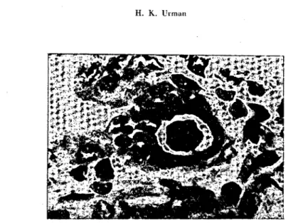
Fig. ı - Haevy periduetal mononuelear eell infiltration. An exIolieted ecU within the duet lumen, and two duet epithelium eeııs wİth intrenuelear inelusion bodies.
H. and E. stain XI25
Fig. 2 - Intranuelear and intraeytoplasmie inelusion oodies in duet eeııs, and peri-duetal mononuelear eel i infiltration.
i
i
i,
H. K. Urrnan
Fig. 3 - Intranudear and intraeytojllasmie incilision bodics in a hypertrophie epithe-!ium eel\.