of corneal epithelium healing time after 2 different surface
ablation methods
Mustafa Eliaçik, MD, Hüseyin Bayramlar, MD, Sevil K. Erdur, MD, Yunus Karabela, MD, Göktuğ Demirci, MD, İbrahim G. Gülkilik, MD, Mustafa Özsütçü, MD.
ABSTRACT -سا يأ( يماملاا يرصبلا ءزجلل يمحلاتلا سارفلما مادختسا :فادهلأا حيحصت دعب يراهظلا ءاشغلا مائتللا مزلالا نمزلا ةنراقلم )يت يس وأ عاطتقاو )كيزلالا( ةيراهظلا ةقبطلا ىلع رزيللا قيبطتب ينعلا ةينرق بدتح .)يك رآ يب( يئوضلا راسكنلاا ةقيرطب ةينرقلا نمضتت ةلسلسلما تلااحلل ةبقترلما ةيلخادتلا ةساردلا هذه :ةقيرطلا مسق يف ةيراسكنلاا رزيللا ةحارج اوقلت نيذلا ًاضيرم 28 نم ًانيع 56 ،لوبنطسا ،لوب يديم ةعماج بطلا ةيلك ىفشتسم يف نويعلا ضارمأ عضخ ضيرم لك .م2014 ويام م2014 سرام ينب ام ةرتفلا يف ،ايكرت يب( ةقيرطب ىرخلاا ينعلاو )كيزلالا( ةقيرطب هينيع ىدحلأ ةيلمعلل ضيرم لك صحف تم .ةيئاوشع ةقيرطب يننيعلا ينب رايتخلاا تم .)يك رآ قرغتسلما نمزلا باسح تمو ،مايأ ةسمخ ةدلمو يرود لكشب ةيلمعلا دعب نودب )يت يس وأ -سا يأ ( ـلا مادختساب ةينرقلل يراهظلا ءاشغلا ءافشل نع جاعزنلاا تاجرد لدعم باسح تم .ةيوادلما ةقصلالا تاسدعلا ةلازا رفصلا نم طاقنب ينعلا عمدت و ءوضلا باهر ،مللأا نع ضيرلما لاؤس قيرط .5 ىلا )دجوي لا ىنعبم( -سا يأ ( ـلا ةقيرطب ساقلما نرهظتلا ةدوعل ينمزلا طسوتلما :جئاتنلا و )يك رآ يب(ـلا ةعومجم يف موي 3.07±0.64 :تناك )يت يس وأ ناك ينتعومجلما ينب قرفلاو )كيزلالا( ـلا ةعومجم يف 3.55±0.54 ناك يعوضولما ريغلا يجاعزنلاا طسوتلما .)0.03 =يب( ًايئاصحإ ًاحضاو 2.85±0.44 و )يك رآ يب( ـلل تعضخ يتلا نويعلا يف 4.42±0.50 تلادعلما .صحفلل لولأا مويلا يف )كيزلالا( ـلل تعضخ يتلا نويعلا يف تناك ثلاثلاو يناثلا ينمويلا يف جاعزنلاا ةجردل اهيلع لوصلحا تم يتلا .)يلاوتلا ىلع 0.03=يب و 0.024 =يب( ،ًايئاصحا ةحضاو ًاضيأ ينتعومجلما ينب ًايئاصحا ةحضاو تاجرد رهظي مل سمالخاو عبارلا ينمويلا .يلاوتلا ىلع )0.1=يب و 0.069=يب( يراهظلا ءاشغلا مائتللا رصقأ انمز )يك رآ يب( ـلا ةقيرط ترهظأ :ةتمالخا جاعزنا ةجرد ترهظأ هسفن تقولا يف هنكلو ًايئاصحإ ًاحضاو ناك يذلا .ةيلمعلا دعب ثلاثلا مويلا ىتح رثكأ
Objectives: To compare epithelial healing time following laser epithelial keratomileusis (LASEK) and photorefractive keratectomy (PRK) with anterior segment optic coherence tomography (AS-OCT).
Methods: This prospective interventional case series study comprised 56 eyes of 28 patients that underwent laser refractive surgery in the Department of Ophthalmology, Medipol University Medical Faculty, Istanbul, Turkey, between March 2014 and May 2014. Each patient was randomized to have one eye operated on with PRK, and the other with LASEK. Patients were examined daily for 5 days, and epithelial healing time was assessed by using AS-OCT without removing therapeutic contact lens (TCL). Average discomfort scores were calculated from ratings obtained from questions regarding pain, photophobia, and lacrimation according to a scale of 0 (none) to 5.
Results: The mean re-epithelialization time assessed with AS-OCT was 3.07±0.64 days in the PRK group, 3.55±0.54 days in the LASEK group, and the difference was statistically significant (p=0.03). Mean subjective discomfort score was 4.42±0.50 in the PRK eyes, and 2.85±0.44 in the LASEK eyes on the first exam day (p=0.001). The score obtained on the second (p=0.024), and third day (p=0.03) were also statistically significant. The fourth (p=0.069), and fifth days scores (p=0.1) showed no statistically significant difference between groups.
Conclusion: The PRK showed a statistically significant shorter epithelial healing time, but had a statistically significant higher discomfort score until the postoperative fourth day compared with LASEK.
Saudi Med J 2015; Vol. 36 (1): 67-72 doi: 10.15537/smj.2015.1.9983 From the Department of Ophthalmology (Eliaçik, Erdur, Karabela, Demirci, Gülkilik,Özsütçü), Medipol University Medical Faculty, and the Department of Ophthalmology (Bayramlar), Medeniyet University Medical Faculty, Istanbul, Turkey.
Received 8th August 2014. Accepted 30th October 2014.
Address correspondence and reprint request to: Dr. Mustafa Eliaçik, Department of Ophthalmology, Medipol Kosuyolu Hospital, Lambaci Sok Koşuyolu Kadiköy 34718, Istanbul, Turkey. Tel. +902 (16) 5446634. E-mail: [email protected]
A
nterior segment optical coherence tomography (AS-OCT) is a valuable, non-invasive and non-contact method to evaluate anterior segment structures quantitatively. It provides high resolution images by using a long wavelength (1.310 nm) of light; it offers rapid quantitative analysis of various structures. AS-OCT has demonstrated good repeatability and reproducibility with low intra-observer and inter-observer variability.1-3 Since laser in situ keratomileusis(LASIK) was introduced in the mid-1990’s, it has become the predominant refractive surgery for the correction of myopia, hyperopia, and astigmatism.4
However, LASIK has unique risks, including problems related to corneal flap creation and iatrogenic ectasia. Despite these factors, where possible, surface ablation procedures still advised by most refractive surgeons. Photorefractive keratectomy (PRK) is one of the surface ablation procedures, performed for treat refractive errors in patients not candidates for LASIK. However, eyes with PRK have some notable adverse effects like severe pain, discomfort and prolonged visual recovery due to the process of epithelial wound healing, which usually starts on the day of surgery and may last up to 4 days after surgery. In 2003, Camellin introduced a new technique called laser-assisted sub-epithelial keratectomy (LASEK) which would minimize discomfort, speed visual recovery time and reduce the incidence of corneal haze with PRK by preserving the epithelium as a flap and replacing it following the laser treatment.5,6 Faster re-epithelialization decreases the risk
factor for postoperative infections and facilitates earlier visual rehabilitation, and reduces patient discomfort. Several studies report conflicting results in epithelial healing time after LASEK versus PRK because the different methods were used. Some comparative studies about epithelial healing time have reported no benefits of LASEK over PRK. There are also some clinical studies have demonstrated controversial results.7-9 The aim of
the current study is to document the healing process of the cornea after LASEK and PRK quantitatively using a non-invasive, non-contact, and a more sophisticated method AS-OCT without removing therapeutic contact lenses (TCL).
Methods. This was a prospective, randomized, single-center study. All surgeries were performed by one surgeon. Approval from the Institutional Review Board
of Medipol University was obtained for the study. Twenty-eight patients (56 eyes) participated in this study conducted at Medipol University of Medicine Eye Clinic, Istanbul, Turkey between March 2014 and May 2014. Patients were included if they were at least 18 years old, and had spherical equivalent manifest refraction between -1.00 and -5.00 diopters (D), and astigmatism less than -2.00 D, stable refraction at least 12 months before surgery, and a minimum follow-up of 3 months. No one had signs of keratoconus, uncontrolled glaucoma, untreated retinal abnormalities, or previous intraocular or corneal surgery. The exclusion criteria were; older than 40 years, diabetes, and history of herpetic keratitis, and previous intraocular, or corneal surgery, pregnancy, nursing, collagen vascular diseases, or dry eye. Pre-operative assessments included uncorrected and best corrected visual acuity, cycloplegic refraction, pachymetry with AS-OCT, examination of cornea with Orbscan and slitlamp biomicroscopic examination of both anterior and posterior segments. Tear function was assessed using a Schirmer I test and the tear film breakup time (TBUT) was determined. Soft contact lens was discontinued for a minimum of 7 days before examination and treatment. Contralateral eyes in each patient were subject to random allocation through which, PRK surgery was carried out on one eye, and LASEK was carried out for the other eye of same patient by the same surgeon. All patients agreed to participate and gave their informed consent forms after the nature of the procedure had been explained. This study was conducted according to the declaration of Helsinki and relevant laws/regulations.
Surgical procedures. All procedures were performed with the VISX Star S4 excimer laser using a standard protocol by the same surgeon. The aim of the surgery was emmetropia. Preoperatively 10% povidone-iodine was used to clean the eyelids and periocular area for 1 minute, and then the eye was washed out with 20 ml of a balanced salt solution. All treatments were performed using topical anesthesia with one drop of 0.5% proparacaine (Alcaine, S.A. Alcon-Couvreur, Puurs, Belgium). A closed-loop lid speculum is used to retract the lids of the eye to be treated, and the other eye was occluded.
The PRK. A 7.0-mm optical zone marker was
applied to the cornea, centered over the entrance pupil. The epithelial cell layer of the cornea was debrided with a crescent knife and then photoablation was performed on the corneal stroma. The corneal surface and entire conjunctiva were irrigated with a balanced salt solution and the excess fluid was removed with another cellulose sponge.
Disclosure. Authors have no conflict of interests, and the
The LASEK. An alcohol solution cone (J2908, Janach, Como, Italy) with a 10 mm diameter was applied to the corneal surface, and a 20% ethyl alcohol solution diluted with distilled water was filled inside the cone and was left for approximately 40 seconds. At the end of the time a merocel sponge (Medtronic Ophthalmics Inc, Jacksonville, FL) was used to soak up the alcohol in the cone and the entire ocular surface was carefully washed off with a balanced salt solution. An epithelial microhoe (Janach J2916A) was used to detach the flap edge and then the flap was shifted as one intact sheet toward the 12:00 position using an epithelial flap peeler (Janach J2930A). After the denuded corneal surface was ablated, the epithelial flap was washed with balanced salt solution and gently repositioned with using a spatula (Janach J2920A). For all treatment groups, at the end of surgery a therapeutic contact lens (TCL) (Purevision; base curve of 8.3 mm, 0 diopter (D), diameter of 14 mm; Bausch Lomb, Rochester, NY) was fitted to the treated eye with contact lens applanator (Janach J2935) after one drop of both 0.3% ciprofloxacin and 0.1% diclofenac were instilled on the surgical site.
Postoperative Medication: All patients were examined daily during 5 days follow up. Postoperative treatment included topical diclofenac and tobramycin drops 4 times daily during the reepithelization period. A preservative-free sodium hyaluronate 0.1% drops were applied every hour during first 48 hours postoperatively and 4 times in a day for 1 month. After re-epithelialization, 0.3% tobramycin (Tobrex, Alcon-Couvreur, Puurs, Belgium) and 0.1% deksametazon (Maxidex ,Alcon-Couvreur, Puurs, Belgium) were administered four times daily for 10 days and twice in a day for 15 days.
Postoperative examinations. All patients were examined every day for 5 days, then at the 10th day, and 1, and 3 months. During first 5 days, specifically epithelial healing was followed up. Epithelial wound healing was assessed using Visante OCT Anterior Segment Imaging (Carl Zeiss Meditec, Inc, Berlin, Germany) by imaging the cornea with TCL in-situ. OCT images taken immediately after surgery and then every 24 hours until complete epithelial healing had occurred. Horizontal and vertical radial images through the central cornea were obtained for each eye on central fixation position to show sections of migrating epithelial tissue. The TCLs were removed when complete epithelialization was determined. All patients were interviewed in standardized conditions with prior information to ensure valid reliable responses. Pain, photophobia, and lacrimation were each given a score from 1 to 5. Patients were asked for each complaint separately during 5 days thereafter surgery until
complete healing had occurred. At the 10th day, 1 and 3 months after surgery, uncorrected and best spectacle-corrected visual acuity, manifest refraction, slit-lamp microscopy, corneal topography with Orbscan were performed by one investigator. Subepithelial stromal haze was evaluated with the slit lamp microscope using broad tangential illumination, and graded as 0 to 4+ by 2 authors at different times, and different examination rooms. In our study, each patient served as him or her own control subject.
The Statistical Package for Social Sciences software version 17 (SPSS Inc, Chicago, IL, USA) was used for statistical analysis. The normality of the distribution of each of the parameters was checked using the normal Kolmogorov-Smirnov test. Visual acuity data were converted from Snellen chart values to LogMAR notation for statistical analysis. The Mann-Whitney U test was used to compare discomfort scores and reepithelization, and chi-squared test was used postoperative uncorrected distance vision. The significance level was set to α = 0.95 and p<0.05 was considered significant.
Results. The average age of the patients was 26.39 ± 4.99 years (range: 18-34 years). There were 16 men (57.1%) and 12 women (42.9%). Follow-up rates were 100% up to 3 months.
Epithelial healing and haze. The time required for reepithelialization was 3.07 ± 0.64 days in the PRK group, and 3.55 ± 0.54 days in the LASEK group, and the difference was statistically significant (p=0.03) (Figures 1-3). Up to 3 months postoperatively, subepithelial haze was graded separately by 2 masked practitioners. There was no difference in the subepithelial opacity between the 2 groups (p=0.244). Almost all patients showed a subepithelial opacity below, or equal to grade 1. However only one eye in the PRK group showed opacity grade 2.
Discomfort score. Mean subjective discomfort
score was 4.42 ± 0.50 in PRK eyes, and 2.85 ± 0.44 in LASEK eyes on the first exam the day after surgery. In the PRK eyes, there were no significant decrease in pain until third day, and in LASEK eyes discomfort scores were stable during this session. At the fourth day both groups’ scores were nearly same (PRK eyes: 2.1 ± 0.42, LASEK eyes: 1.67 ± 0.6). After the third day, no statistical differences were found between these groups (Figure 4).
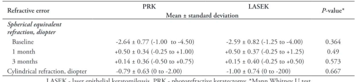
Refractive outcomes. The mean preoperative manifest spherical equivalent refraction and refractive cylinder in the eyes that received each of the procedures are shown in Table 1. Baseline spherical equivalent refraction and refractive cylinder were not statistically different in both groups (p=0.364 [PRK], and p=0.667 [LASEK]). The
Figure 1 - Optical coherence tomography images from patient No. 12’s
right eye (laser-assisted sub-epithelial keratectomy [LASEK]) showing: A) at the first hour of surgery over central cornea vertex, showing LASEK flap under therapeutic contact lenses (TCL); B) first post-operative day over temporal limbus, showing migrating epithelial layer under TCL (arrow); C) second post-operative day over central corneal vertex; and D) fourth post-operative day, showing complete epithelial layer under TCL.
Figure 3 - Images from eye of patient no. 12. Fourth postoperative day
after removing therapeutic contact lenses (TCL) and staining with florescein sodium. A, B) laser-assisted sub-epithelial keratectomy (LASEK) eye, and C, D) photorefractive keratectomy (PRK) eye.
Figure 4 - Mean discomfort scores after photorefractive keratectomy
(PRK) and laser subepithelial keratomileusis (LASEK).
Figure 2 - Optical coherence tomography images from patient No. 12’s
left eye (photorefractive keratectomy [PRK]) showing: A) at the first hour of surgeryover central cornea vertex, showing absence of epithelial layer and edges of epithelial edges under therapeutic contact lenses (TCL); B) first post-operative day over temporal limbus, showing edge of epithelial layer (arrow);
C) second post-operative day over showing edges of epithelial layer (arrows); and D) third post-operative day, showing complete epithelial layer under TCL.
mean spherical equivalent refractions of both groups at 1 and 3 months after surgery are summarized in Table 1. There were no statistically significant differences between refraction in the 2 groups at the mentioned visits (p=0.490 [PRK], and p=0.573 [LASEK]).
Visual outcomes. Baseline uncorrected visual acuity and best spectacle-corrected visual acuity were not statistically different among the eyes that received each of the procedures. The number and percent of eyes with uncorrected visual acuity at the first week, 1, and 3 months after surgery are shown in Table 2. There were
no statistically significant differences in uncorrected visual acuity after LASEK and PRK during follow-up. All patients had 20/20 visual acuity at the end of the examinations; no eyes needed spectacle-correction for better visual acuity.
Discussion. Anterior segment optical tomography has become a widely used diagnostic technique for examination of the cornea since 2006.10,11 The
AS-OCT is applicable to measure and visualize the anterior chamber, cornea and surrounding areas. It provides sensitive and detailed observation, and quantitative measurements of the changes. It has several advantages over other techniques. It is a light-based system that rapidly provides high-resolution images. Its non-contact nature ensures patient comfort and allows for rapid image acquisition in the sitting position. It also allows quantitative and dynamic data analysis with high reproducibility and repeatability.12 The present
study demonstrated progression of epithelial healing in situ in patients with TCL lenses following PRK and
LASEK as measured quantitatively by AS-OCT. The second aim of this paper was to compare the primary (best corrected vision, manifest refractive spherical equivalent) and secondary outcomes (pain, haze) after 2 different surgical procedures.
Although evaluation of the epithelial healing process with using OCT is not a new idea, in this study the effects of 2 different surface ablation techniques were compared quantitatively by AS-OCT.10,11 We
attempted to verify previous studies’ preliminary results by changing methods and using AS-OCT. In previous studies, the size of the remaining epithelial defect was visualized with the use of fluorescein, which is generally contraindicated in the presence of a TCL due to lens staining. The TCL removal is thus, required to enable fluorescein staining to assess the epithelium, but carries
the risk of traumatizing new epithelium, which is easily detachable during the early phase of healing, and which may further delay full epithelialization. A non-invasive method for monitoring epithelium healing under the TCL will be useful to detect the appropriate time point for TCL removal.
Surface ablation techniques have become increasingly important for low to moderate myopia since LASIK complications like flap creating and managing problems and corneal ectasia. Refractive surgeons need to find out the answer of which surface ablation technique is better than the others. Since mid-2000’s, studies have been conducted to find this answer become popular. The published literature as to which method is the best is somewhat confusing.No statistically significant difference was noted regarding the primary and secondary outcomes of LASEK and PRK by 2 meta-analysis conducted between 2008 and 2010.7,9 Similary
Ghoreishi et al13 found same results after comparing
alcohol-assisted versus mechanical epithelium removal in PRK in 1,250 eyes. In other studies, Epi-LASIK and Transepithelial PRK seemed more preferable methods when they comparing conventional techniques as LASEK and PRK.4-6,14-16 In contrast to these findings. Ghamen
reported a statistically significant shorter epithelial healing process after PRK than butterfly LASEK.8,17
Recently, Einollahi18 demonstrated the adverse effects
of mechanical epithelial debridement on anterior keratocyte density and anterior stromal reflectivity using confocal microscopy. They also recommended alcohol-assisted epithelial debridement as the procedure of choice for epithelial removal in patients with mild myopia. The results of these studies, while conflicting, all report excellent visual and refractive outcomes with surface ablation techniques. The first step and common feature of all these techniques is removing corneal epithelium over ablation area using different methods. The corneal epithelium is highly resistant to pathogen penetration and epithelium tries to restore its protective barrier as quickly and efficiently as possible after surface refractive procedures. Faster reepithelialization provides
Table 1 - Refractions before and after LASEK and PRK in 28 patients (56 eyes).
Refractive error PRK LASEK P-value*
Mean ± standard deviation
Spherical equivalent refraction, diopter
Baseline -2.64 ± 0.77 (-1.00 to -4.50) -2.59 ± 0.82 (-1.25 to -4.00) 0.364 1 month +0.50 ± 0.34 (-0.25 to +1.00) +0.50 ± 0.37 (-0.25 to +1.25) 0.49 3 months +0.14 ± 0.36 (-0.50 to +0.75) +0.15 ± 0.40 (-0.25 to +0.50) 0.573 Cylindrical refraction, diopter -0.79 ± 0.63 (0 to -2.00) -1.00 ± 0.74 (0 to -200) 0.667
LASEK - laser epithelial keratomileusis, PRK - photorefractive keratectomy, *Mann Whitney U test
Table 2 - Refraction and visual acuity changes during follow-up.
Refractive error PRK LASEK P-value*
n (%)
Spherical equivalent refraction after surgery within ± 0.50 diopter
1 month 17 (63.0) 18 (67.0) >0.05 3 months 26 (92.0) 25 (89.0) >0.05
Spherical equivalent refraction after surgery within ± 1.00 diopter
1 month 27 (96.0) 28 (100) >0.05 3 months 28 (100) 28 (100) >0.05
Uncorrected visual acuity ≥20/40 1 week 22 (78.0) 24 (86.0) >0.05 1 month 28 (100) 28 (100) >0.05 3 months 28 (100) 28 (100) >0.05 ≥20/20 1 week 10 (36.0) 12 (43.0) >0.05 1 month 22 (78.0) 23 (82.0) >0.05 3 months 28 (100) 28 (100) >0.05
PRK - photorefractive keratectomy, LASEK - laser epithelial keratomileusis, UCVA - uncorrected visual acuity. *Chi-squared test
an earlier visual rehabilitation, reduction of discomfort and quicker restoration of barrier.
Since LASEK was introduced mid-1990s, 20% alcohol is commonly used to create an epithelial flap during this surgical procedure.4 In 2012, Zhang et al19
demonstrated ocular toxicity of 18-20% alcohol over corneal epithelial cells. Due to their study, alcohol could delay corneal epithelium restoration after surgery during the early postoperative period by slowing down the establishment of epithelial tight junctions. Likewise in this study, we found the mean epithelial healing time shorter in PRK eyes than LASEK eyes. Similar to other studies, we found no statistically significant difference between compared techniques in uncorrected visual acuity and haze 3 months after surgeries, but mean discomfort scores was higher in PRK group, and it was statistically significant.
All researchers have simply quantified epithelial defect by using slit-biomicroscopic evaluating and using fluorescein staining. The time of TCLs removal was decided when there was no observable remaining epithelial defect. There was no additional explanation about iatrogenic trauma induced by actions to assess the wound area. Also the debris on contact lenses could affect the objectivity of investigators’ observation on corneal wound area. As a result of this, determination have arisen doubts about the objectivity of the all the results that were indicated by previous studies. The question to be asked is if delays in tight junction formation make corneal epithelium more vulnerable to iatrogenic trauma achieves by TCLs removing. We controlled the factors that could affect the objectivity of this study by changing methods and using AS-OCT.
In summary, AS-OCT demonstrates the ability to monitor the corneal epithelial healing under a TCL and provides information of clinical value regarding the healing process after surface ablation techniques. This technology has revolutionized our ability to examine and will eventually useful than more of the applications currently performed with other technologies. Major limitation of this study is small number of cases. Furthermore, randomized clinical trials including more cases using AS-OCT are necessary to investigate the differences between surface ablation techniques to provide the right choice for patients.
References
1. Nolan WP, See JL, Aung T, Friedman DS, Chan YH, Smith SD, et al. Changes in angle configuration after phacoemulsification measured by anterior segment optical coherence tomography. J Glaucoma 2008; 17: 455-459.
2. Nolan W. Anterior segment imaging: ultrasound biomicroscopy and anterior segment optical coherence tomography. Curr Opin Ophthalmol 2008; 19: 115-121.
3. Nolan W. Anterior segment imaging: identifying the landmarks. Br J Ophthalmol 2008; 92: 1575-1576.
4. O’Brart DP. Excimer laser surface ablation: a review of recent literature. Clin Exp Optom 2014; 97: 12-27.
5. Huang PY, Huang PT, Astle WF, Ingram AD, Hebert A, Huang J, et al. Laser-assisted subepithelial keratectomy and photorefractive keratectomy for post-penetrating keratoplasty myopia and astigmatism in adults. J Cataract Refract Surg 2011; 37: 335-340.
6. Reilly CD, Panday V, Lazos V, Mittelstaedt BR. PRK vs LASEK vs Epi-LASIK: a comparison of corneal haze, postoperative pain and visual recovery in moderate to high myopia. Nepal J Ophthalmol 2010; 2: 97-104.
7. Zhao LQ, Wei RL, Cheng JW, Li Y, Cai JP, Ma XY. Meta-analysis: clinical outcomes of laser-assisted subepithelial keratectomy and photorefractive keratectomy in myopia. Ophthalmology 2010; 117: 1912-1922.
8. Ghanem VC, Kara-Jose N, Ghanem RC, Coral SA. Photorefractive keratectomy and butterfly laser epithelial keratomileusis: a prospective, contralateral study. J Refract Surg 2008; 24: 671-684.
9. Cui M, Chen XM, Lu P. Comparison of laser epithelial keratomileusis and photorefractive keratectomy for the correction of myopia: a meta-analysis. Chin Med J (Engl) 2008; 121: 2331-2335.
10. Kaluzny BJ, Szkulmowski M, Bukowska DM, Wojtkowski M. Spectral OCT with speckle contrast reduction for evaluation of the healing process after PRK and transepithelial PRK. Biomed Opt Express 2014; 5: 1089-1098.
11. Pang CE, M V, Tan DT, Mehta JS. Evaluation of Corneal Epithelial Healing Under Contact Lens with Spectral-Domain Anterior Segment Optical Coherence Tomography (SD-OCT). Open Ophthalmol J 2011; 5: 51-54.
12. Kim M, Park KH, Kim TW, Kim DM. Changes in anterior chamber configuration after cataract surgery as measured by anterior segment optical coherence tomography. Korean J Ophthalmol 2011; 25: 77-83.
13. Ghoreishi M, Attarzadeh H, Tavakoli M, Moini HA, Zandi A, Masjedi A, et al. Alcohol-assisted versus Mechanical Epithelium Removal in Photorefractive Keratectomy. J Ophthalmic Vis Res 2010; 5: 223-227.
14. Sia RK, Ryan DS, Edwards JD, Stutzman RD, Bower KS. The U.S. Army Surface Ablation Study: comparison of PRK, MMC-PRK, and LASEK in moderate to high myopia. J Refract Surg 2014; 30: 256-264.
15. Korkmaz S, Bilgihan K, Sul S, Hondur A. A Clinical and Confocal Microscopic Comparison of Transepithelial PRK and LASEK for Myopia. J Ophthalmol 2014; 2014: 784185. 16. Celik U, Bozkurt E, Celik B, Demirok A, Yilmaz OF. Pain,
wound healing and refractive comparison of mechanical and transepithelial debridement in photorefractive keratectomy for myopia: Results of 1 year follow-up. Cont Lens Anterior Eye 2014; 37: 420-426.
17. Ghanem VC, Souza GC, Souza DC, Viese JM, Weber SL, Kara-Jose N. PRK and butterfly LASEK: prospective, randomized, contralateral eye comparison of epithelial healing and ocular discomfort. J Refract Surg 2008; 24: 591-599. 18. Einollahi B, Baradaran-Rafii A, Rezaei-Kanavi M, Eslani M,
Parchegani MR, Zare M, et al. Mechanical versus alcohol-assisted epithelial debridement during photorefractive keratectomy: a confocal microscopic clinical trial. J Refract Surg 2011; 27: 887-893.
19. Zhang P, Liu M, Liao R. Toxic effect of using twenty percent alcohol on corneal epithelial tight junctions during LASEK.Mol Med Rep 2012; 6: 33-38.
![Figure 1 - Optical coherence tomography images from patient No. 12’s right eye (laser-assisted sub-epithelial keratectomy [LASEK]) showing: A) at the first hour of surgery over central cornea vertex, showing LASEK flap under therapeutic contact lenses](https://thumb-eu.123doks.com/thumbv2/9libnet/5425169.103342/4.955.494.832.119.333/figure-optical-coherence-tomography-assisted-epithelial-keratectomy-therapeutic.webp)