Radyoloji / Radiology
OLGU SUNUMU / CASE REPORT
https://doi.org/10.31067/0.2019.110
ACU Sağlık Bil Derg 2019; 10(1):101-103
101
Correspondence:
Cenk Ersavaş
Medipol University School of Medicine, Department of General Surgery, Istanbul, Turkey Phone: +90 505 314 53 08 E-mail: [email protected] Received : March 17, 2017 Revised : April 26, 2017 Accepted : May 01, 2017
1Medipol University School of Medicine, Radiology, Istanbul, Turkey 2Medipol University School of Medicine, Department of General Surgery, Istanbul, Turkey 3Acıbadem University School of Medicine, Department of Radiology, Istanbul, Turkey
Gülhan Ertan Akan Cengiz Erol Cenk Ersavaş Sıla Ulus
A Case of Splenic Artery Aneurysm
Presenting With İntra-Abdominal
Bleeding
Gülhan Ertan Akan
1, Cengiz Erol
1, Cenk Ersavaş
2, Sıla Ulus
3İNTRABDOMINAL KANAMA İLE ORTAYA ÇIKAN BİR SPLENİK ARTER ANEVRİZMASI OLGUSU ÖZET
Visseral arter anevrizmaları nadirdir. Splenik arter anevrizmaları en sık görülen visseral arter anevrizmalarıdır. Rüp-tür ve ölümcül kanama riski taşırlar. Tromboze olduklarında görüntüleme metodları ile gözden kaçabilirler. Bu yazıda batın içi kanama ile prezente olan, tromboze olması nedeniyle ancak tekrarlayan bilgisayarlı tomografi (BT) incele-melerde saptanabilen ve anjiografik incelemede yine tromboze olması nedeniyle tespit edilemeyen splenik arter anevrizmasının, BT ile korele olarak körlemesine embolizasyon ile tedavi edilmesini sunmayı amaçladık.
Anahtar sözcükler: Splenik arter anevrizması, tromboz, rüptür, bilgisayarlı tomografi ABSTRACT
Visceral artery aneurysms are rare pathologies. Splenic Artery Aneurysms (SAA) are the most common visceral artery aneurysms accounting for 60% to71% of them, with an incidence of 0.7% in the normal population, even though rates as high as 10% have been found on necropsy studies. This aneurysm has a risk of rupture resulting in death. It can be overlooked (missed) on the imaging modalities when it is thrombosed. Here, we present a case of splenic artery aneurysm, which could be diagnosed with repetitive computed tomography (CT) examinations because it was partially thrombosed and overlooked in the 1st CT. The aneurysm was treated with blind embolization in correlation with CT.
Keywords: Splenic artery aneurysm, thrombosis, rupture, computed tomography
V
isceral artery aneurysms are usually rare pathologies, and among these Splenic Artery Aneurysms (SAA) They are the most common (1,2). They are usually asymptomatic.; however, due to the risk of rupture and fatal bleeding, early diagnosis and treatment is essential. Typical clinical presentation of a ruptured SAA includes abdominal pain and hemodynamic instability. Here, we present a case of splenic artery aneurysm, which could have been diagnosed with repetitive computed tomography (CT) examinations because due to the fact that it was thrombosed. The aneurysm was treated with blind embolization in correlation with CT.Intraabdominal Bleeding Due to Splenic Artery Aneurysm
102 ACU Sağlık Bil Derg 2019; 10(1):101-103
Case report
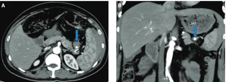
A previously healthy 38-year-old male presented to the emergency department with left sided abdominal pain. No significant past history of medical illness was stated. The patient had a pulse rate of 125/min. and blood pres-sure of 80/65 mm Hg. Hemoglobin level was 10 gm/dL, and liver function tests were normal. Ultrasonography (US) examination revealed free fluid in the abdomen. Triphasic, contrast enhanced multidedector multidetec-tor CT (MDCT) of abdomen and pelvis was performed with a 256 slice scanner (Brilliance iCT; Philips Healthcare, Cleveland, OH). In that examination, the free abdominal fluid had high density and was accepted as hemoperito-neum. No source of bleeding could have been identified with CT. The patient was treated with Ringer’s lactate solution (2000 cc) and iron preparations (100 mg/per day) intravenously, for 3 days. No significant decrease was not-ed of the Hbg levels in the 3 day-period. After treatment, blood pressure and pulse rate were in the normal levels. The follow-up abdominal US was also normal. The patient was discharged from the hospital since his condition was stable. 15 days later, the patient presented with left sided abdominal pain once more. Triphasic, contrast enhanced CT examination was repeated and in this 2nd examina-tion, a partially thrombosed pseudoaneurysm was noted in the distal segment of the lower pole splenic artery. It was 2 cm in diameter; the contrast filled part of the aneu-rysm was 7 mm and the remaining part was thrombosed (Figure 1 a-b). There was also high density intra-abdom-inal free fluid. When the previous CT was evaluated ret-rospectively, a totally thrombosed small aneurysm could barely have been perceptible in this localization. It was assumed that the imaging findings became clearer or distinct because of recurrent thrombosis. 2 days after the
2nd CT scan, celiac artery and selective splenic artery an-giography was performed. The aneurysm could not have been visualized since it was totally thrombosed again. The polar artery feeding the lower pole of the splenic ar-tery was embolised with glue injection blindly, with the aid of a microcatheter, in correlation with the location in CT using CT to locate the aneurysm. 3 days later, control abdominal CT-Angiography (CTA) revealed that the low-er pole splenic artlow-ery and its branches wlow-ere embolized and the pseudoaneurysm was totally thrombosed. Infarct sites were noted in the lower pole of the spleen due to the procedure. Intra-abdominal bleeding had regressed remarkably. Follow-up in 1 year revealed no aneurysm re-occurrence.
Discussion
Visceral artery aneurysms are rare; however, they are im-portant to recognize due to the possibility of rupture. Among these, SAA are the most common one (1,2). It has an incidence rate of 0.7%,; however, on necropsy studies rates are as high as 10% have been reported (3). Atherosclerosis, portal hypertension, chronic liver disease, ischemia, and fibromuscular dysplasia are included in the risk factors (2,4). Asymptomatic cases can be detected in-cidentally during diagnostic imaging performed for other indications (5). The most important complication is rup-ture. It can be asymptomatic and it can be the only and initial symptom. The reported rate of rupture is 2–9%, with an associated mortality of 36% (6). The risk for rup-ture reaches 50% in pregnant patients. When the diam-eter of the aneurysm exceeds 2 cm, likelihood of a rupture increases (7). The physical signs of a rupture most often include sudden and intense abdominal pain, most com-monly in the left upper quadrant or epigastrium. Nausea
A
B
Ertan Akan G et al.
103
ACU Sağlık Bil Derg 2019; 10(1):101-103
References
1. Abbas MA, Stone WM, Fowl RJ, Gloviczki P, Oldenburg WA, Pairolero PC, et al. Splenic artery aneurysms: two decades experience at Mayo clinic. Ann Vasc Surg 2002;16:442–9. [CrossRef]
2. Trastek VF, Pairolero PC, Bernatz PE. Splenic artery aneurysms. World J Surg 1985;9:378–83. [CrossRef]
3. Goshayeshi L, Vosoghinia H, Rajabzadeh F, Ahadi M, Asadi Sakhmaresi T, Farzanehfar MR. Splenic artery aneurysm as an unusual cause of new onset ascites: a case report. Middle East J Dig Dis 2014;6:37–41. 4. Mentzel HJ, Seidel J, Vogt S, Vogt L, Kaiser WA. Vascular complications (splenic and hepatic artery aneurysms) in the occipital horn syndrome: report of a patient and review of the literature. Pediatr Radiol 1999;29:19–22. [CrossRef]
and vomiting can also accompany the pain. In our case, the patient presented to the emergency department with left upper quadrant abdominal pain. It is possible to estab-lish a diagnosis with US, CT, and angiography. However, as in our case, it can be difficult to diagnose if the aneurysm is thrombosed.
It is not recommended to treat small and asymptomatic aneurysms. Treatment options for aneurysms with a di-ameter larger than 2 cm and all symptomatic cases are
surgical interventions, transcutaneous embolization or laparoscopic ligation (8).
In conclusion, splenic artery aneurysm rupture is a chal-lenging diagnosis and needs to be considered in patients presenting with abdominal pain and intra-abdominal bleeding. The need for repetitive and multi-modality ra-diologic work-up should be in mind on account of throm-bosis. Blind embolization can be used in correlation with CT in thrombosed aneurysms.
5. Dolar E, Uslusoy H, Kiyici M, Gurel S, Nak SG, Gulten M, et al. Rupture of the splenic arterial aneurysm due to Behcet’s disease. Rheumatology 2005;44:1327–28. [CrossRef]
6. Messina LM, Shanley CJ. Visceral artery aneurysms. Surg Clin North Am 1997;77:425–42. [CrossRef]
7. Pescarus R, Montreuil B, Bendavid Y. Giant splenic artery aneurysms: case report and review of the literature. J Vasc Surg 2005;42:344–7.
[CrossRef]
8. Merlo M, Cumino A, Pecchio A, Carignano G, Mioli PR, Cassolino P. Splenic artery aneurysm. Two successfully operated cases. Minerva Cardioangiol 1998;46:123–6.