References
1. Kolvekar SK, Chaubey S, Firmin R. Floating thrombus in the aorta. Ann Thorac Surg 2001; 72: 925-7.
2. Berneder S, van Ingen G, Eigel P. Arch thrombus formation in an apparently normal aorta as a source for recurrent peripheral embolization. Thorac Cardiovasc Surg. 2006; 54: 548-9.
3. Choi JB, Choi SH, Kim NH, Jeong JW. Floating thrombus in the proximal aortic arch. Tex Heart Inst J 2004; 31: 432-4.
4. Qanadli SD, Rangheard AS, Lacombe P. Intraluminal thrombus of the ascending aorta with systemic embolism detected by spiral CT. Clin Radiol 2000; 55: 75-7.
5. Laperche T, Laurian C, Roudaut R, Steg PG. Mobile thromboses of the aortic arch without aortic debris. A transesophageal echocardiographic finding associated with unexplained arterial embolism. Circulation 1997; 96: 288-94.
Address for Correspondence: Dr. Bilgin Emrecan, Yunusemre Mah. 6440 sok No 7/3 K›n›kl› 20070 Denizli, Turkey
Phone: +90 505 488 99 16 Fax: +90 258 213 49 22 E-mail: [email protected]
Thrombosed giant right
coronary artery aneurysm
Tromboze olmufl dev sa¤ koroner arter anevrizmas›
A 50-year-old male patient, without previous history, presented with prolonged precordial pain following moderate exercise. He was transferred to a local hospital, where acute coronary syndrome (ACS) was diagnosed and was treated with conventional (aspirin,
β-blockers, nitrates, heparin) treatment. His recovery was uncomplicated and he left the intensive care unit 48 hours later. He was discharged from the hospital 7 days later and the following treatment was prescribed: β-blocker, angiotensin converting enzyme inhibitor, statin, and aspirin. The patient was a smoker (60 packs per year), presented dyslipidemia and had no positive family history of coronary artery disease. After his release from the hospital, he was admitted to our hospital for cardiac catheterization without any symptoms. He was hospitalized as planned, two weeks following the ACS and underwent coronary angiography.
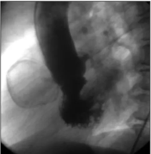
Coronary angiography showed normal left main coronary artery, left anterior descending artery and left circumflex artery. The right coronary artery (RCA) was totally occluded, with a mass at proximal segment (Fig. 1, 2. Video 1, 2. See corresponding video/movie images at www.anakarder.com). Transthoracic and transesophageal echocardiography showed no abnormalities of cardiac valves or con-tractile function and the ascending aorta as well.
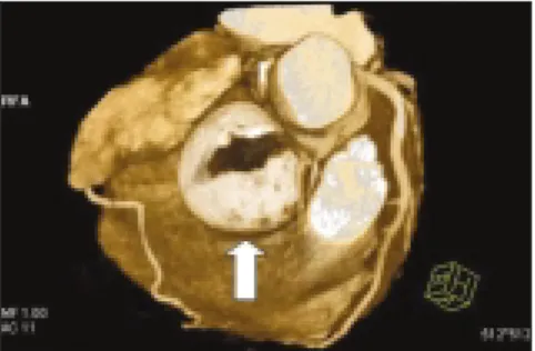
The patient was then transferred to radiodiagnostic unit to investigate this mass with multislice computed tomography (MSCT) (Fig. 3. Video 3. See corresponding video/movie images at www.anakarder.com) and cardiac magnetic resonance imaging (MRI) (Fig. 4. Video 4. See corresponding video/movie images at www.anakarder.com). Blood examination and chest X-ray showed no significant pathological findings. For certain diagnosis and treatment, surgical resection of the mass was our preferred treatment modality, with the agreement of cardiovascular surgery department. As the patient refused the surgical intervention; a course of conservative treatment was decided upon and it was recommended that the patient follows a pharmaceutical treatment. Follow-up MSCT was planned at the end of six months and the patient was discharged.
Coronary artery aneurysm is an abnormal dilatation of focal or diffuse segments of coronary artery. The incidence of coronary aneurysm among coronary artery disease is about 1.5% to 5% (1-3). It may be congenital, or secondary to other diseases, such as atherosclerosis, trauma, previous coronary intervention, mycotic emboli, Kawasaki's disease or systemic lupus erythematosus (4). In this case, our patient presented with ACS. Coronary angiography displayed total occlusion of the RCA and a giant aneurysm. Abnormal flow pattern in the aneurysm may lead to thrombus formation with
Anadolu Kardiyol Derg 2007; 7: 348-57 Orijinal Görüntüler
Original Images
350
Figure 2. View of evacuated aortic thrombi
Figure 1. Selective right coronary angiography shows that the right coronary artery is totally occluded from the beginning and a residual trace of contrast agent in the entrance of giant aneurism
Figure 2. Right anterior oblique view of coronary angiogram shows a giant right coronary artery aneurysm at the proximal portion
subsequent vessel occlusion, distal thromboembolization or even myocardial infarction (4). The aneurysm may also present as an intracardiac mass once it is thrombosed (5). New imaging techniques as MSCT and MRI are going to be well-established and widely used methods to evaluate such abnormalities nowadays.
Hüseyin Çelebi, Cihan Duran*, Alp Burak Çatakoglu, Murat Gülbaran**, Cemflid Demiroglu, Vedat Aytekin** From Departments of Cardiology and *Radiology,
Florence Nightingale Hospital **Department of Cardiology, Faculty of Medicine, ‹stanbul Bilim University -
Florence Nightingale Hospital, ‹stanbul, Turkey
References
1. Robinson FC. Aneurysms of the coronary arteries. Am Heart J 1985; 109: 129-35.
2. Syed M, Lesch M. Coronary artery aneurysm: a review. Prog Cardiovasc Dis 1997; 40: 77-84.
3. Robertson T, Fisher L. Prognostic significance of coronary artery aneurysm and ectasia in the Coronary Artery Surgery Study (CASS) registry. In: Shulman ST, editor. Kawasaki disease. Proceedings of the second international symposium. New York: A.R. Liss; 1987. p.325-39.
4. Bruce F, Waller. Non-atherosclerotic coronary heart disease. In: Fuster V, Alexander RW, Rourke RA, editors. Hurst's the Heart. 10th ed. Philadelphia: Mcgraw-Hill Book Company; 2001. p.1168-70.
5. Otsuka M, Minami S, Hato K, Suto Y, Kajiwara K, Yamagishi H, et al. Acute myocardial infarction caused by thrombotic occlusion of a coronary artery aneurysm. Cathet Cardiovasc Diagn 1997; 41: 423-5. Address for Correspondence: Prof. Dr. Vedat Aytekin, ‹stanbul Bilim Üniversitesi T›p Fakültesi-Florence Nightingale Hastanesi, Kardiyoloji AD,
Abide-i Hürriyet Cad.No:290 fiiflli/‹stanbul 80220, Türkiye
Fax: 296 52 22 Gsm: 0532 236 84 87 E-mail: [email protected]
Multiple and bilateral coronary
fistulas resulting in myocardial
ischemia due to significant stealing of
coronary artery blood flow
Koroner arterden önemli miktarda kan çal›nmas›
sonucu miyokard iskemisine
neden olan iki tarafl› ve çoklu koroner fistül
A coronary artery fistula is a direct communication between a coronary artery and one of the cardiac chambers or vessels around the heart. The incidence of congenital coronary artery fistulas was reported to be 0.08% in Turkish adults who underwent diagnostic cardiac angiography (1). If myocardial ischemia is documented in case of coronary fistulas, one of the following therapeutic options should be chosen: surgical procedure involving closure of the openings of the fistulas from inside the pulmonary trunk, covered stent implantation and percutaneous transluminal embolisation involving closure of the fistulas from the proximal portion or mid-portion (2-4).
Anadolu Kardiyol Derg 2007; 7: 348-57
Orijinal Görüntüler
Original Images
351
Figure 4. Coronal T2-weighted image of the thorax demonstrates the large spherical mass
Figure 3. Multislice computed tomography demon-strates a giant aneurysm strongly suspected to be originated from right coronary artery. A three dimensional reconstruction (using volume render-ing techniques or VRT) shows the lumen of the aneurysm incompletely filled with mural thrombus.
Figure 1. Two small coronary-pulmonary fistulas from the left main coronary artery and one coronary-pulmonary fistula from the left anterior descending artery were detected on coronary angiography (arrows)
Figure 2. Two coronary fistulas originated from the proximal and distal portion of the right coronary artery (RCA) are seen on coronary angiography (arrows). The fistula arose from the proximal RCA drains in to the pulmonary artery like fistulas seen in Figure 1