DOI: 10.5455/annalsmedres.2019.11.730 2020;27(1):312-8
Allergic sensitization in patients with pruritus sine materia
in the absence of a somatic cause
Erkin Pekmezci
Istanbul Medipol University, Faculty of Medicine, Department of Dermatology, Istanbul, Turkey Copyright © 2020 by authors and Annals of Medical Research Publishing Inc.
Abstract
Aim: Pruritus sine materia in the absence of a somatic cause is generally attributed to psychogenic factors, but it is also well known
that psychological stress may affect skin through its impacts on immune response. In this three-year retrospective study we aimed to reveal the allergic sensitization of patients with chronic pruritus without a somatic cause.
Material and Methods: Three hundred and sixty five male and female patients with widespread chronic pruritus between 6 and
75 years old were retrospectively recruited. In addition to routine hematologic examination, skin prick test and serum total IgE measurement were performed on each patient.
Results: In cutaneous examination, the patients had no visible manifestation except self excoriations in some, and all had normal
values in hematologic laboratory examination. Altogether 10% of patients had positive results for both skin prick test and serum total IgE, and 42% of patients had positive results for either skin prick test and/or serum total IgE, which may be regarded as a presentation of immediate type hypersensitivity.
Conclusion: Considering the quite high positive ratios obtained for both skin prick test and serum total IgE, it may be encouraging to
perform these tests routinely on the patients with pruritus sine materia, both for revealing a probable allergic tendency and for better handling the treatment modalities.
Keywords: Pruritus sine materia; skin prick test; IgE; allergy
Received: 13.11.2019 Accepted: 29.01.2020 Available online: 19.02.2020
Corresponding Author: Erkin Pekmezci, Istanbul Medipol University, Faculty of Medicine, Department of Dermatology, Istanbul, Turkey E-mail: [email protected]
INTRODUCTION
Pruritus, the Latin word for itch, is a discomforting and subjective complaint that may deeply worsen the quality of the patient’s life. It is a well known symptom of numerous dermatologic and systemic diseases (1). One-third of the general population experiences pruritus at least once a week. Even minor and ordinary life events have been demonstrated to be linked with increased levels of pruritus in general population, as well as in the patients with skin diseases (2). In general practice the referrals for pruritus in female patients were reported to be 1.2 to 1.5 times greater than the males (1). Although pruritus can be measured on different scales like visual analogue scale, ratio scale or generalized labelled magnitude scale, all of these procedures are problematic due to highly subjective nature of the symptom. Pruritus may be acute or chronic, localized or generalized (3). It can be classified aetiologically or clinically, and may be
with or without skin lesions (1). The intensity of pruritus can be mild, moderate or severe with increased irritability, disturbance of daily activities or sleep disorders (3). Pruritus is a complex process involving pain sensors and the autonomic nervous system (1). It is provoked or enhanced by various compounds such as histamine, serotonin, bradykinin, endorphins, prostaglandins, proteases, cytokines and neuropeptides (1,3). While some of these substances act directly on the free nerve endings, some others perform their effects through mastocytes. Factors increasing the sensation of pruritus include dryness of the epidermis, anoxia of skin tissues, capillary dilation, superficial irritating stimuli, and psychogenic responses. Also scratching may enhance the sensation of pruritus, creating an itch-scratch-itch cycle (3). In a five-year retrospective study on 2,100 geriatric patients at a dermatology clinic, the most common disorder was pruritus lacking pathological skin signs,
which is also called pruritus sine materia (4), probably due to abovementioned increasing factors. But in clinical practice, the information about the frequency of patients with pruritus sine materia is far from being satisfactory. Pruritus is generally not recorded or ignored as one of several symptoms of the relevant diseases in the literature. In a few studies, the rate of unexplained pruritus approximately ranged from 2 to 20%. In case of prolonged, generalized and unexplained pruritus, systemic diseases should be considered as differential diagnoses (1). Apart from a dermatological disease, pathogenesis of pruritus associated with underlying disease states is varied. Malignant, hepatic, and renal diseases are thought to produce pruritus by circulating toxic substances. It can also be a side effect of a variety of drugs. This may be the result of a direct action on skin structures, or indirectly through iatrogenic hepatotoxicity or nephrotoxicity. Subclinical sensitivity to any drug may cause pruritus (3). It has been shown that sensory, motor and affective areas are activated in the brain during pruritus, reflecting the fact that ‘it is the brain that itches, rather than the skin’. The demonstration of the role of central nervous system in the pathogenesis supports the presence of a psychogenic component in every case of pruritus and also a possibility of a specific psychogenic pruritus as a clinical entity (5). Emotional stress, psychogenic trauma, anxiety, depression, and psychoses can enhance all forms of pruritus (3). Many studies have found a positive association between pruritus and depression scores (2). Of course, cutaneous and systemic causes must be excluded before a diagnosis of psychogenic or psychiatric pruritus can be made (3). Pruritus sine materia in the absence of a somatic cause was classified by different authors as psychological disorders responsible for skin sensations (6), psychogenic pruritus (7), or conditions in which strong psychogenic factors are ascribed (8). Although this disorder is generally attributed to psychogenic factors, it is crucial to comprehend that psychological stress affects skin through its direct and indirect effects on immune response, cutaneous neuropeptide expression, and skin barrier function. It should be considered that during chronic stress various hormones and neuropeptides are released leading to immunological changes with imbalance between T-helper 1 and T-helper 2 cells, in favor of the latter (9). Therefore the lack of allergic signs should not interfere the attemps for searching the atopic tendency of these patients at least in an initial level. In this three-year retrospective study we aimed to reveal the allergic sensitization of patients with chronic pruritus in which no somatic cause was identified.
MATERIAL and METHODS
Three hundred and sixty five Caucasian male and female patients between 6 and 75 years old, who referred to our hospital dermatology outpatient clinic with widespread chronic pruritus lasting more than two months, in the years 2016-2018 were retrospectively recruited. The patients declared no visible skin change in the course of disease which had a waxing and waning feature in an unexpected, unregulated manner. All of the patients
declared no probable cause for pruritus. In cutaneous examination, they all had no visible manifestation except self excoriations in some, and they all had normal values in hematologic laboratory examination consisting complete blood count, fasting glucose, blood urea nitrogen, creatinine, aspartate transaminase, alanine transaminase, γ-glutamyl transpeptidase, alkaline phosphatase, total bilirubin, direct bilirubin, low density lipoprotein, triglyceride, thyroid stimulating hormone and ferritin levels. All of the recruited patients had also two additional screening tests for a probable unidentified allergy: Skin prick test (SPT) and serum total IgE (tIgE). SPT was performed with eight common individual and group of aeroallergens (Stallergenes Greer) consisting Alternaria alternata, Cladosporium mix, Hevea brasiliensis (Latex), Dermatophagoides farinae (Df), Weed mix, Dermatophagoides pteronyssinus (Dp), Penicillium mix and Grass mix, together with the positive control (histamine, 1% w/v) and negative control. The patients were medication free at least for one week on the test day. The pricks were performed with sterile pinpoint plastic applicators (Yılmaz Medikal, Turkey) on each test material after one drop on volar surface of left forearm. The responses were evaluated 20 minutes later. All the included patients had adequate responses for both positive and negative controls, i.e. wheal size at least 3 mm for positive control and less than 3mm for negative control. The results were accepted as positive/(+) if the wheal of allergen was the same with or greater than the diameter of wheal caused by positive control and otherwise it was accepted as negative/(-). The severity of reaction was ignored in both cases whether the accepted result was (+) or (-). If the patients had (+) result for only one allergen it was recorded as 1, if the outcome was positive for 2 allergens it was recorded as 2, and so on. For further analysis, as its standard differs according to age groups, the result of tIgE measurement (Elecsys IgE II electrochemiluminescence immunoassay, Roche Diagnostics) was explained in terms of fold value which was obtained by having the ratio of ‘test result to maximum standard value’ for each patient. In this context, results greater than the maximum standard value were accepted as (+) and the results lower than or the same with the maximum standard value were accepted as (-). In order to express in numerical values, (-) was considered as 0, and (+) was assigned according to abovementioned formula, e.g. if the test result was 1.7 fold of the maximum standard value for a patient and 2.5 fold for another one, test results for tIgE were accepted as 1.7 and 2.5 respectively.
For the purpose of detailed comparison, first we classified the total patient population into four subgroups (G) as G1(SPT+, tIgE+), G2(SPT+, tIgE-), G3(SPT-, tIgE+), G4(SPT-, tIgE-); and later composed two more subgroups as G5(all SPT+ patients ignoring the tIgE results) and G6(all tIgE+ patients ignoring the SPT results) to detect the significancy dynamics among SPT, tIgE and age. Student’s t test (two-tailed, unpaired, homoscedastic) was used for statistical analyses.
RESULTS
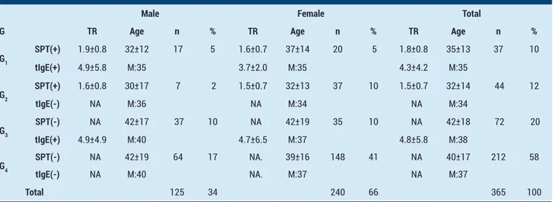
The test results, demographic values and distributions of all patients are depicted in Table 1. Here it should be drawn attention that 10% of all patients have (+) test results for both SPT and tIgE, and 32% of the patients have (+) results for either SPT or tIgE. Totally, 42% of all patients have (+) test results, for either SPT and/or tIgE, as an indicator of immediate type hypersensitivity. The ratio of total female to male patients is 1.9.
The distribution of positive pricks according to each allergen for both male and female patients are depicted in Figure 1. Here it can be seen that positive prick counts for Df and Dp, the two house dust mites, are greater than the other allergens. In Table 2 it is demonstrated the test results and demographic distributions of all SPT(+) and all tIgE(+) patients without paying attention whether they are (+) or (-) for the other test. In this manner, G5 is the sum of G1 and G2, and G6 is the sum of G1 and G3 depicted
Table 1. The test results, demographic values and distributions of patients classified according to SPT and tIgE values
Male Female Total
G TR Age n % TR Age n % TR Age n %
G1 SPT(+) 1.9±0.8 32±12 17 5 1.6±0.7 37±14 20 5 1.8±0.8 35±13 37 10 tIgE(+) 4.9±5.8 M:35 3.7±2.0 M:35 4.3±4.2 M:35 G2 SPT(+) 1.6±0.8 30±17 7 2 1.5±0.7 32±13 37 10 1.5±0.7 32±14 44 12 tIgE(-) NA M:36 NA M:34 NA M:34 G3 SPT(-) NA 42±17 37 10 NA 42±19 35 10 NA 42±18 72 20 tIgE(+) 4.9±4.9 M:40 4.7±6.5 M:37 4.8±5.8 M:38 G4 SPT(-) NA 42±19 64 17 NA. 39±16 148 41 NA 40±17 212 58 tIgE(-) NA M:40 NA. M:37 NA M:37 Total 125 34 240 66 365 100
G: Group, SPT: Skin prick test, tIgE: Total IgE, TR: Test result, ±: Standard deviation, M: Median, NA: Not applicable
Table 2. The test results and demographic distributions of SPT(+) and tIgE(+) patients
Male Female Total
G TR Age n % TR Age n % TR Age n %
G5 SPT(+) 1.8±0.8 31±13 24 6 1.5±0.7 34±14 57 16 1.6±0.7 33±13 81 22 M:35 M:34 M:34 G6 tIgE(+) 4.9±5.2 39±16 54 15 4.3±5.4 40±17 55 15 4.6±5.3 39±17 109 30 M:36 M:36 M:36
G: Group, SPT: Skin prick test, tIgE: Total IgE, TR: Test result, ±: Standard deviation, M: Median
in Table 1. Here it can be seen that totally 81 patients are SPT(+) with a mean 1.6 positive pricks for each single patient. For the tIgE(+) patients, although the distribution is not normal (i.e. standard deviation, ±5.3, is bigger than the mean), the mean value is 4.6 which denotes that it is 4.6 times higher than the maximum standard value and should be regarded as quite high. The uneven distribution suggests that, a minor part of the patients have extremely high tIgE values. Although the results are not shown here, the comparisons of SPT, tIgE and age values of either G5
or G6, between male and female patients were all found non-significant statistically.
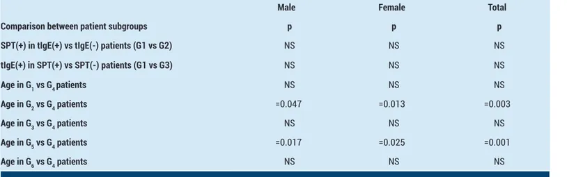
The broader scale comparisons performed to detect the significancy among SPT, tIgE, and age of the patients, and their p values are shown in Table 3. The comparison of SPT positivity between tIgE(+) and tIgE(-) patients, and tIgE positivity between SPT(+) and SPT(-) patients are both found non-significant, regarding that there is no connection between the positivities of SPT and tIgE.
When we compare the age values of SPT(+) and/or tIgE(+) patients (i.e. G1,2,3,5,6) to SPT(-) and tIgE(-) patients (i.e. G4); the results are statistically significant for only G2 and G5. G2 and G5 are the subgroups comprised of patients with SPT(+) values in which there is null and decreased effects of tIgE(+) patients respectively. Considering both the significant and non-significant results in comparison of age values, it can be said that while SPT positivity decreases with increasing age, there is no effect of age in tIgE positivity.
Figure 1. The distribution of positive pricks according to each
allergen for both male and female patients
DISCUSSION
Pruritus sine materia in the absence of a somatic cause, functional itch disorder or psychogenic pruritus; although all are the definitions of the same disorder, they are literally ill-defined. According to the diagnostic criteria of the French Psychodermatology Group (5), there are three major and seven minor criteria among which all the majors and at least three of the minors must be present for this disorder. The major or compulsory criteria are localized or
generalized pruritus without any dermatologic symtpom, chronic pruritus lasting more than six weeks, and the absence of a somatic cause. The minor or additional criteria are an increase in pruritus with the life events that have psychological impacts, changes in intensity accompanied with daily stress, nocturnal alterations, predominance during rest or inactivation, associated psychological disorders, pruritus that might be alleviated by psychotropic drugs, and pruritus that could be relieved by psychotherapy. Although the included patient profiles in our study were in accordance with the suggested criteria and the patients lack the manifest history, signs and symptoms of allergic diseases; regarding the psycho-neuro-immunologic connections (9), we applied a general screening test comprising SPT and serum tIgE, for probable allergic sensitizations.
SPT is the standard method to evaluate IgE-mediated (Type-I) sensitization worldwide (10) without appraising the ability of the relevant protein to cause inflammation by way of other mechanisms (11). It is generally the first line screening test to disclose probable agents in patients with symptoms entailing allergy. In order to obtain consistent data in long term investigations, SPT should be performed with standardized allergens in the way that the same commercially available extracts should be used in all patients and the test should be performed according to the same standard operating procedure (10). Although it is a first line diagnostic procedure, the reports are contradicting about the sensitivity and specifity of this test. There are reports suggesting that, while negative SPT response has a good anticipating value for excluding an IgE-mediated reaction, an isolated positive response should not be considered as a proof of a consistent allergy, and sensitizations are not constantly present with clinical symptoms (10,12). But there are also studies emphasizing the high sensitivity and low specificity of SPT, regarding patient history (11). Allergic sensitization to a protein can also be detected by allergen-specific
Table 3. The comparisons between subgroup pairs regarding SPT, tIgE and age of the patients
Male Female Total
Comparison between patient subgroups p p p
SPT(+) in tIgE(+) vs tIgE(-) patients (G1 vs G2) NS NS NS
tIgE(+) in SPT(+) vs SPT(-) patients (G1 vs G3) NS NS NS Age in G1 vs G4 patients NS NS NS Age in G2 vs G4 patients =0.047 =0.013 =0.003 Age in G3 vs G4 patients NS NS NS Age in G5 vs G4 patients =0.017 =0.025 =0.001 Age in G6 vs G4 patients NS NS NS
IgE (sIgE) level in serum (11,13). As the presence of sIgE antibody is necessary but not always sufficient for the diagnosis or management of an allergic disease, most clinical laboratories present some additional serologic tests, including tIgE, that can be useful in the diagnosis of patients with Type-I hypersensitivity. Due to its age-dependent concentration, serum tIgE level must be examined considering its nonatopic age-adjusted reference interval (13). When compared to sIgE, higher tIgE levels in atopic conditions are suggested to be an indicator of greater impairment in immune response (14). There are studies implying strong association of tIgE with history of wheeze, asthma, allergic rhinitis, and recommending the measurement of tIgE level as an essential test for atopic patients (15,16).
Asymptomatic skin sensitization is reported as a familiar reaction affecting 8-30% of the population when using a local standard panel of aeroallergens (12). In our study 22% of the patients are found SPT(+) with a mean 1.6 positive pricks for each single patient (Table 2). Although our patients have chronic pruritus and can not be considered asymptomatic, whether these positivities are the result of true allergy or due to asymptomatic skin sensitization coexisting with functional itch disorder should be discussed. The comparison of SPT(+) patients between tIgE(+) and tIgE(-) ones, and tIgE(+) patients between SPT(+) and SPT(-) ones are both found non-significant (Table 3), regarding that there is no connection between the positivities of SPT and tIgE. While this finding may suggest our patients have asymptomatic skin sensitization, it was reported that tIgE was related to allergic symptoms mostly in subjects with sensitivity to higher number of allergens and at substantially higher levels, compared to sIgE positivity (14). Likewise, in another study (17) it was reported that the mean tIgE increased with the number of positive sIgE results. In our study, the positivity of tIgE is found 30% in total patient population and the mean tIgE value is 4.6 fold of the maximum standard value (Table 2). Also regarding the uneven distribution (i.e. standard deviation, ±5.3, that is greater than the mean) which suggests at least some of the patients have extremely high tIgE values, we can say a considerable amount of tIgE(+) patients, whether they are SPT(+) or not, have true allergy. Albeit a positive SPT in an asymptomatic subject indicates a risk of developing allergy in the future is unknown to a great extent, some prospective studies indicate that 30-60% become allergic, depending on allergens and follow-up period (12). In a 23-year prospective study (18), the risk of rhinitis was found more than two times greater in asymptomatic SPT(+) than in SPT(-) subjects.
The epidermis of patients with atopic dermatitis (AD) allows allergens to penetrate into the skin and enhance sensitization to airborne proteins as a consequence of impaired barrier function, mucosal absorption, and sometimes increased local inflammation. In AD, airborne proteins have the ability to increase the severity of
inflammation through inherent proteolytic enzyme activity, activation of proteinase-activated receptors-2, and IgE binding (11). Our patients, though lack the advanced inflammatory signs of AD, might have partially weakened epidermal barrier due to chronic scratching, which leaded to the penetration of airborne proteins into the skin. Atopic patients are reported more likely to be sensitized to house dust mites than the majority of other proteins in their environment (11,14,19). In our study also positive prick counts for Df and Dp, the two house dust mites, are found greater than the other allergens (Figure 1).
The ratio of total female to male patients in our study is found 1.9, which is approximate to referrals reported for pruritus in general practice (1). Although we could not find a gender associated difference for both SPT and tIgE positivity, there are contradicting reports for this topic. While no difference was shown between male and female subjects in a study with prick test to common relevant environmental allergens (20), in another study it was reported that females were associated with reduced reactivity to histamine pricks (21). The findings for gender differences in tIgE in the literature are not consistent also. Some authors have observed higher tIgE levels in men than in women, whereas others did not find any difference. Also, some authors reported that the higher tIgE levels in men could be explained by higher prevalences of smoking (17).
In our study we have found that while SPT positivity decreases with increasing age, there is no effect of age in tIgE positivity (Table 3). Several studies have reported decreasing prevalence of atopy with increasing age (20,22-26). In a study to evaluate probable changes in sensitivity of skin test and severity of symptoms in allergic rhinitis twenty years after primary testing, both of the parameters revealed significant decreases in the last assessment (23). In another study with allergic rhinitis patients to appraise the differences fifteen years after primary assessments of SPT, sIgE, tIgE, sIgE/tIgE ratio, and nasal eosinophils; all parameters including rhinitis symptoms showed a decreasing trend in older age groups (24). But sometimes there are discrepancies between the decrease of sIgE and tIgE evaluations through years, as reported in a study (17) on the connection of age and IgE levels. In the mentioned study; while sIgE positivity decreased in older age groups, tIgE had no relationship with age. The reported decrease of sIgE/tIgE ratio (24) and the decrease of only sIgE without any change in tIgE in older age groups (17) may justify the suggestion that tIgE may be regarded as an indicator of greater dysregulation of the immune response in atopic conditions (14). In a large scale study (27), consisting more than 16,000 subjects who were performed SPT to eight allergens, while 30% of individuals between 12 and 24 years old had at least one positive SPT; this ratio was 8% in subjects aged between 65 and 74. Atopy was assessed for connection with age in another large cross-sectional study (28) of healthy subjects between 18 and 60 years old, and it was revealed a reduced rate of atopy
in the advanced ages, such as significant decreases in the prevalence of positive sIgE, SPT and allergic manifestations with every 10-year increase in age. Overall, population-based cross-sectional studies have shown that the prevalence of sensitization is higher in younger than in older age groups, and it is hypothesized that the chronological decrease in sensitization might reflect aging-related immunosenescence (29). Supporting this hypothesis, the skin of older people goes through atrophy and results in decreased cellularity and collagen amounts. The substantial reduction in blood vessels and mast cells offer fewer binding sites for allergens and produce less histamine to cause wheal and flare reactions (25).
In our study, it should be emphasized that 10% of all patients have positive results for both SPT and tIgE, and 32% of the patients have positive results for either SPT or tIgE. In the final analysis, totally 42% of all patients have positive test results, for either SPT and/or tIgE, which may be regarded as a presentation of immediate type hypersensitivity (Table 1). Pruritus sine materia in the absence of a somatic cause is a greatly challenging condition for both the patient and physician because of its chronicity and resistance to treatment. While there are contradicting reports on the sensitivity of SPT, the high ratio of tIgE positivity in our patients and its relative high mean value make us to think that the majority of SPT(+) patients may have true allergy. The greater positivity of SPT to house dust mites and decreasing positivity with increasing age, in accordance with the literature data on atopic patients, may also support our implications. Although pruritus sine materia in the absence of a somatic cause is broadly reported to ensue as the result of psychogenic causes, regarding the quite high positive ratios we found for both SPT and tIgE, and their relative low costs, it may be encouraging to perform these tests routinely on the patients with this clinical entity. In case of positivity, the treatment may be focused on antiallergic or sometimes immunosuppressant modalities as a clinical approach, besides the classical psychotropic and topical treatments. Also, even some of the positive patients have no true allergy while the tests are being performed; they should be better followed up for the risk of developing an allergic disease in the future.
Study limitations
This is a retrospective study with idiocratic limitations. As the patients were referred to our outpatient clinic for a routine dermatologic examination we could only perform a small number of screening tests because of both ethical and financial concerns. Therefore a similar prospective study may be designed with widening the scope of SPT and also including serum sIgE measurement. Also a psychiatric consultation might be of help to disclose the connection between allergy and emotional stress in these patients.
CONCLUSION
Considering the quite high positive ratios we found for both SPT and tIgE, it may be encouraging to perform
these tests routinely on the patients with pruritus sine materia in the absence of a somatic cause, both to reveal a probable allergic tendency and to conduct the treatment modalities better.
Financial Disclosure: There are no financial supports.
Ethical approval: This article that has been written regarding the three-year retrospective data obtained at our routine dermatology outpatient clinic. The relevant and applicable ethical principles (i.e. Declaration of Helsinki) are considered during the writing process.
Erkin Pekmezci ORCID: 0000-0003-1469-2557
REFERENCES
1. Frese T, Herrmann K, Sandholzer H. Pruritus as reason for encounter in general practice. J Clin Med Res 2011;3:223-9.
2. Misery L, Dutray S, Chastaing M, et al. Psychogenic itch. Transl Psychiatry 2018;8:52.
3. Yonova D. Pruritus in certain internal diseases. Hippokratia 2007;11:67-71.
4. Rubegni P, Poggiali S, Nami N, et al. Skin diseases in geriatric patients: our experience from a public skin outpatient clinic in Siena. G Ital Dermatol Venereol 2012;147:631-6.
5. Misery L, Alexandre S, Dutray S, et al. Functional itch disorder or psychogenic pruritus: suggested diagnosis criteria from the French psychodermatology group. Acta Derm Venereol 2007;87:341-4.
6. Misery L, Chastaing M. Joint consultation by a psychiatrist and a dermatologist. Dermatol Psychosom 2003;4:160-4.
7. Weisshaar E, Fleischer AB, Bernhard JD, Cropley TG. Pruritus and dysesthesia. In: Bolognia JL, Jorizzo JL, Schaffer JV, eds. Dermatology. 3rd edition. New Haven: Elsevier Saunders; 2012. p.111-25.
8. Koblenzer CS. Psychosomatic concepts in dermatology. Arch Dermatol 1983;119:501-12.
9. Solomon I, Ilie MA, Draghici C, et al. The impact of lifestyle factors on evolution of atopic dermatitis: an alternative approach. Exp Ther Med 2019;17:1078-84. 10. Haahtela T, Burbach GJ, Bachert C, et al. Clinical
relevance is associated with allergen specific wheal size in skin prick testing. Clin Exp Allergy 2014;44:407-16.
11. Hostetler SG, Kaffenberger B, Hostetler T, et al. The role of airborne proteins in atopic dermatitis. J Clin Aesthet Dermatol 2010;3:22-31.
12. Burbach GJ, Heinzerling LM, Edenharter G, et al. GA(2)LEN skin test study II: clinical relevance of inhalant allergen sensitizations in Europe. Allergy 2009;64:1507-15.
13. Hamilton RG. Clinical laboratory assessment of immediate-type hypersensitivity. J Allergy Clin Immunol 2010;125:284-96.
14. Droste JH, Kerhof M, de Monchy JG, et al. Associaton of skin test reactivity, specific IgE, total IgE, and eosinophils with nasal symptoms in a community-based population study. The Dutch ECRHS group. J Allergy Clin Immunol 1996;97:922-32.
15. Bener A, Safa W, Abdulhalik S, et al. An analysis of skin prick test reactions in asthmatics in a hot climate and desert environment. Allerg Immunol (Paris) 2002;34:281-6.
16. Ramos E, Orea Solano M, Flores Sandoval G, et al. [Correlation between the concentrations of total serum IgE and skin tests]. Rev Alerg Mex 1994;41:94-7.
17. Kerkhof M, Droste JH, de Monchy JG, et al. Distribution of total serum IgE and specific IgE, to common aeroallergens by sex and age, and their relationship to each other in a random sample of the Dutch general population aged 20-70 years. Allergy 1996;51:770-6. 18. Settipane RJ, Hagy GW, Settipane GA. Long-term risk
factors for developing asthma and allergic rhinitis: a 23-year follow-up study of college students. Allergy Proc 1994;15:21-5.
19. Hon KL, Wang SS, Wong WL, et al. Skin prick testing in atopic eczema: atopic to what and at what age. World J Pediatr 2012;8:164-8.
20. Barbee RA, Lebowitz MD, Thompson HC, et al. Immediate skin-test reactivity in a general population sample. Ann Intern Med 1976;84:129-33.
21. Song WJ, Lee SM, Kim MH, et al. Histamine and allergen skin reactivity in the elderly population: results from the Korean longitudinal study on health and aging. Ann Allergy Asthma Immunol 2011;107:344-52. 22. Nam YH, Lee SK. Comparison between skin prick
test and serum immunoglobulin E by CAP system
to inhalant allergens. Ann Allergy Asthma Immunol 2017;118:608-13.
23. Simola M, Holopainene E, Malmberg H. Changes in skin and nasal sensitivity to allergens and the course of rhinitis; a long-term follow-up study. Ann Allergy Asthma Immunol 1999;82:152-6.
24. Di Lorenzo G, Leto-Barone MS, La Piana S, et al. Clinical course of rhinitis and changes in vivo and in vitro of allergic parameters in elderly patients: a long-term follow-up study. Clin Exp Med 2013;13:67-73. 25. Scichilone N, Callari A, Agugliaro G, et al. The impact
of age on prevalence of positive skin prick tests and specific IgE tests. Respir Med 2011;105:651-8.
26. Karakaya G, Kalyoncu AF. The natural course of atopy determined by skin prick tests in patients with bronchial asthma and/or rhinitis. Allergol Immunopathol (Madrid) 2006;34:257-62.
27. Gergen PJ, Turkeltaub PC, Kovar MG. The prevalence of allergic skin test reactivity to eight common aeroallergens in the US population: results from the second national health and nutrition examination survey. J Allergy Clin Immunol 1987;80:669-79.
28. Wüthrich B, Schindler C, Medici TC, et al. IgE levels, atopy markers and hay fever in relation to age, sex and smoking status in a normal adult Swiss population. SAPALDIA (Swiss study on air pollution and lung diseases in adults) team. Int Arch Allergy Immunol 1996;111:396-402.
29. Amaral AFS, Newson RB, Abramson MJ, et al. Changes in IgE sensitization and total IgE levels over 20 years of follow-up. J Allergy Clin Immunol 2016;137:1788-95.