Journal of Clinical and Analytical Medicine | 1
DOI: 10.4328/JCAM.4599 Received: 01.05.2016 Accepted: 23.06.2016 Printed: 01.09.2016 J Clin Anal Med 2016;7(5): 753-6 Corresponding Author: Yeşim Altay, 48. Cadde No: 33/31 Cukurambar, 06520, Ankara, Turkey.
GSM: +905324902298 E-Mail: [email protected]
Ipsilateral Rotational Autokeratoplasty
Yeşim Altay Department of Ophthalmology, Ufuk University Faculty of Medicine, Ankara, Turkey
İpsilateral Rotasyonel Otokeratoplast
Özet
Kornea skarı tüm dünyada monooküler körlüğün önde gelen nedenlerinden bi-ridir, kornea transplantasyonu ise dünyada en çok yapılan solid organ trans-plantasyonudur. Kornea skarlarının tedavisi için uygulanan keratoplasti tek-nikleri lameller allokeratoplasti ve penetran allokeratoplastiyi kapsar. İpsila-teral rotasyonel otokeratoplasti ise kornea skarı olan bazı hastalar için stan-dart penetran keratoplastiye etkin bir alternatif olabilir. Bu işlem hastanın kendi korneasının, skarlaşmayı uzaklaştırarak şeffaf korneayı görme ekseni-ne getirecek şekilde, döndürülmesini içerir. Rotasyoekseni-nel keratoplasti için hasta seçiminde dikkate alınması gereken önemli bir konu, korneal skar boyutudur. İpsilateral otokeratoplasti sonrası daha fazla astigmatizma ve daha az kor-neal pupiller şeffaf bölge bulunmasına bağlı olarak penetran allokeratoplasti kadar iyi görme kalitesi sağlanamayabilir. Ancak pediatrik hastalar veya vas-kularize kornealı hastalar gibi allogreft rejeksiyon riski yüksek olan hastalar-da etkin bir alternatif cerrahi teknik olabilir. Ayrıca bu tekniğin kullanılması, gelişmekte olan ülkelerdeki donor kornea dokusu kısıtlılığına en azından kıs-mi çözüm sağlayabilir.
Anahtar Kelimeler
Kornea; Kornea Nakli; Kornea Opasitesi
Abstract
Corneal opacity is a leading cause of monocular blindness, and corneal trans-plantation is the most commonly performed solid organ transtrans-plantation in the world. Keratoplasty techniques for corneal opacities include lamellar al-lokeratoplasty and penetrating alal-lokeratoplasty. Ipsilateral rotational auto-keratoplasty can be an effective alternative to penetrating alloauto-keratoplasty for some patients with corneal scars. This procedure involves a rotation of the patient’s own cornea to move opacity out of the visual axis. An important consideration when selecting cases for rotational autokeratoplasty is the di-mensions of the corneal scar. Although ipsilateral autokeratoplasty may not provide as good a quality of vision as penetrating allokeratoplasty because of higher astigmatism and reduced corneal pupillary clear zone, these disad-vantages are often outweighed when the risk of allograft rejection is high, as in pediatric patients and those with vascularised corneas. This technique would at least partially resolve the issue of scarcity of donor corneal tissue in developing countries.
Keywords
Cornea; Corneal Transplantation; Corneal Opacity
Ipsilateral Rotational Autokeratoplasty
| Journal of Clinical and Analytical Medicine Ipsilateral Rotational Autokeratoplasty
2
Introduction
Corneal opacity is a leading cause of monocular blindness worldwide. The definitive treatment of visually-impairing cor-neal opacity is penetrating allogenic keratoplasty using donor corneal tissue. Although corneal scar and keratitis are the most frequent indications for keratoplasty in developing countries, corneal edema and keratoconus constitute the majority of pa-tients undergoing penetrating keratoplasty in developed coun-tries. The reported success rate for penetrating corneal grafts is 73% at 5 years, and 62% at 10 years [1]. However, endothe-lial rejection rates are 15-20% in adults and 10-50% in pediat-ric grafts [2,3]. Rejection rates are higher in pediatpediat-ric corneal transplantation because of the more active immune systems of younger patients [4]. In addition to rejection, late corneal failure is anticipated due to the continuing loss of donor corneal en-dothelial cells with time [5]. More importantly, the accessibility to corneal tissue for corneal grafting is a major limiting factor. Because of the increasing gap between demand and supply of donor corneal tissue, alternative techniques have been devised. One of them is rotational autokeratoplasty, which is indicated for patients with a limited corneal opacity involving the visual axis. This procedure involves a rotation of the patient’s own cornea to move opacity out of the visual axis and to replace it with clear cornea [4,6-9].
Several authors have suggested the use of different graft shapes for ipsilateral autokeratoplasty, such as a triangle or a figure eight [9,10]. However, none has replaced the standard circular graft with an eccentric center [11-15].
Like any other surgical technique, appropriate patient selec-tion is required for successful rotaselec-tional autokeratoplasty. Usu-ally rotational autokeratoplasty is chosen for nonprogressive corneal scars following blunt and penetrating corneal trauma, postinfectious keratitis scar, chemical injuries, and idiopathic or postherpetic lipid keratopathy [6,8,9,16,17]. These patients are at high risk of allograft corneal rejection if subjected to a conventional keratoplasty. Other than the usual indications, Cunha et al. described ipsilateral penetrating autokeratoplasty associated with a crescent-shape resection of 0.5 mm of the inferior cornea as an alternative method to corneal transplan-tation for keratoconus. They reported reduction of visual acuity with increased astigmatism during the follow-up period [18]. An important consideration during selection of cases for rota-tional autokeratoplasty is the dimensions of the corneal scar. A four to five mm of clear peripheral cornea is required to obtain at least 3 mm of central clear cornea, free of suture track scars. Surgical Technique
The surgical technique performed is identical to that of con-ventional penetrating keratoplasty, with the exception that the host cornea is eccentrically cut and then rotated before sutur-ing. Because of the more rapid loosening of sutures that occurs when they are placed into the anterior sclera, interrupted su-tures are recommended for rotational autokeratoplasty. Several authors have described methods for determining the size and location of the trephination. McDonnell et al. [9] rec-ommended the following guidelines:
“1. Use the maximum area of available clear cornea (this usu-ally meant making the peripheral edge of the graft very near the limbus).
2. Try to ensure that the opacity to be rotated is as near the edge of the graft as possible; this allows maximum movement of the opacity by the rotation and its replacement with the
maximum area of the clear cornea.
3. Make the central edge of the graft at least 3 mm away from the visual axis. This ensures clear cornea at the visual axis and minimizes the affect [sic] of the suture.
4. If possible, rotate the opacity under the upper lid.”
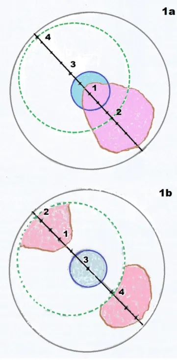
In guidelines formalised by Roa and Lam [14], the center of the cornea is marked and a point 2.5 mm away from the center is marked in the opacity. A third mark is made 2.0 mm away from the central mark in line with the second mark, and a fourth mark is made 3.5 mm away from the third. The distance between marks 2 and 4 is taken as the trephine size to be used [14] (Figure 1).
The mathematical formulas for determining the size of the do-nor and host trephination have been described by several au-thors. The two most useful are described by Bourne and Bru-baker [19] and Jonas et al. [7]. In Bourne and BruBru-baker’s method [19], two measurements were taken. First, the diameter of the
Figure 1. Schematic picture of cornea with opacity involving pupillary area. The center of the cornea is marked (1), and a point 2.5 mm away from center is marked in the opacity (2), third mark is made 2.0 mm away from the central mark, in line with the second mark; a fourth mark is made 3.5 mm away from the third (a). Depiction of rotation of calculated corneal graft (b).
| Journal of Clinical and Analytical Medicine
754
| Journal of Clinical and Analytical Medicine Ipsilateral Rotational Autokeratoplasty
3
largest circle of the clear cornea was measured. Second, the shortest distance from the edge of this circle to the geomet-ric center of the cornea was assessed (this was considered positive if the opacity involved the center of the cornea and negative if the opacity was within the largest area of the clear cornea). The required trephine size was obtained with the fol-lowing equation: 1.5 multiplied by the diameter of the largest clear circle added to the shortest distance from the circle to the corneal center [19]. The other formula, by Jonas et al. [7], utilises a trephine size of 0.75 of the overall corneal diameter, when the scar lies at the geometric center of the cornea, and adjusts this according to extent of the scar. Both the Jonas et al. [7] and Bourne and Brubaker formulas [19] give similar results, with the Jonas et al. [7] formula resulting in a slightly larger trephine size.
Another approach to postoperative simulation of rotational au-tokeratoplasty uses digital imaging. Agarwal et al. [15] have de-scribed the use of digital image manipulation software (Adobe Photoshop, version 5.0) to plan the size, location, and rotation of the graft. Using the drawing tool, a circle approximating the pupil was drawn on a new transparent layer created on the orig-inal image of the eye. Subsequently, a circle was drawn on the original photograph of the cornea to simulate corneal trephi-nation. The corneal area within this circle was rotated on the image to simulate rotational autokeratoplasty. This step was repeated using varying sizes and positions of the larger circle, which simulated the corneal trephine, to include the maximum area of the clear cornea within the pupillary axis [15].
Outcomes
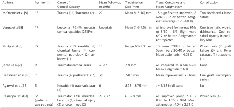
Several case series have been reported, as presented in Table 1. In previously published studies of autokeratoplasty, graft sur-vival was noted to be ≥ 90%. In a study by Murty et al. [6], only 3 of 22 grafts failed, two because of prolonged operative time and one because of uncontrolled glaucoma. Ramappa et al. [4] reported that the success rate was 81.25% over a mean follow-up of 29.43 months in pediatric patients. Bertelmann et al. [18] reported only one failure out of seven cases, which they attrib-uted to a triple procedure and preoperative poor endothelial cell count (1200 cells/mm2). Continued endothelial loss is the major cause of graft failure following allograft corneal
transplanta-tion. Although long-term results are unknown, Bertelmann et al. [20] showed that mean endothelial cell loss at 1 year was 15% in rotational autografts compared with 40% in homografts. The published case series in the literature have reported signifi-cant number of patients achieving good visual outcomes. In a report by McDonnell and Falcon [9], 2 patients did not have any astigmatic correction and four other patients had correction ranging from 1.25 to 4 D cyl. Murty et al. [6] reported 1 to 2.5 D cyl in 58.8% of patients, 3 to 5.5 D cyl in 23.5%, and 6 to 9 D cyl in 29.4%. Only Jonas et al. [7] compared visual outcomes af-ter rotational autokeratoplasty with those of a nonrandomized control group of penetrating allografts, and found significantly lower postoperative visual acuity and higher corneal astigma-tism in patients having rotational autokeratoplasty. The eccen-tric trephination, disparity of corneal thickness between the pe-ripheral clear cornea and the central scarred cornea into which it is sutured, and the proximity of one edge of the trephination to the center of the cornea are potential reasons for increased astigmatism after rotational autokeratoplasty [7].
The most common postoperative complication reported after rotational autokeratoplasty was wound leak [6]. This was prob-ably because of the difficulties in tissue apposition while su-turing due to differences in the thickness of the central and peripheral cornea as well as scarred versus normal tissue. Also, the cosmetic outcome of autokeratoplasty is inferior to that of conventional penetrating keratoplasty. The corneal opacity is visible postoperatively, although it seems smaller because it is bisected during trephination.
Although rotational autokeratoplasty does not provide as high a level of best-corrected visual acuity as penetrating keratoplasty due to higher astigmatism, there are several advantages that outweigh these disadvantages in many patients whose risk of allograft rejection is higher than normal, such as pediatric pa-tients and those with vascularised cornea:
1. This technique does not require a donor cornea and there is no risk of transmission of infection such as human immunode-ficiency virus or hepatitis.
2. Most importantly, this technique obviates the lifelong risk of allograft rejection. This can be a very important factor in some patients, such as those with traumatised eyes with extensive corneal vascularisation, in children or young adults, and in those
Table 1. Summary of studies giving outcomes for ipsilateral rotational autokeratoplasty Authors Number (n) Cause of
Corneal Opacity
Mean Follow-up (months)
Trephination Size (mm)
Visual Outcomes and Mean Astigmatism
Complication McDonnel et al.[9] 16 Trauma (14) Trachoma (2) 27 Mean 8 (7-10) mm 13 significantly improved, 8
were 6/12 or better. Astig-matism range (1.25-4.0 D)
Two developed a loose suture
Verma et al.[8] 17 Leucoma (76.4%) macular corneal opacities (23.5%)
Uncertain Mean 7 (6-7.5) mm All improved from preop HMs to 5/60 – 6/9. Eight were 6/12 or better. Astigmatism not reported
One traumatic wound dehiscence. One re-sidual opacity in pupil-lary zone
Murty et al.[6] 27 Trauma (12) keratitis (8) chemical burns (4) con-genital pathology (2) un-known (1)
12 Range 6.5-9.0 mm 13 were 20/80 or better. Seven were 20/40 or better. Mean astigmatism 4.25 D
Wound leak (7) graft failure (3) ant. Polar cataract (1) glaucoma (1)
Jonas et al.[7] 9 Traumatic corneal scars 31.27 7-9 mm All improved to mean 0.28. Mean astigmatism 6 D
None
Bertelman et al.[18] 7 Trauma (4) postkeratitis (3) 39 7-8.5 mm Mean improvement 3.5 lines One graft decompen-sation
Agarwal et al.[15] 5 Keratitis (4) traumatic scar
(1) 6 8.25 – 8.75 mm >= 6/18 in all cases No
Ramappa et al.[4] 33 (pediatric age patients)
Traumatic (20) microbial keratitis (8) chemical injury (3) undetermined (2)
27 ± 37 6.5 – 8 mm All improved preop 2.05 ± 0.96 to 1.25 ± 0.84. Mean astigmatism 4.04 ± 2.21 D
Wound leak (4)
Journal of Clinical and Analytical Medicine | 755
| Journal of Clinical and Analytical Medicine Ipsilateral Rotational Autokeratoplasty
4
where follow-up is difficult.
3. Another advantage is reduced healing time, which allows ear-ly suture removal and reduction in the dose and duration of cor-ticosteroid use postoperatively, thus avoiding steroid-induced complications such as cataract and glaucoma.
Conclusions
Ipsilateral rotational autokeratoplasty can be a viable alterna-tive to conventional penetrating keratoplasty in some patients whose risk of allograft rejection is higher than normal. It avoids the risk of allograft rejection or long-term use of corticoste-roids and obviates the waiting period for high-quality donor cornea tissues. Careful selection of patients can yield encour-aging results with the use of this alternative technique. Competing interests
The authors declare that they have no competing interests.
References
1. Williams KA, Lowe MT, Bartlett CM, Kelly L, Coster DJ. The Australian Corneal Graft Registry 2007 Report. Adelaide: Flinders University Press; 2007.p.114-8. 2. Claesson M, Armitage WJ, Fagerholm P, Stanovi U. Visual outcome in corneal grafts: a preliminary analysis of the Swedish Corneal Transplant Register. Br J Ophthalmol 2002;86(2):174-80.
3. Wagoner MD, Al-Ghamdi AH, Al-Rajhi AA. Bacterial keratitis after primary pedi-atric penetrating keratoplasty. Am J Ophthalmol 2007;143(6):1045-7.
4. Ramappa M, Pehere NK, Murty SI, Chaurasia S, Rao HL, Sangwan VS. Rotation-al autokeratoplasty in pediatric patients for nonprogressive paracentrRotation-al corneRotation-al scars. Ophthalmol 2012;119(12):2458-62.
5. Armitage WJ, Dick AD, Bourne WM. Predicting endothelial cell loss and long-term corneal graft survival. Invest Ophthalmol Vis Sci 2003;44(8):3326-31. 6. Murty S, Bansal AK, Sridhar MS, Rao GN. Ipsilateral rotational autokeratoplas-ty. An alternative to penetrating keratoplasty in nonprogressive central corneal scars. Cornea 2001;20(5):455-7.
7. Jonas JB, Rank RM, Budde WM. Autologous ipsilateral rotating penetrating kera-toplasty. Am J Ophthalmol 2001;131(4):427-30.
8. Verma N, Melengas S, Garap JA. Ipsilateral rotational autokeratoplasty for the management of corneal opacities. Aust N Z J Ophthalmol 1999;27(1):21-5. 9. McDonnell PJ, Falcon MG. Rotational autokeratoplasty. Eye 1989;3(5):576-80. 10. Wilson RS. ‘Figure 8’ ipsilateral autokeratoplasty: A modification using stan-dard corneal trephines. Arch Ophthalmol 1976;94(1):121-2.
11. Arnalich-Montiel F, Dart JK. Ipsilateral rotational autokeratoplasty: a review. Eye 2009;23(10):1931-8.
12. Jhanji V, Sharma N, Agarwal T, Vajpayee RB. Alternatives to allograft corneal transplantation. Curr Opin Ophthalmol 2010;21(4):301-9.
13. Afshari NA, Duncan SM, Tanhehco TY, Azar DT. Optimal size and location for corneal rotational autografts. A simplified mathematical model. Arch Ophthalmol 2006;124(3):410-3.
14. Rao SK, Lam D SC. Calculating graft size and position in rotational corneal autografting: A simplified approach. Indian J Ophthalmol 2008;56(3):233-5. 15. Agarwal T, Sharma N, Jhanji V, Vajpayee RB. Computer simulation-assisted rotational autokeratoplasty with pupillary enlargement for management of cases with partial corneal opacification. Br J Ophthalmol 2010;94(1):24-5.
16. Sah WJ, Myoung YW, Hahn TW, Kim JH. Rotational autokeratoplasty in ad-vanced lipid keratopathy. Ophthalmic Surg Lasers 1997;28(12):1020-4. 17. Gunes A, Kansu Bozkurt T, Unlu C, Sezgin Akcay BI, Bayramlar H. Ipsilateral rotational autokeratoplasty for the management of traumatic corneal scar. Case Rep Ophthalmol Med 2012; 2012: 853584. doi: 10.1155/853584.
18. Cunha FJ, Lira W, Brandt CT. Refractive and topographic changes of ipsilat-eral rotational autokeratoplasty with a crescent-shape 0.5 mm resection of the inferior cornea in keratoconus after a 24 months follow-up. Arq Bras Oftalmol 2011;74(3):184-9.
19. Bourne WM, Brubaker RF. A method for ipsilateral rotational autokeratoplasty. Ophthalmology 1978;85(12):1312-6.
20. Bertelmann E, Hartmann C, Scherer M, Rieck P. Outcome of rotational kerato-plasty: comparison of endothelial cell loss in autografts vs allografts. Arch Oph-thalmol 2004;122(10):1437-40.
How to cite this article:
Altay Y. Ipsilateral Rotational Autokeratoplasty. J Clin Anal Med 2016;7(5): 753-6.
| Journal of Clinical and Analytical Medicine
756