Non-surgical minimally invasive rhinoplasty: tips and tricks from the
perspective of a dermatologist
Ali Sahan
1, Funda Tamer
2 ✉1Doctor Al-Sa Aesthetic & Cosmetic Dermatology Clinic, Ankara, Turkey. 2Department of Dermatology, School of Medicine, Ufuk University, Ankara, Tur-key. ✉Corresponding author: [email protected]
101
2017;26:101-103
doi: 10.15570/actaapa.2017.29
Introduction
Non-surgical rhinoplasty with dermal fillers can be easily per-formed for most patients that have cosmetic concerns about the appearance of their nose. Proposed indications include patients that are not appropriate candidates for surgery or patients that do not want to have surgery and revision nose surgery. It is a fast, simple, safe, and cost-effective procedure without tissue damage. Therefore, non-surgical nose augmentation with dermal fillers is becoming more popular among specialists, including plastic sur-geons. However, it is important to be familiar with the anatomical structure and the features of the fillers to attain successful results (1). Serious adverse effects including ophtalmic artery emboli fol-lowing nasal augmentation with calcium hydroxylapatite filler, central retinal artery occlusion, visual acuity loss, and brain in-farctions after nasal filler injection of hyaluronic acid (HA) have been reported (2, 3). Pressure necrosis of the nasal tip due to over-injection or ischemic necrosis of the nasal area due to arterial em-bolism should also be kept in mind (4). It has been suggested that skin necrosis of the nasal region is mainly caused by hydrophilic features of HA dermal fillers (5). Recently, Signorini et al. classi-fied the filler complications as early reactions and late reactions. Early reactions include pain, ecchymosis, erythema, bruising, bleeding, vascular infarction, soft tissue necrosis, infection, hy-persensitivity, inappropriate placement, and distant spread. Late reactions include inflammatory reactions such as granuloma, nodules, dyspigmentation, and displacement of fillers (6).
Cosmetic dermatologists perform cosmetic procedures, includ-ing facial filler injection. However, the glabella and nasal region are regarded as high-risk areas for skin necrosis and emboliza-tion. Therefore, many dermatologists avoid treating these areas. This article presents our experience of non-surgical rhinoplasty
with dermal fillers by detailing our technique.
Methods
Medical records of 35 patients that had dermal filler rhinoplasty at the Al-Sa dermatology clinic between January 2016 and January 2017 were examined retrospectively. Photos were taken before and immediately after the injections. Prior nose surgery, coexistence of nasal obstruction or nasal deformity, previous cosmetic proce-dures, complications, and patient satisfaction were analyzed. All of the patients were followed for 6 months. Patient satisfaction was determined on a scale of 0 to 5 (0 = dissatisfied, 5 = very satis-fied) (7).
We used 5% lidocaine cream (Emla) for local anesthesia 30 minutes before the injection. We used a combination of 0.1% octe-nidine dihydrochloride and 2% 2-phenoxyethanol (Octenisept®) to disinfect the skin.
The procedure can be explained in two steps:
Step 1: A 22 gauge and 70 mm blunt cannula is used for filler
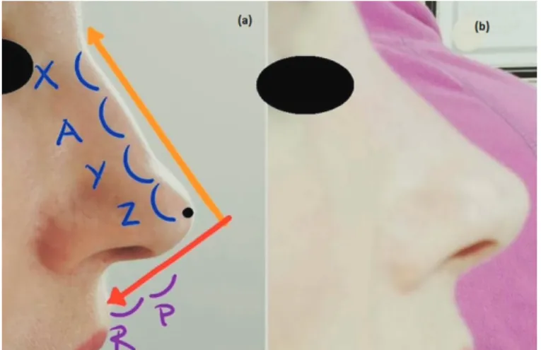
injection. The entry point is made with a 21-gauge sharp needle on the tip of the nose. The cannula is slowly moved forward in the supraperiostal layer until it reaches the radix. The cannula is kept parallel to the nasal spine. The skin of the nasal dorsum is raised gently with the thumb and second finger during cannula move-ment. After the cannula reaches the radix, the filler is injected into the nasal dorsum in the supraperiostal layer using a retro-grade linear threading technique. A 0.15 to 0.25 cc volume of 15 mg/ml HA dermal filler with 0.3% lidocaine (Juvéderm Volbella®) is placed at point X and a 0.1 to 0.2 cc volume of 15 mg/ml HA is placed at point Y. Point A is the nasal hump and point Z is the na-sal tip. The injection volume is 0.05 to 0.1 cc for the nana-sal tip (Fig. 1). The cannula is removed following the tip injection.
Abstract
Introduction: Rhinoplasty is one of the most common aesthetic procedures performed by plastic surgeons all over the world. In
recent years, a non-surgical nose job has become the preferred option for most patients and specialists. Minimally invasive tech-niques already result in high satisfaction rates. Moreover, patients do not need anesthesia and there is no post-operative recovery period.
Methods: Between January 2016 and January 2017, 35 patients (33 female, 2 male) that had undergone nonsurgical rhinoplasty
with dermal fillers were included in the study. Clinical and demographic features of the patients, our technique, satisfaction rates, and complications were discussed.
Results: We recorded an extremely high satisfaction rate and did not observe any complications. All of the patients returned to
their normal daily activities immediately after the procedure.
Conclusion: Non-surgical nose augmentation with dermal fillers is an easy, safe, and comfortable technique. It appears that
der-matologists need to assume a more active role in rhinoplasty. Keywords: cosmetic techniques, dermal fillers, rhinoplasty
Acta Dermatovenerologica
Alpina, Pannonica et Adriatica
Acta Dermatovenerol APA
102
Acta Dermatovenerol APA | 2017;26:101-103
A. Sahan et al.
Step 2: Afterwards, the same cannula is inserted
perpendicu-lar to the nasal tip through the same entry hole. Then it is gen-tly moved forward from the tip to the columella and nasolabial angle. After the cannula reaches the nasolabial angle, the filler is injected to point P and point R in the supracartilaginous layer using a retrograde linear threading technique. Herein, we used a 24 mg/ml HA dermal filler with 0.3% lidocaine (Juvéderm Ultra 4®). The injection volume is 0.1 to 0.15 cc for point P and 0.1 to 0.2 cc for point R (Fig. 1). Finally, the cannula is removed from the tip of the nose.
The patients were advised not to wear glasses and to avoid ex-ercise for a week after the rhinoplasty.
Results
Between January 2016 and January 2017, 35 patients (33 female, 2 male) were treated with non-surgical minimal invasive rhinoplas-ty at the Al-Sa dermatology clinic. The mean age of the patients was 27 (range: 20–35).
None of the patients had prior nose surgery or a non-surgical nose job, nasal obstruction, or nasal deformity. However, all of the patients had had prior botulinum toxin injections for facial wrinkles, and 13 patients (39.3%) had had prior lip augmentation with HA fillers.
The patient satisfaction score was 5 for 32 patients; the satisfac-tion score was 4 for two patients and 3 for one patient. We sought to achieve the most natural appearance of the nose and the face (Figs. 2–4). However, three patients had very high and unrealistic expectations. Nevertheless, a very high patient satisfaction rate was noted in our study.
No complications were observed during the procedure or the 6-month follow-up. The patients were informed that the results could last approximately 8 months after the injection.
Discussion
Rhinoplasty is one of the most common cosmetic procedures per-formed by plastic surgeons. However, non-surgical nose jobs with a dermal filler are becoming increasingly popular in the world. Filler rhinoplasty has become an advantageous choice for pa-tients that are afraid of surgery or general anesthesia. It is a fast, safe, simple, and effective method when compared with surgical rhinoplasty. Youn et al. evaluated the anthropometric analysis of 242 patients that had undergone HA filler rhinoplasty. Transient complications such as bruising, headache, swelling, and
erythe-ma were reported in erythe-many patients. However, arterial occlusion was observed in only three patients. Needles were used instead of cannulas to perform filler injections in these patients. They sug-gested that filler rhinoplasty is as effective as surgical augmenta-tion rhinoplasty based on anthropometric measures (8). Rho et al. used three-dimensional imaging to analyze facial anthropometric measures of 40 patients that had undergone filler rhinoplasty. Na-sal volume increase, nose height increment, increase in nasola-bial, nasofrontal, and nasomental angle, decrease in nose length, and nasofacial angle were observed. The results were similar to previously published studies including patients with surgical rhi-noplasty (9).
Surgical rhinoplasty is associated with many complications, such as breathing difficulties as a result of reduced nasal airway size, hyposmia, numbness, infection and dislocation of implants, atrophy, skin necrosis, fibrosis, subcutaneous cysts, and granu-lomas. Moreover, serious post-operative deformities including pollybeak deformity characterized by a deep radix and promi-nent cartilaginous dorsum, asymmetries, deviations, overresec-tion and irregularities of the nasal dorsum, alar collapse, slit-like nostrils, wide nasal tip or bridge, pinched tip, and overshortened nose have been reported. Therefore, 5 to 15% of patients undergo revision rhinoplasty following a primary surgical nose job (10).
Non-surgical rhinoplasty with fillers can be used to treat post-surgical rhinoplasty contour asymmetries. Bray et al. used Rest-ylane-Lidocaine® to correct post-rhinoplasty complications such as right upper lateral cartilage disruption and tip scar. The filler was injected supraperiosteally with a 30-gauge needle using lin-ear threading, serial puncture, and/or fanning techniques. They Figure 1 | Schematic view of filler rhinoplasty. The patient before (a) and
im-mediately after (b) the procedure.
103
Acta Dermatovenerol APA | 2017;26:101-103 Non-surgical rhinoplasty
reported slight complaints such as swelling, tenderness, and red-ness lasting for a few days (4).
HA fillers are biocompatible and biodegradable; however, com-plications such as foreign body reactions and delayed hypersen-sitivity can be observed with their injection. Moreover, Chae et al. reported the migration of injected HA filler from the nose to fore-head, clinically presenting as two soft, skin-colored, subcutane-ous nodules (11). Tissue necrosis is the most severe complication of fillers. One of the most dangerous area of the face for filler injec-tion is the nose. The dorsal nasal arteries usually lie bilaterally to the nasal dorsum; however, they are not constant. Tansatit et al. classified dorsal nasal artery courses into five patterns; bilateral dorsal nasal arteries with unequal sizes, random distribution, a single dorsal nasal artery anastomosis with contralateral/ipsilat-eral latcontralateral/ipsilat-eral nasal artery, or bilatcontralateral/ipsilat-eral latcontralateral/ipsilat-eral nasal arteries. They reported the nasal tip as a high-risk area, and they suggested that necrosis risk decreased at the upper sides of the nasal dorsum. However, patients with a single dorsal nasal artery had a high ne-crosis risk for all parts of the nose. Therefore, safety rules and us-ing recommended techniques may not always prevent necrosis or ocular complications (12). Nevertheless, having full knowledge of the anatomical features of the nose, slow injection, avoiding ex-cess pressure, using a small needle, and aspiration before injec-tion minimizes the risk of complicainjec-tions (6, 13). We used a 15 mg/ ml HA dermal filler on the nasal dorsum of our patients to reduce the risk of vessel compression and pressure necrosis.
On the other hand, HA filler rhinoplasty can be completely re-versed with hyaluronidase when needed. Signorini et al. recom-mend an injection of 10 to 20 U hyaluronidase for areas less than 2.5 mm and two to four injections of 10 to 20 U hyaluronidase for areas greater than 2.5 mm (6).
Appropriate patient selection is crucial for successful out-comes. Hence, all the patients should be examined carefully to decide on the procedure. Redaelli et al. recommended evaluat-ing patients with five photographs taken from the front and the
left and right lateral sides. The antero-posterior view of the nose, nose-tip and alar symmetry, and distances between the nose, eyes, and mouth should also be considered. They used fillers to correct the nasal spine starting at the root of the nose (13). Kim et al. reported that they began the procedure with filler injection at the columella-labial angle. Afterwards, augmentation was per-formed on the entire nasal dorsum from radix to tip. Then, addi-tional filler was injected into the radix. Finally, small-volume bo-luses were injected into the nasal tip (14). Amore et al. described an Italian technique of modifying the nasal tip with fillers. The medial crura was pulled forward and down to subluxate the alar cartilage and stretch the fibrous septum. Approximately 0.22 ml of HA was injected into the lower third of the space created. Then, 0.1 to 0.3 ml of HA was injected deeply through the interdomal space (15).
People of different races have different facial features. For in-stance, Asians usually have a flat nose with a wide alar base, wide nasal tip, and retracted columella, whereas Caucasians have a prominent dorsum and nasal tip. Therefore, the procedure var-ies depending on patients’ expectations (16). Here we report our experience with filler rhinoplasty in 35 Turkish patients and de-tail how we performed it. Understanding patients’ expectations and explanation of the possible results of the procedure helped us achieve high patient satisfaction rates. We prefer to perform filler rhinoplasty on patients that have not had previous nose surgery to reduce certain risks, and there were no complications during the procedure and 6-month follow-up.
In conclusion, rhinoplasty with HA filler is an easy, quick, and cost-effective procedure when compared with surgical rhinoplas-ty. The procedure can be performed safely in dermatology outpa-tient clinics. Appropriate paoutpa-tient selection and a proper injection technique is crucial for reducing serious complications. We have shared the way we performed non-surgical rhinoplasty with our colleagues interested in cosmetic dermatology.
References
1. Kurkjian TJ, Ahmad J, Rohrich RJ. Soft-tissue fillers in rhinoplasty. Plast Reconstr Surg. 2014;133:121–6.
2. Cohen E, Yatziv Y, Leibovitch I, Kesler A, Cnaan RB, Klein A, et al. A case report of ophthalmic artery emboli secondary to calcium hydroxylapatite filler injection for nose augmentation: long-term outcome. BMC Ophthalmol. 2016;16:98. 3. Lin YC, Chen WC, Liao WC, Hsia TC. Central retinal artery occlusion and brain
infarctions after nasal filler injection. QJM. 2015;108:731–2.
4. Bray D, Hopkins C, Roberts DN. Injection rhinoplasty: non-surgical nasal aug-mentation and correction of post-rhinoplasty contour asymmetries with hyalu-ronic acid: how we do it. Clin Otolaryngol. 2010;35:227–30.
5. Sung HM, Suh IS, Lee HB, Tak KS, Moon KM, Jung MS. Case reports of adipose-derived stem cell therapy for nasal skin necrosis after filler injection. Arch Plast Surg. 2012;39:51–4.
6. Signorini M, Liew S, Sundaram H, De Boulle KL, Goodman GJ, Monheit G, et al. Global aesthetics consensus: avoidance and management of complications from hyaluronic acid fillers: evidence- and opinion-based review and consensus recommendations. Plast Reconstr Surg. 2016;137:961e–71e.
7. Bukhari AA. Non-surgical epicanthoplasty and rhinoplasty: epicanthorhinoplas-ty. JKAU: Med. Sci. 2010;17:3–9.
8. Youn SH, Seo KK. Filler rhinoplasty evaluated by anthropometric analysis. Der-matol Surg. 2016;42:1071–81.
9. Rho NK, Park JY, Youn CS, Lee SK, Kim HS. Early changes in facial profile follow-ing structured filler rhinoplasty: an anthropometric analysis usfollow-ing a 3-dimen-sional imaging system. Dermatol Surg. 2017;43:255–63.
10. Rettinger G. Risks and complications in rhinoplasty. GMS Curr Top Otorhi-nolaryngol Head Neck Surg. 2007;6:Doc08.
11. Chae SY, Lee KC, Jang YH, Lee SJ, Kim DW, Lee WJ. A case of the migration of hya-luronic acid filler from nose to forehead occurring as two sequential soft lumps. Ann Dermatol. 2016;28:645–7.
12. Tansatit T, Apinuntrum P, Phetudom T. Facing the worst risk: confronting the dor-sal nador-sal artery, implication for non-surgical procedures of nador-sal augmentation. Aesthetic Plast Surg. 2017;41:191–8.
13. Redaelli A, Limardo P. Minimally invasive procedures for nasal aesthetics. J Cu-tan Aesth Surg. 2012;5:115–20.
14. Kim P, Ahn JT. Structured nonsurgical Asian rhinoplasty. Aesthetic Plast Surg. 2012;36:698–703.
15. Amore R, Donnamaria D, Marini F, Amuso D, Leonardi V, Di Vito Francesco M. Nonsurgical rhinoplasty with injectable fillers: Italian technique for reshaping the tip of the nose. AJCS. 2015;32:172–7.
16. Liew S, Scamp T, de Maio M, Halstead M, Johnston N, Silberberg M, et al. Ef-ficacy and safety of a hyaluronic acid filler to correct aesthetically detracting or deficient features of the Asian nose: a prospective, open-label, long-term study. Aesthet Surg J. 2016;36:760–72.