Abstract. Hepatocellular carcinoma (HCC) is the fifth most common male-predominant type of cancer worldwide. There is no effective treatment regimen available for advanced‑stage disease and chemotherapy is generally ineffective in these patients. The number of studies on the prevalence of K‑Ras mutations in HCC patients is currently limited. A total of 58 patients from 6 comprehensive cancer centers in 4 metro-politan cities of Turkey were enrolled in this study. Each center committed to enroll approximately 10 random patients whose formalin‑fixed paraffin‑embedded tumor tissues were available for K‑Ras, exon 2 genotyping. Two methods were applied based on the availability of adequate amounts of tumor DNA. In the first method, the samples were processed using TheraScreen. The genomic DNA was further used to detect the 7 most frequent somatic mutations (35G>A; 35G>C; 35G>T; 34G>A; 34G>C; 34G>T and 38G>A) in codons 12 and 13 in exon 2 of the K‑Ras oncogene by quantitative polymerase chain reaction (PCR). In the second method, the genomic DNA was amplified by PCR using primers specific for K‑Ras exon 2 with the GML SeqFinder Sequencing System's KRAS kit. The identified DNA sequence alterations were confirmed by sequencing both DNA strands in two independent experiments with forward and reverse primers. A total of 40 samples had adequate tumor tissue for the mutation analysis. A total of 33 (82.5%) of the investigated samples harbored no mutations in exon 2. All the
mutations were identified via a direct sequencing technique, whereas none were identified by TheraScreen. In conclusion, in our patients, HCC exhibited a remarkably low (<20%) K‑Ras mutation rate. Patients harboring K‑Ras wild‑type tumors may be good candidates for treatment with epidermal growth factor inhibitors, such as cetuximab.
Introduction
Hepatocellular carcinoma (HCC), as one of the most common malignancies worldwide (the fifth most frequent neoplastic disease in men and seventh in women), represents a global health concern. HCC accounts for >80% of all primary liver cancers, with a strong male predominance (2‑4 times more frequent in men compared with women). Globally, HCC accounts for 4.6% of all cancers and has a mortality rate of 94%, leading to approximately one million deaths annually. Over the last 4 decades, the incidence of HCC has been on the increase in the developed world. For example, in the United States, the incidence has doubled since the 1970s and the mortality rate from HCC has increased by 41% over this time period (1‑6).
The vast majority of the burden of HCC is concentrated in the developing world, accounting for 84% of the total worldwide incidence and 83% of total deaths. HCC follows a rather distinct geographic pattern and >80% of HCC cases worldwide occur in sub‑Saharan Africa and Eastern Asia, with incidence rates of >20/100,000. Southern European countries, including Spain, Italy and Greece, report incidence levels of 10.0‑20.0/100,000, whereas North America, South America, Northern Europe and Oceania report incidence levels of <5.0/100,000 individuals. This distribution and incidence disparity is very similar to the global distribution pattern of hepatitis B virus (HBV) and hepatitis C virus (HCV) infection: The HCC rate is clearly highest in regions endemic for HBV and HCV. Even variations in the age‑, gender‑ and race‑specific rates of HCC
Prevalence of K-Ras mutations in hepatocellular carcinoma:
A Turkish Oncology Group pilot study
NAZIM SERDAR TURHAL1, BERNA SAVAŞ2, ÖZNUR ÇOŞKUN2, EMINE BAŞ3, BÜLENT KARABULUT4, DENIZ NART5, TANER KORKMAZ6, DILEK YAVUZER7,
GÖKHAN DEMIR8, GÜLEN DOĞUSOY9 and MEHMET ARTAÇ10
1Department of Medical Oncology, Marmara University, Faculty of Medicine, 34722 Istanbul; 2Department of Pathology,
Ankara University, Faculty of Medicine, 06100 Ankara; 3Department of Pathology, Marmara University, Faculty of Medicine, 34899 Istanbul; Departments of 4Medical Oncology and 5Pathology, Ege University, Faculty of Medicine, 35100 Izmir;
6Department of Medical Oncology, Acibadem University, Faculty of Medicine, Maslak Hospital, 34662 Istanbul; 7Department of Pathology, Kartal Training and Research Hospital, 34890 Istanbul; Departments of 8Medical Oncology
and 9Pathology, Bilim University, Faculty of Medicine, 34340 Istanbul; 10Department of Medical Oncology, Necmettin Erbakan University, Meram Faculty of Medicine, 42080 Konya, Turkey
Received March 6, 2015; Accepted August 17, 2015 DOI: 10.3892/mco.2015.633
Correspondence to: Dr Mehmet Artaç, Department of Medical Oncology, Necmettin Erbakan University, Meram Faculty of Medicine, Beysehir Street, 42080 Konya, Turkey
E-mail: [email protected]
Key words: hepatocellular carcinoma, K‑Ras expression, mutation analysis, cetuximab
in various geographical regions are suggested to be associated with the different prevalence rates of hepatitis viruses in these regions. For example, HCC is rarely encountered before the age of 40 years, with the exception of the regions where HBV infection is hyperendemic. In rather higher-risk populations, such as the Chinese, the mean age range for a diagnosis of HCC is 55‑59 years, but it is 63‑65 years in Europe and North America (7,8). Turkey is considered to be an HBV‑endemic country, with a carrier rate of 5‑10%, whereas the incidence rate is 1.5% for HCV. The Turkish Ministry of Health has reported the incidence of HCC to be 0.83/100,000 in 2003, which has remained approximately the same between 2000 and 2003. According to the Turkiye Hepatitis Prevalence's 2010 data, the rate of HCV carriage is 0.95% in Turkey (9‑11).
Despite the advances in the understanding of the molecular pathogenesis of HCC, as well as its associated diagnostic techniques and novel therapies, including targeted therapies, HCC remains a dismal diagnosis. Unresectable HCC is an aggressive neoplasm; in the case of intermediate disease, the median survival is 16‑20 months, whereas it is only 6 months for advanced‑stage untreated patients. Several factors, including high tumor multiplicity rate, extent of vascular invasion and coexistent cirrhosis, are HCC characteristics that contribute to these unsatisfactory outcomes. Furthermore, the late detection of HCC is a factor contributing to the poor outcome of HCC patients, as over two‑thirds of patients are diagnosed at advanced stages of the disease. However, in the subpopulation of patients who are diagnosed at an early disease stage and who receive potentially curative treatment, such as liver transplantation, surgical resection and tumor ablation, a 5‑year survival rate of 40‑70% may be expected (1‑6).
Similar to the majority of other cancers, HCC results from a combination of environmental factors, such as infection with HBV, and specific genetic alterations, although the precise molecular pathogenesis of HCC remains unknown. Ras proteins are a family of small guanosine triphosphate‑regu-lated molecular switches, which convey signals from the cell membrane to the nucleus, and activate several molecular path-ways involved in proliferation, transformation and malignant progression. The Ras family of proteins includes H‑Ras, N‑Ras and K‑Ras (12‑14). The transcription factor Fos is the main nuclear target of the Ras/Raf/mitogen‑activated protein kinase kinase (MEK)/mitogen‑activated protein kinase (MAPK) pathway. Fos forms a heterodimer with Jun, which yields the active AP1 complex that is involved in tumor proliferation (13). A number of single‑point mutations of the Ras gene have led to the constitutive activation of a Ras protein with impaired GTPase activity, which results in the continuous stimulation of cellular proliferation. The frequency of these gene mutations is tumor type-dependent: They are occasionally observed in breast, ovarian, esophageal, prostate, and gastric cancers, but they are ubiquitous in pancreatic adenocarcinomas and present in half of colon and thyroid cancers. In total, ~30% of all human tumors harbor a Ras mutation, which occurs most often in the K‑Ras gene (13). K‑Ras mutations have been iden-tified in 7% of human liver cancers, whereas H-Ras and N-Ras mutations are also observed at lower rates. The activation of Ras signaling has been universally observed in human HCC samples, and Ras activation has been shown to lead to hepato-cellular proliferation and transformation (12,13).
The epidermal growth factor receptors (EGFRs) are reported to be frequently expressed in human HCC, most likely contributing to hepatocellular carcinogenesis and tumor aggressiveness. Thus, targeting the EGFR function has been suggested as a promising strategy in the treatment of advanced HCC. Cetuximab is a monoclonal antibody against EGFR and is approved for the treatment of K‑Ras wild‑type advanced colorectal and head and neck cancer patients. Cetuximab has also been used in the treatment of advanced HCC patients, with promising although conflicting results. As it has been suggested by several studies on the role of K‑Ras mutations in the prediction of response to cetuximab in colon cancer, it is possible that the K‑Ras mutation status may also predict the response to cetuximab in HCC patients (2,3,5,6,15).
Considering the significance of the prediction of response to therapy in HCC patients who are considered for cetuximab therapy, we designed the present pilot study to estimate the K‑Ras mutation rate in Turkish HCC patients.
Patients and methods
Patients and sampling. A total of 73 HCC patients from
6 comprehensive cancer centers in 4 metropolitan cities of Turkey were enrolled in this study. This study was approved by the Ethics Committee of Marmara University, Faculty of Medicine (Istanbul, Turkey). Each center committed to participate with approximately 10 random patients, whose formalin‑fixed paraffin‑embedded (FFPE) tumor tissues were available for K‑Ras genotyping. Hematoxylin and eosin-stained slides were prepared from FFPE tumor tissue blocks and re‑evaluated by an expert pathologist (B.S.) for confirmation of the diagnosis and evaluation of the adequacy of the tumor cell content of the blocks for further molecular analysis. A total of 58 patients whose FFPE tumor tissue blocks were found to be suitable for molecular analysis were included in this study.
K‑Ras mutation analysis. The most important K‑Ras
muta-tions in exon 2 were assessed. FFPE tumor tissues of patients were used for K‑Ras genotyping. Genomic tumor DNA was extracted from 6-µm unstained FFPE sections. Tumor tissue was manually scraped from deparaffinized unstained slides into microcentrifuge tubes. Following manual microdis-section of tumor tissue under a light microscope, DNA was isolated with a QIAamp® DNA FFPE Tissue kit (Qiagen Inc.,
Valencia, CA, USA) according to the manufacturer's instruc-tions. Appropriate controls were included in each assay. Two methods were applied based on the availability of adequate amounts of tumor DNA. In the first method, the samples were processed using TheraScreen® KRAS RGQ PCR kit
(catalogue no. 870001; Qiagen Manchester, Ltd., Manchester, UK) according to the manufacturer's instructions. The genomic DNA was further used to detect 7 somatic muta-tions (35G>A; 35G>C; 35G>T; 34G>A; 34G>C; 34G>T and 38G>A) in codons 12 and 13 in exon 2 of the K‑Ras oncogene by quantitative polymerase chain reaction (PCR) using a LightCycler® 480 II instrument (Roche, Penzberg, Germany)
with both Scorpions and Amplification Refractory Mutation System Technologies. In the second method, the genomic DNA was amplified by PCR using primers specific for K‑Ras
exon 2 with GML® SeqFinder Sequencing System's KRAS
kit (catalogue no. 106025.v.3.0; GML Corporation, Wallerau, Switzerland). The PCR conditions were as follows: 10 sec at 95˚C, 40 cycles of 30 sec at 95˚C, 1 min at 59˚C, 1 min at 72˚C and 7 min at 72˚C using GeneAmp® PCR System 9700
(Applied Biosystems, Foster City, CA, USA). The ampli-fied PCR products were then cleaned up using ExoSAP‑IT®
(GML Corporation). Sequencing of the amplified fragments was performed using a sequencing PCR reaction consisting of 25 cycles of 10 sec at 94˚C, 5 sec at 50˚C and 4 min at 60˚C using the BigDye® Terminator v3.1 Cycle Sequencing kit
(catalogue no. 4336917; Applied Biosystems) according to the manufacturer's instructions. These samples were processed using the ABI 310 Automated Sequencer (Applied Biosystems). The identified DNA sequence alterations were confirmed by sequencing both DNA strands in two independent experiments with forward and reverse primers.
Results
Geographic distribution of enrolled patients. In total,
58 patients were randomly selected from 6 comprehensive cancer centers in Turkey. Tumor samples from the following cancer centers were found to be suitable for mutation analysis: Marmara University, Kartal Training and Research Hospital, Ege University and Bilim University.
Prevalence of K‑Ras mutations in HCC patients. The total
prevalence of the investigated K‑Ras mutations based on the different detection methods is presented in Table I. Of the 58 samples initially retrieved for this study, 40 had adequate tumor tissue for mutation analysis. The majority of these samples (33/40; 82.5%) had no mutations at exon 2 (wild-type). The observed mutations were only detected in the samples
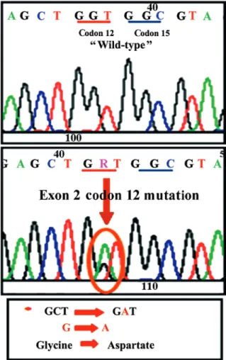
processed via the direct sequencing technique. A representa-tive image of a mutation detected by the direct sequencing analysis (replacement of glycine by aspartate in codon 12 of exon 2 of the K‑Ras gene) is shown in Fig. 1. The distribution of the detected mutations based on the contributing centers is described in Table II.
In our study, TheraScreen did not identify any mutations, while direct sequencing identified 7 mutations in the investi-gated patient population. Due to technical considerations (the starting amount of DNA), we were unable to perform direct Table I. Distribution of K‑Ras mutant and wild-type tumors
based on the mutation analysis method.
Methods Wild-type Mutant Total
TheraScreen 22 0 22
Direct sequencing 11 7 18
Figure 1. Representative image of a mutation detected by direct sequencing analysis. Glycine was replaced by aspartate in codon 12 of exon 2 in the K‑Ras gene.
Table II. Distribution of the K‑Ras wild‑type and mutant tumor samples based on the clinical sites of the study.
Direct Direct
TheraScreen TheraScreen sequencing sequencing Inadequate Wild‑type, Center No. exon 2 wild‑type exon 2 mutated exon 2 wild‑type exon 2 mutated tissue %
Marmara 7 5 - 1 - 1 85
University
Kartal Training and 13 8 ‑ 1 2 2 69
Research Hospital
Ege University 13 5 ‑ 6 2 ‑ 84
Bilim University 10 4 ‑ 3 3 ‑ 70
sequencing on all samples. A number of previous studies demonstrated the high sensitivity of TheraScreen in identi-fying K‑Ras mutations (approaching 100%) (16‑18); thus, we considered that the results of two mutation analysis methods are valid and may be pooled together.
Discussion
HCC is a difficult disease to treat, particularly when presenting at an advanced or non‑resectable stage. Chemotherapy with traditional cytotoxic agents (doxorubicin, cisplatin, or 5‑fluoro-uracil) is associated with a low response rate (<10%) and offers little overall survival benefit. The most common comorbidities, particularly cirrhosis and liver failure, lead to poor tolerance of chemotherapy in these patients, as well as an unpredict-able clinical course, considering that these drugs are mainly metabolized by the liver. Other potential treatment options, such as interferon therapy, anti‑androgens, or tamoxifen, have been proven to be essentially ineffective. Recent advances in the identification of molecules involved in hepatocarcinogenesis has led to the identification of possible therapeutic targets for the targeted therapy of HCC, such as growth factors and neoan-giogenesis factors, as well as their receptors, namely tyrosine kinase intracellular enzymatic pathways and intracellular signal transmission molecules. The focus of targeted therapy for HCC has been largely on tyrosine kinase inhibitors and monoclonal antibodies. The most widely investigated pathways, which are involved in the process of enzymatic activation of growth‑ and proliferation‑promoting intracellular signals, include the Ras/Raf/MEK/MAPK pathway, the phosphoinositol 3‑kinase pathway and the Wnt/catenin pathway (2,5). Thus, studies that assist in selecting patients to receive novel treatments, such as cetuximab, are pivotal, as such therapies may prove curative for these patients. Cetuximab is considered to be a promising agent for advanced‑stage HCC patients, as nearly all HCC cells exhibit increased expression of EGFR. The results of the few small‑sized studies may be summarized as follows: Cetuximab may be effective in the control of advanced HCC, prolonging patient survival; however, combination regimens of cetuximab with oxaliplatin and gemcitabine are particularly effective. The advantage of cetuximab monotherapy in the treatment of advanced HCC is that it exhibits a tolerable toxicity profile, although combination therapy also has an acceptable toxicity profile (2,3,5,6,15,19).
In our pilot study, we found that ~80% of HCC cancer patients carry the wild‑type K‑Ras gene. This is similar to the findings of previous studies, which reported that the K‑Ras mutation is an uncommon or even rare event in HCC. This is in contrast to the frequent presence of K‑Ras mutations in chemical models of HCC in animals (1,19‑24). Considering the small sample size of the previous and present studies, the true prevalence of K‑Ras mutations in HCC has yet to be determined. The discrepancy between various studies may be attributed to the involved etiological factors, as well as to the geographic origins of the patient populations (20‑24). The majority of K‑Ras genes are located on exon 2 and mutations in codons 12, 13, 146 and 154 are the most frequent; >80% of mutations occur in codons 12 and 13 (25-27). Thus, we may hypothesize that our results comprehensively represent K‑Ras mutations in the investigated group of patients. Additionally,
we acknowledge that the clinical characteristics of our patients may be beneficial; however, these data were not added to this pilot study. Our aim was to design the present pilot study to estimate the K‑Ras mutation rate using novel technologies in Turkish HCC patients.
The majority of the previous studies on the role of K‑Ras mutations in response to cetuximab therapy have been performed on colon and head and neck cancer patients. Activating K‑Ras gene mutations are detected in ~15‑30% of non‑small‑cell lung cancer patients and 40‑45% of colorectal cancer patients. Several studies have demonstrated that K‑Ras mutations negatively predict the lack of response to EGFR tyrosine kinase inhibitors in non‑small‑cell lung cancer patients and to cetuximab or panitumumab in colorectal cancer patients. Thus, the general consensus is that patients with K‑Ras mutations do not benefit from cetuximab treat-ment. Patients with wild‑type K‑Ras, in contrast to the mutant type, may even benefit from cetuximab dose escalation and the overall survival time is twice as long in K‑Ras wild‑type patients treated with cetuximab compared with patients harboring K‑Ras mutations (26‑32). In addition to extensive studies on the role of the K‑Ras mutation status in deter-mining the response to cetuximab in colon cancer patients, the same phenomenon has been observed in several other types of cancer, including advanced rectal cancer, gastric cancer, inoperable biliary tract cancer and squamous cell carcinoma of the skin (33‑36).
In conclusion, our results demonstrated that, in a popula-tion of HCC patients referred to 6 major comprehensive cancer centers in Turkey, K‑Ras mutations are a rather uncommon finding. Thus, it may be feasible to initiate in these centers clinical trials using cetuximab for the treatment of unresect-able or advanced‑stage HCC patients.
References
1. Taketomi A, Shirabe K, Muto J, Yoshiya S, Motomura T, Mano Y, Ikegami T, Yoshizumi T, Sugio K and Maehara Y: A rare point mutation in the Ras oncogene in hepatocellular carcinoma. Surg Today 43: 289‑292, 2013.
2. Rossi L, Zoratto F, Papa A, Iodice F, Minozzi M, Frati L and Tomao S: Current approach in the treatment of hepatocellular carcinoma. World J Gastrointest Oncol 2: 348‑359, 2010. 3. Poggi G, Montagna B, Melchiorre F, Quaretti P, Delmonte A,
Riccardi A, Tagliaferri B, Sottotetti F, Di Cesare P, Stella MG, et al: Hepatic intra‑arterial cetuximab in combi-nation with 5‑fluorouracil and cisplatin as salvage treatment for sorafenib‑refractory hepatocellular carcinoma. Anticancer Res 31: 3927-3933, 2011.
4. Dhanasekaran R, Limaye A and Cabrera R: Hepatocellular carcinoma: Current trends in worldwide epidemiology, risk factors, diagnosis, and therapeutics. Hepat Med 4: 19‑37, 2012. 5. Corey KE and Pratt DS: Current status of therapy for
hepato-cellular carcinoma. Therap Adv Gastroenterol 2: 45‑57, 2009. 6. Zhu AX, Stuart K, Blaszkowsky LS, Muzikansky A,
Reitberg DP, Clark JW, Enzinger PC, Bhargava P, Meyerhardt JA, Horgan K, et al: Phase 2 study of cetuximab in patients with advanced hepatocellular carcinoma. Cancer 110: 581‑589, 2007. 7. Mittal S and El‑Serag HB: Epidemiology of hepatocellular
carcinoma: Consider the population. J Clin Gastroenterol 47 (Suppl): S2‑S6, 2013.
8. El‑Serag HB: Epidemiology of viral hepatitis and hepatocellular carcinoma. Gastroenterology 142: 1264‑1273.e1, 2012.
9. Alacacioglu A, Somali I, Simsek I, Astarcioglu I, Ozkan M, Camci C, Alkis N, Karaoglu A, Tarhan O, Unek T, et al: Epidemiology and survival of hepatocellular carcinoma in Turkey: Outcome of multicenter study. Jpn J Clin Oncol 38: 683‑688, 2008.
10. Dogan E, Yalcin S, Koca D and Olmez A: Clinicopathological characteristics of hepatocellular carcinoma in Turkey. Asian Pac J Cancer Prev 13: 2985‑2990, 2012.
11. Can A, Dogan E, Bayoglu IV, Tatli AM, Besiroglu M, Kocer M, Dulger AC, Uyeturk U, Kivrak D, Orakci Z, et al: Multicenter epidemiologic study on hepatocellular carcinoma in Turkey. Asian Pac J Cancer Prev 15: 2923‑2927, 2014.
12. Ye H, Zhang C, Wang BJ, Tan XH, Zhang WP, Teng Y and Yang X: Synergistic function of Kras mutation and HBx in initiation and progression of hepatocellular carcinoma in mice. Oncogene 33: 5133‑5138, 2014.
13. Adjei AA: Blocking oncogenic Ras signaling for cancer therapy. J Natl Cancer Inst 93: 1062‑1074, 2001.
14. Kim JH, Kim HY, Lee YK, Yoon YS, Xu WG, Yoon JK, Choi SE, Ko YG, Kim MJ, Lee SJ, et al: Involvement of mitophagy in oncogenic K‑Ras‑induced transformation: Overcoming a cellular energy deficit from glucose deficiency. Autophagy 7: 1187‑1198, 2011.
15. Asnacios A, Fartoux L, Romano O, Tesmoingt C, Louafi SS, Mansoubakht T, Artru P, Poynard T, Rosmorduc O, Hebbar M, et al: Gemcitabine plus oxaliplatin (GEMOX) combined with cetuximab in patients with progressive advanced stage hepatocellular carcinoma: Results of a multicenter phase 2 study. Cancer 112: 2733-2739, 2008.
16. Vallée A, Le Loupp AG and Denis MG: Efficiency of the TheraScreen® RGQ PCR kit for the detection of EGFR mutations in non‑small cell lung carcinomas. Clin Chim Acta 429: 8‑11, 2014.
17. Adams JA, Post KM, Bilbo SA, Wang X, Sen JD, Cornwell AJ, Malek AJ and Cheng L: Performance evaluation comparison of 3 commercially available PCR‑based KRAS mutation testing platforms. Appl Immunohistochem Mol Morphol 22: 231‑235, 2014.
18. Chang SC, Denne J, Zhao L, Horak C, Green G, Khambata‑Ford S, Bray C, Celik I, Van Cutsem E and Harbison C: Comparison of KRAS genotype: TheraScreen assay vs. LNA‑mediated qPCR clamping assay. Clin Colorectal Cancer 12: 195‑203.e2, 2013. 19. Sanoff HK, Bernard S, Goldberg RM, Morse MA, Garcia R,
Woods L, Moore DT and O'Neil BH: Phase II study of capecitabine, oxaliplatin and cetuximab for advanced hepato-cellular carcinoma. Gastrointest Cancer Res 4: 78‑83, 2011. 20. Colombino M, Sperlongano P, Izzo F, Tatangelo F, Botti G,
Lombardi A, Accardo M, Tarantino L, Sordelli I, Agresti M, et al: BRAF and PIK3CA genes are somatically mutated in hepato-cellular carcinoma among patients from South Italy. Cell Death Dis 3: e259, 2012.
21. Lord PG, Hardaker KJ, Loughlin JM, Marsden AM and Orton TC: Point mutation analysis of ras genes in spontaneous and chemically induced C57Bl/10J mouse liver tumours. Carcinogenesis 13: 1383‑1387, 1992.
22. Weihrauch M, Benick M, Lehner G, Wittekind M, Bader M, Wrbitzk R and Tannapfel A: High prevalence of K‑ras‑2 mutations in hepatocellular carcinomas in workers exposed to vinyl chloride. Int Arch Occup Environ Health 74: 405‑410, 2001. 23. Weihrauch M, Benicke M, Lehnert G, Wittekind C, Wrbitzky R
and Tannapfel A: Frequent k‑ras‑2 mutations and p16(INK4A) methylation in hepatocellular carcinomas in workers exposed to vinyl chloride. Br J Cancer 84: 982‑989, 2001.
24. Bai F, Nakanishi Y, Takayama K, Pei XH, Inoue K, Harada T, Izumi M and Hara N: Codon 64 of K‑ras gene mutation pattern in hepatocellular carcinomas induced by bleomycin and 1‑nitro-pyrene in A/J mice. Teratog Carcinog Mutagen 23 (Suppl 1): 161‑170, 2003.
25. Ciardiello F and Tortora G: EGFR antagonists in cancer treatment. N Engl J Med 358: 1160‑1174, 2008.
26. Yarom N and Jonker DJ: The role of the epidermal growth factor receptor in the mechanism and treatment of colorectal cancer. Discov Med 11: 95‑105, 2011.
27. Douillard JY, Oliner KS, Siena S, Tabernero J, Burkes R, Barugel M, Humblet Y, Bodoky G, Cunningham D, Jassem J, et al: Panitumumab‑FOLFOX4 treatment and RAS mutations in colorectal cancer. N Engl J Med 369: 1023‑1034, 2013.
28. Garassino MC, Borgonovo K, Rossi A, Mancuso A, Martelli O, Tinazzi A, Di Cosimo S, La Verde N, Sburlati P, Bianchi C, et al: Biological and clinical features in predicting efficacy of epidermal growth factor receptor tyrosine kinase inhibitors: A systematic review and meta‑analysis. Anticancer Res 29: 2691‑2701, 2009. 29. Yen LC, Uen YH, Wu DC, Lu CY, Yu FJ, Wu IC, Lin SR and
Wang JY: Activating KRAS mutations and overexpression of epidermal growth factor receptor as independent predictors in metastatic colorectal cancer patients treated with cetuximab. Ann Surg 251: 254‑260, 2010.
30. Folprecht G, Gruenberger T, Bechstein W, Raab HR, Weitz J, Lordick F, Hartmann JT, Stoehlmacher‑Williams J, Lang H, Trarbach T, et al: Survival of patients with initially unresectable colorectal liver metastases treated with FOLFOX/cetuximab or FOLFIRI/cetuximab in a multidisciplinary concept (CELIM study). Ann Oncol 25: 1018‑1025, 2014.
31. Ji JH, Park SH, Lee J, Kim TW, Hong YS, Kim KP, Kim SY, Baek JY, Kang HJ, Shin SJ, et al: Prospective phase II study of neoadjuvant FOLFOX6 plus cetuximab in patients with colorectal cancer and unresectable liver‑only metastasis. Cancer Chemother Pharmacol 72: 223-230, 2013.
32. Jiang Y, Kimchi ET, Staveley‑O'Carroll KF, Cheng H and Ajani JA: Assessment of K‑ras mutation: A step toward person-alized medicine for patients with colorectal cancer. Cancer 115: 3609‑3617, 2009.
33. Rubovszky G, Láng I, Ganofszky E, Horváth Z, Juhos E, Nagy T, Szabó E, Szentirmay Z, Budai B and Hitre E: Cetuximab, gemcitabine and capecitabine in patients with inoperable biliary tract cancer: A phase 2 study. Eur J Cancer 49: 3806‑3812, 2013. 34. Shi M, Shi H, Ji J, Cai Q, Chen X, Yu Y, Liu B, Zhu Z and Zhang J:
Cetuximab inhibits gastric cancer growth in vivo, independent of KRAS status. Curr Cancer Drug Targets 14: 217‑224, 2014. 35. Sun PL, Li B and Ye QF: Effect of neoadjuvant cetuximab,
capecitabine and radiotherapy for locally advanced rectal cancer: Results of a phase II study. Int J Colorectal Dis 27: 1325‑1332, 2012.
36. Uribe P and Gonzalez S: Epidermal growth factor receptor (EGFR) and squamous cell carcinoma of the skin: Molecular bases for EGFR‑targeted therapy. Pathol Res Pract 207: 337‑342, 2011.