Ultrastructural Changes in Rat Thyroid Tissue After
Acute Organophosphate
Poisoning and Effects of
Antidotal Therapy with Atropine and Pralidoxime:
A Single-Blind, Ex Vivo Study
Deniz Satar, MD1; Salim Satar, MD2; Ufuk Ozgu Mete, MD3; Jeffrey R. Suchard, MD4; Metin Topal, MD2; Emre Karakoc, MD5; and Mehmet Kaya, PhD 3
1Pathology
Department,
Adana Numune
Education
ant] Research
Hospital,
Adana, Turkey;
2Department
of Emergency
34edicine,
Cukurova
University
School
of 34edicine,
Adana, Turkey;
3Department of
Histology
and Embryology,
Cukurova University
School
of 34edicine,
Adana,
Turkey;
4Department
of Emergency
Medicine,
University
of California Irvine 34edical
Center,
Orange,
California; and 5Department
of k4edical
Intensive
Care, Cukurova University
School
of k4edicine,
Adana, Turkey
A B S T R A C T
B A C K G R O U N D : Organophosphate (OP) insecticides are widely used in both agri- cultural and landscape pest control, and the potential for human exposure to these com- pounds is significant.
O B J E C T I V E S : The aims of this study were to investigate the effects of acute poi- soning with the OF methamidophos and the effects of antidotal therapy with atropine and pralidoxime on rat thyroid tissue ultrastructure.
M E T H O D S : In this single-blind, ex vivo study, male Wistar albino rats weighing 220 to 230 g were divided into 4 treatment groups. Group 1 received a median lethal dose of methamidophos (30 mg/kg) via oral gavage. Group 2 received saline via oral gavage and served as the control group for group 1. Group 3 received methamidophos (30 mg/kg) via oral gavage, and after 8 minutes atropine 0.05 mg/kg and pralidoxime chloride (2-FAM) (40 mg/kg) were administered intraperitoneally (IF). Atropine was titrated to reverse signs of cholinergic excess. Group 4 received saline via oral gavage followed by IF injections and served as the control for group 3. Rat thyroid tissues were examined using electron microscopy, and the histologic changes were examined by a histopathologist who was blinded to treatment. All rats were euthanized by intracardiac blood collection. The rats in groups 1 and 2 were euthanized 8 minutes after treatment. The rats in groups 3 and 4 were euthanized 96 hours after treatment.
R E S U L T S : Thirty-four male rats (aged 16 weeks) were included in the study. The rats were grouped accordingly: group 1 (n 10); group 2 (n 7); group 3 (n 10); and group 4 (n 7). The mean (SD) pseudocholinesterase (FCE) activity was significantly lower in the methamidophos-treated rats (group 1) compared with the corresponding control group (group 2) (32.6 [17.0] vs 579.4 [59.0] U/L, respectively;
Accepted~r publication April 7, 2008.
© 2008 Excerpta Medica Inc. All rights reserved.
doi:10.1016/j.curtheres.2008.07.001 0011-393X/$32.00
D. S A T A R E T A L .
P < 0.001). PCE activity was significantly higher in rats treated with atropine and 2-PAM (group 3) (392.5 [39.4] U/L; P < 0.001) compared with those not receiving antidotal therapy (group 1). Group 1 experienced changes in thyrocytes and organelles that were not detected in the antidote-treated rats in group 3. These changes included follicular cell nuclei exhibiting an increase in chromatin content, pyknotic nuclei, mitochondrial degeneration, dilated granular endoplasmic reticulum cisternae, re- duced microvilli, and intraluminal cellular debris. Within follicular ceils, formation of vacuoles filled with fine granular material was noted.
C O N C L U S I O N : Acute OP poisoning was associated with histopathologic effects in rat thyroid tissue that appeared to be mitigated by antidotal therapy in this small animal study. More extensive studies using immunohistochemical methods are needed.
(Cuff Thef Res C/in Exp. 2008;69:334 342) © 2008 Excerpta Medica Inc.
K E Y W O R D S : methamidophos, organophosphate, poisoning, thyroid, ultrastructure.
I N T R O D U C T I O N
Organophosphate (OP) insecticides are widely used in both agricultural and landscape pest control, and the potential for human exposure to these compounds is significant. The primary toxicity associated with acute exposure to OP insecticides is cholinergic crisis resulting from inhibition of acetylcholinesterase activity) Additional potential effects of OPs include delayed polyneuropathy, immunotoxicity, carcinogenesis, and endocrine, developmental, and reproductive toxicities. 2,3
Depressed thyroid hormone concentrations have been described in critical illness as well as in association with a number of drugs; however, thyroid function returned to normal when the nonthyroidal illness resolved) This temporary, illness-associated hypothyroidism has been referred to as euthyroid sick sy~ldrome. In a previous single-blind in vivo experimental study, 5 we found that acute OP poisoning in rats induced a hypo- thyroid state consistent with euthyroid sick syndrome. Some authors have postulated that this condition might be related to altered cytokines, reduced pulsatile secretion of thyroid-stimulating hormone (TSH), decreased peripheral conversion of thyroxine (T4) to triiodothyronine (T3), and perhaps to an attempt by the body to reduce energy expenditure in times of critical illness. 6
There is evidence that acetylcholine is involved in regulating pituitary functions, 7 and it is well known that the symptoms after poisoning with OP compounds occur due to the effects on acetylcholine receptors. Numerous studies have found thyroid dysfunc- tion during critical illness. 6
The aims of this study were to investigate ultrastructural changes in rat thyroid tis- sue after administration of a single median lethal dose (LD50) of the highly toxic OP pesticide methamidophos, and whether antidotal therapy with atropine and pralidoxime chloride (2-PAM) mitigated or altered these changes.
M A T E R I A L S A N D M E T H O D S
This study was approved by the Cukurova University Animal Research Ethics Com- mittee (Adana, Turkey). Male Wistar albino rats were obtained from Cukurova
University Medical Sciences Experimental Research Center (Adana, Turkey). The rats weighed between 220 and 230 g. They were housed 5 to a cage, with a room tempera- ture of 22 (+2)°C and a light-dark cycle of 12 hours on and 12 hours off. Food and water were available ad libitum.
In preparation for the study, the animals were anesthetized with 75 mg/kg ketamine and 5 mg/kg xylazine IM. Methamidophos (O,S-dimethyl phosphoramidothioate) with a purity of 99.1% was diluted in tap water to 30 mg/kg 1. m/L 1 and administered via oral gavage using a 20-gauge feeding tube. Atropine sulfate and 2-PAM were dissolved in 0.9% saline and injected intraperitoneally (IP) with a 23-gauge needle. Each rat was treated and examined separately. Rats were euthanized by intracardiac blood collection. Blood samples were centrifuged at 1000 cycles/min for 10 minutes, and the plasma was stored at ~ 0 ° C until analyzed for pseudocholinesterase (PCE) activity. PCE was assayed using an enzymatic colorimetric method (S-butyrylthiocholine iodide; Cobas Integra ® 800, Roche, Oberkochen, Germany).
Thyroid tissues were fixed in 5% glutaraldehyde in Millonig's phosphate buffer (Electron Microscopic Science, Hatfield, Pennsylvania) at p H 7.4 for 4 hours and post- fixed in 1% osmium tetroxide in phosphate buffer at p H 7.4 for 2 hours at 4°C. Tissues were dehydrated in graded ethanol and embedded in araldite (Electron Microscopic Science). Thin sections were cut using a Reichert Ultracut S ultramicrotome (Leica Microsystems, Vienna, Austria), stained with uranyl acetate and lead citrate, and then examined with a Zeiss EM 10B electron microscope (Carl Zeiss Inc., Thornwood, New York). The histopathologic changes were examined by a histopathologist who was blinded to treatment.
The rats were divided into 4 treatment groups. Group 1 received 30 mg/kg meth- amidophos (the LD50 of this compound in rats) via oral gavage. 7 Group 2 received an equivalent volume of 0.9% saline via oral gavage. Cholinergic signs (eg, muscle fascicu- lations, bronchorrhea, bradycardia, seizures) were noted to begin within 5 to 8 minutes of treatment with methamidophos. Following the same protocol in our previous study, the rats in groups 1 and 2 were euthanized 8 minutes after treatment. 5
Group 3 received 30 mg/kg methamidophos via oral gavage. The signs and symp- toms of methamidophos poisoning began within 5 to 8 minutes after treatment, as in group 1. Eight minutes after treatment, group 3 was treated with atropine and 2-PAM IE The atropine was titrated to reverse signs of cholinergic excess, especially bronchor- rhea. The 2-PAM was administered as a bolus dose of 40 mg/kg. Group 4 was admin- istered initially via oral gavage with 0.9% saline in equivalent volumes to group 3 and then received IP injections of 0.9% saline in equal numbers, volumes, and times to those in group 3. The animals in groups 3 and 4 were euthanized after 96 hours because the intermediate syndrome after severe OP poisoning usually occurs within 96 hours) Our main concern was to make sure that the animals that were administered the anti- dotes were cured; 96 hours were sufficient to show whether the antidote was effective.
After cholinergic signs and symptoms were identified in group 1, intracardiac blood samples were drawn and placed in ethylenediaminepentaacetic acid blood-collection tubes and thyroid biopsies were obtained. The rats in groups 3 and 4 were allowed to emerge from anesthesia and were returned to their cages. Four days after the initial
D. S A T A R E T A L .
interventions, these rats were reanesthetized and euthanized by intracardiac blood col- lection, and thyroid biopsies were obtained.
S T A T I S T I C A L A N A L Y S I S
Statistical analyses were performed using the statistical package SPSS version 10.0 (SPSS Inc., Chicago, Illinois). The Mann-Whitney U test was used for between-group comparisons. The Bonferroni adjustment was used when multiple comparisons were performed. P < 0.05 was considered significant. Data were reported as mean (SD).
R E S U L T S
P S E U D O C H O L I N E S T E R A S E A C T I V I T Y
Thirty-four male rats (aged 16 weeks) were included in the study. The rats were grouped accordingly: group 1 (n 10); group 2 (n 7); group 3 (n 10); and group 4 (n 7). The mean (SD) PCE activity was significantly lower in the methamidophos- treated rats (group 1) compared with the control group (group 2) (32.6 [17.0] vs 579.4 [59.0] U/L; P < 0.001). PCE activity was significantly higher in rats treated with atropine and 2-PAM (group 3) (392.5 [39.4] U/L; P < 0.001) compared with those not receiving antidotal therapy (group 1). PCE activity in antidote-treated rats (group 3) was significantly lower compared with the control group (group 4) (616.3 [54.3] U/L; P < 0.001) 96 hours (4 days) after initial treatment.
T H Y R O I D T I S S U E U L T R A S T R U C T U R A L F I N D I N G S
In group 1, the nuclei of follicular ceils arranged around the colloid rested on the basal lamina and exhibited an increase in chromatin content. In some follicles, pyknotic nuclei, degenerated mitochondria, dilated granular endoplasmic reticulum cisternae, and reduced microvilli were found (Figures 1 and 2). Intraluminal cellular debris was also found. Vacuoles filled with fine granular material within the cytoplasm of follicu- lar cells and indented heterochromic nuclei were noted (Figure 3).
In group 2, the structure of the thyroid follicles was normal. The follicular cell nuclei were spherical and contained prominent nucleoli. Short microvilli on the apical sur- faces of the ceils, numerous profiles of rough endoplasmic reticula, and well-developed Golgi complexes were seen. Parafollicular ceils within the follicular epithelium were located near the basal lamina and contained small, membrane-bound secretory granules (Figure 4).
In group 3, the nuclei and cytoplasmic organelles of the follicular ceils were generally normal. Numerous rough endoplasmic reticulum cisternae, short microvilli extending to the follicular lumen, well-developed Golgi complexes, and small secretory granules were observed. The connective tissue surrounding the follicles contained capillaries (Figure 5).
In group 4, follicular and parafollicular ceils appeared normal. Nuclei and cytoplas- mic organdies of the epithelial ceils exhibited normal histologic structures (Figure 6).
D I S C U S S I O N
Acute depression of the pituitary-thyroid axis during concurrent illness has been inter- preted teleologically as an attempt by the body to reduce energy expenditure, at least
' ,. ::' ;./~ ' ",~. ~t... ~,'.- c. ~ ~.-. • ~,.~
,., ,,
• ~ ' ~ t ~ ' ~ ~ - ~ ~ ~
• •: ~ • '•.~- ~ •,-~-~ ,•.
• ..._/~ ..~, ~ ,..:~.~-. _
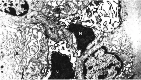
Figure 1. Electron micrograph of the thyroid tissue of the methamidophos-treated rats (group 1) showing pyknoUc nuclei (N), degenerated mitochondria (M), and dilated granular endoplasmic reticulum (GER) cisternae (uranyl acetate and lead citrate; original magnification 8837X).
Figure 2. Electron micrograph of the thyroid tissue of the methamidophos-treated rats
(group 1) showing reduction of microvilli (Mv), degenerated mitochondria (M), and dilated granular endoplasmic reticulum (GER) cistemae (uranyl acetate
and lead citrate; original magnification 10,850X).
D. S A T A R E T A L .
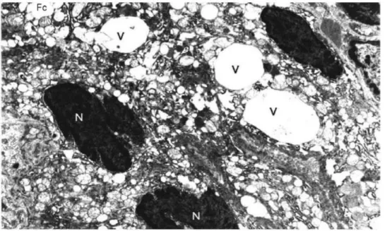
Figure 3. Electron micrograph of the thyroid tissue of the methamidophos-treated rats (group 1) showing vacuoles filled with fine granular material (V) in the cytoplasm of follicular cells (Fc) and indented heterochromic nuclei (N) (uranyl acetate and lead citrate; original magnification 7087X).
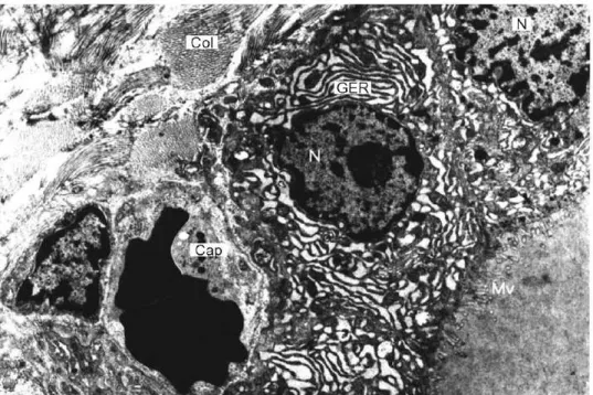
Figure 4. Electron micrograph of the thyroid tissue of the control group for group 1 (group 2) showing follicular cells (Fc) containing numerous granular endoplasmic reticula (GER), nuclei (N), colloid (C), and a capillary (Cap) (uranyl acetate and lead citrate; original magnification 8837X).
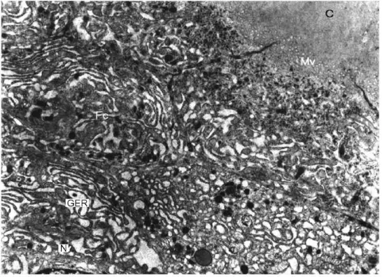
Figure 5. Electron micrograph of the thyroid tissue of rats that were treated with atropine and pralidoxime chloride 8 minutes after receiving methamidophos (group 3). The nuclei of the follicular cells containing normal chromatin (N), numerous granular endoplasmic reticulum (GER) cisternae, microvilli (Mv), a capillary (Cap), and collagen fibers (Col) are visible (uranyl acetate and lead citrate; original magnification 7087X).
when it occurs during starvation, and thus as an appropriate response that does not warrant intervention. 8 Whether this is also applicable to other acute stress conditions (eg, surgery, infection, or the initial phases of critical illness) is still a matter of contro- versy. 9 A previous study 4 found decreased levels ofT3, T4, and TSH in rats after poison- ing with a single LDs0 of methamidophos. The decrease in T3 was statistically signifi- cant, consistent with the development of euthyroid sick syndrome (P < 0.005).
In this study, we observed ultrastructural changes in the thyroid tissue (eg, increased chromatin content, pyknotic nuclei, mitochondrial degeneration, dilated granular endo- plasmic reticulum cisternae, reduced microvilli, intraluminal cellular debris, vacuoles filled with fine granular material within follicular ceils). These changes after a single LD50 of methamidophos might be related to impaired thyrocyte function and the development of biochemical markers of hypothyroidism. It is also possible that accumu- lated acetylcholine was responsible for the observed ultrastructural changes. Changes in thyroid tissue induced by other chemicals have been studied, 1°,11 although it is not clear whether these chemicals aftected thyroid ultrastructure at the receptor level or as a neuroendocrine response of the body.
D. S A T A R E T A L .
Figure 6. Electron microscopic examination of the thyroid tissue of the control group for group 3 (group 4). Follicular cells (Fc) appear normal; short microvilli (Mv), colloid (C), nuclei (N), and granular endoplasmic reticulum (GER) cisternae are visible (uranyl acetate and lead citrate, original magnification 7087X).
L I M I T A T I O N S
There were some limitations of this study. We do not know the reason for the ultrastructural changes that we found in the thyroid tissue. These changes might have been due to the direct toxic effect of OP compounds on thyroid tissue or to effects on the hypothalamic-pituitary-thyroid axis. Immunohistochemical studies of the direct toxic effects of OPs need to be performed. Thyroid-releasing hormone concentrations might also help to explain disruption of hypothalamic functions. Another limitation of this study is that the rats were not randomized to treatment group. The small sample size was also a study limitation. In addition, the animals in the 4 groups were not all euthanized at the same time: groups 1 and 2 were euthanized 8 minutes after treatment, and groups 3 and 4 were euthanized
96
hours after treatment. To add to these, the histologic changes were not scored.CONCLUSION
Acute OP poisoning with methamidophos in rats induced ultrastructural changes in thyroid tissue that appeared to be mitigated by antidotal therapy in this small animal study. More extensive studies using immunohistochemical methods are needed.
A C K N O W L E D G M E N T
This study was funded by Cukurova University Research Project Foundation Project, Adana, Turkey (No. TF 2001 M19).
R E F E R E N C E S
1. Pope CN. Organophosphorous pesticides: Do they all have the same mechanism of toxicity? J Toxico! Environ Health B. 1999;2:161 181.
2. Astroff AB, Freshwater K J, Eigenberg DA. Comparative organophosphate-induced effects observed in adult and neonatal Sprague-Dawley rats during the conduct of multigeneration toxicity studies. Reprod ToxicoL 1998;12:619 645.
3. Sultatos LG. Mammalian toxicdogy of organophosphorus pesticides. J Toxico! Environ Health, 1994;43:271289.
4. Camacho PM, Dwarkanathan AA. Sick euthyroid syndrome. What to do when thyroid function tests are abnormal in critically ill patients. Postgrad k4ed, 1999;105:215219.
5. Satar S, Satar D, Kirim S, Leventerler H. Effects of acute organophosphate poisoning on thyroid hormones in rats. AmJ Ther, 2005;12:238242.
6. Van der Berghe G. Novel insights into the neuroendocrinology of critical illness. EurJEndocrinoL 2000;143:1 13.
7. Tuomisto J, M~innist/5 E Neurotransmitter regulation of anterior pituitary hormones. Pharmaco! Rev, 1985;37:249 332.
8. Gardner DF, Kaplan MM, Stanley CA, Utiger RD. Effect of tri-iodothyronine replacement on the metabdic and pituitary responses to starvation. N EnglJ k4ed, 1979;300:579~84.
9. De Groot LJ. Dangerous dogmas in medicine: The nonthyroidal illness syndrome, jr C//n Endocrino! k4etab, 1999;84:151 169.
10. Iakubovskii MM, Zurnadzhi IuN, Khmel'nitskii OK, et al. Ultrastructural changes in the adenohypophysis-thyroid system in chronic poisoning by the herbicide linuron [in Russian]. Arleh PatoL 1991;53:2~30.
11. Gao TS, Hu FN, Teng WP. Effect of mild and moderate excessive iodine supplementation on thyroid function and morphology in non-iodine deficiency rat model [in Chinese]. Zhonghua Nei Ke Za Zhi, 2003;42:705~08.
A D D R E S S C O R R E S P O N D E N C E TO: Emre Karakoc, MD, Department of Medical Intensive Care, C u k u r o v a University School of Medicine, 01330 Adana, Turkey. E-mail: [email protected]