Yilmaz F et al.
Konuralp Medical Journal 2021;13(1): 1-3
1
LETTER TO
EDITOR
Fahri Yilmaz1 Asuman Kilitci2 Oktay Buyukasik³ 1Sakarya University Faculty of Medicine, Department of Pathology, Sakarya, Turkey 2Kırşehir Ahi Evran University Faculty of Medicine, Department of Pathology, Kırşehir, Turkey 3
Lokman Hekim Hospital, Department of General surgery, Ankara, Turkey
Corresponding Author:
Asuman Kilitci
Kırşehir Ahi Evran University Faculty of Medicine, Department of Pathology, Kırşehir, Turkey mail: [email protected] Phone:+90 5079553256 Received: 16.09.2020 Acceptance: 21.11.2020 DOI: 10.18521/ktd.795732
This article was presented in part at the 25th European Congress of Pathology at Lisbon held at 31 August-4 September, 2013
Konuralp Medical Journal e-ISSN1309–3878
[email protected] [email protected] www.konuralptipdergi.duzce.edu.tr
Primary Cavernous Hemangioma of the Thyroid Gland
ABSTRACTHemangioma of the thyroid gland is a rare diagnosis and less than 35 cases have been previously published in the literature. The pathogenesis is unknown but it is suggested to be a developmental anomaly which is associated with an incapability of angioblastic mesenchyma to form canals. On microscopic examination, it should be distinguished from reactive endothelial hyperplasia, angiosarcoma or hemangiosarcoma. We report a case of a primary thyroid hemangioma, with nonspesific US appearance. Cytologic findings were nondiagnostic and final diagnosis was made at pathologic examination of the hemithyroidectomy specimen.
Keywords: Hemangioma, Cavernous, Thyroid Gland, Histopathology
Tiroid Bezinin Primer Kavernöz Hemanjiyomu
ÖZET
Tiroid bezinin hemanjiyomu nadir bir tanıdır ve literatürde günümüze kadar 35'ten az vaka yayınlanmıştır. Patogenezi tam bilinmemekle birlikte, anjiyoblastik mezenşimin kanal oluşturma yetersizliği ile ilişkili gelişimsel bir anomali olduğu ileri sürülmektedir. Mikroskobisinde, reaktif endotelyal hiperplazi, anjiyosarkom ve hemanjiyosarkomdan ayırt edilmelidir. Biz nonspesifik ultrasonografik görünüme sahip bir primer tiroid kavernöz hemanjiyomu olgusunu sunuyoruz. Sitolojik bulgular tanısal değildi ve kesin tanı spesimeninin patolojik incelemesinde kondu.
Yilmaz F et al.
Konuralp Medical Journal 2021;13(1): 1-3
2 Dear Editor,
Hemangioma is a benign neoplasm of blood vessels that can be found in all organ systems. Hemangioma of the thyroid gland is an extremely rare entity and less than 35 cases have been previously published in the literature (1,2). Here, we report a case of a primary thyroid hemangioma (TH), with nonspesific ultrasonographic appearance. Cytologic findings were nondiagnostic and final diagnosis was made at pathologic examination of the hemithyroidectomy specimen. A 24-year-old euthyroid female presented with a slowly growing neck swelling. She had compression symptoms. She had no history of trauma, fine needle aspiration (FNA) or other surgical procedures. Ultrasonography (USG) revealed a solitary hypoechoic nodule of the right
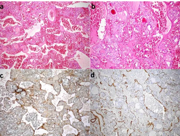
thyroid lobe. Surgery in the form of right hemithyroidectomy was done with a preoperative diagnosis of nodular goiter. On macroscopic examination, most of the right lobe was occupied by a reddish, solid lesion with a diameter of 3.8 cm that proved to be a cavernous TH. Histologically, the tumor was encapsulated with fibrous tissue and contained many cavernous thin walled vessels, some of which were thrombosed (Figure 1a, 1b). The vascular nature of the structures was confirmed by immunohistochemistry: the cells were positive for the endothelial markers CD34, SMA, FactorVIII and negative for TTF-1 (Figure 1c, 1d). No features of malignancy were identified. The definitive diagnosis of a benign and isolated hemangioma of the right thyroid lobe was made.
Figure 1: a, b) The tumor contains many cavernous thin-walled vessels, some of which are thrombosed (H&E, x100; H&E, x200). c) The cells of vascular structures were positive for the endothelial immunohistochemical
markers: FactorVIII related antigene (x200). d) SMA expression in endothelial cells (x100). Secondary hemangiomas have been defined
as pseudomalformations, caused by the organization of a hematoma after a trauma or FNA. Primary TH is well-circumscribed capsulated mass with diameters in 20-40 mm that have a predilection for males, and predominantly affects the left lobe of thyroid (3).
The pathogenesis is unknown but it is suggested to be a developmental anomaly which is associated with an incapability of angioblastic mesenchyma to form canals (1). It is difficult to differentiate cavernous hemangiomas of the thyroid
gland from other typical thyroid diseases due to the similar pattern they exhibit on USG, computed tomography or FNA cytology. Preoperatively, the diagnosis may be overlooked. Definitive diagnosis is reached only with histopathological examination. THs should be distinguished from reactive endothelial hyperplasia, angiosarcoma or hemangiosarcoma. Hemi/total thyroidectomy is the treatment of choice. An effort should be made to dissect the thyroid without rupture of these lesions in order to minimize blood loss during the operation (4).
Yilmaz F et al.
Konuralp Medical Journal 2021;13(1): 1-3
3 REFERENCES
1. Miao J, Chen S, Li Y, Fu L, Li H. A primary cavernous hemangioma of the thyroid gland: A case report and literature review. Medicine. 2017;96(49): e8651.
2. Zehao Li, Jianhua Li, Liwen Li, Hongting Li, Lijun Fu, Xinguang Qiu. One case of cavernous hemangioma of the thyroid gland. Chinese Journal of Endocrine Surgery 2018;12(1):71-3.
3. Dasgupta A, Teerthanath S, Jayakumar M, HS Kıran, Raju M. Primary cavernous haemangioma of the thyroid-a case report. Journal of clinical and diagnostic research. 2014; 8(2):151-2.
4. Michalopoulos NV, Markogiannakis H, Kekis PB, Papadima A, Lagoudianakis E, Manouras A. Primary cavernous hemangioma of the thyroid gland. Southern Medical Journal. 2010;103(7): 674-5.