Experimental Study / Deneysel Çalışma
The effects of subacute deltamethrin exposure on rat liver:
histochemical, immunohistochemical, and biochemical study
Türker Çavuşoğlu,1,2 Muhammet Fatih Aydın,3 Eylem Çağıltay,4 Gürkan Yiğittürk,1 İlker Kızıloğlu,5 Ayfer Meral,6 Kubilay Doğan Kılıç,1 Yiğit Uyanıkgil,1,2 Emel Öykü Çetin Uyanıkgil,7 Oytun Erbaş8
1Department of Histology and Embryology, Medical Faculty of Ege University, İzmir, Turkey 2Ege University, Cord Blood, Cell-Tissue Application and Research Center, İzmir, Turkey 3Department of Gastroentology, Medical Faculty of Kemerburgaz University, İstanbul, Turkey 4Department of Internal Medicine, Endocrine and Metabolism, İzmir Military Hospital, İzmir, Turkey
5Department of General Surgery, İzmir Bozyaka Training and Research Hospital, İzmir, Turkey.
6Department of Biochemistry, Dumlupınar University, Evliya Çelebi Training and Research Hospital, Kütahya, Turkey. 7Department of Pharmaceutical Technology, Ege University, Faculty of Pharmacy, İzmir, Turkey.
8Department of Physiology, Medical Faculty of Bilim University, İstanbul, Turkey.
ABSTRACT
Objectives: This study aims to determine deltamethrin’s effects on early diagnosis of toxicity and liver toxicity markers in an animal model.
Materials and methods: A total of 36 male rats weighing 200-220 gram were separated into four groups. Group 1 rats were given deltamethrin
15 mg/kg/day, group 2 rats were given deltamethrin 30 mg/kg/day, group 3 rats were given deltamethrin 60 mg/kg/day, and group 4 rats (control group) were given 0.9% sodium chloride 1 mL/kg/day for 30 days by oral gavage.
Results: Deltamethrin ingestion resulted in edema, nuclear hypertrophy, and parenchymal and stromal inflammation on the histopathological
examination. Malondialdehyde levels increased and glutathione levels decreased when compared with the control group as the treatment dose increased. Alanine transaminase levels increased significantly when compared with the control group. The changes correlated directly with increasing dose.
Conclusion: Deltamethrin usage causes liver toxicity in a dose dependent manner on rats. This toxicity is observed both biochemically and
histopathologically. Liver biomarkers and oxidative stress parameters can effectively be used for potential toxicity monitorization.
Keywords: Deltamethrin; liver; rat; subacute toxicity.
Sıçan karaciğerinde subakut deltametrin maruziyetinin etkileri:
Histokimyasal, immünohistokimyasal ve biyokimyasal çalışma
ÖZ
Amaç: Bu çalışmada bir hayvan modelinde deltametrinin toksisitenin erken tanısı ve karaciğer toksisitesi göstergeleri üzerindeki etkileri değerlendirildi. Gereç ve yöntemler: İki yüz-iki yüz yirmi gram ağırlığında toplam 36 erkek sıçan dört gruba ayrıldı. Grup 1 sıçanlarına deltametrin 15 mg/kg/gün, grup 2
sıçanlarına deltametrin 30 mg/kg/gün, grup 3 sıçanlarına deltametrin 60 mg/kg/gün ve grup 4 sıçanlarına (kontrol grubu) %0.9 sodyum klorür 1 mL/kg/ gün 30 gün boyunca oral gavaj yolu ile verildi.
Bulgular: Histopatolojik incelemede deltametrin sindirimi ödem, nükleer hipertrofi ve parankimal ve stromal enflamasyon ile sonuçlandı. Kontrol
grubu ile karşılaştırıldığında, tedavi dozu arttıkça malondialdehid düzeyleri arttı ve glutatyon düzeyleri azaldı. Kontrol grubu ile karşılaştırıldığında, alanin transaminaz düzeyleri anlamlı şekilde arttı. Değişiklikler artan doz ile doğrudan ilişki idi.
Sonuç: Sıçanlarda deltametrin kullanımı doza bağımlı olarak karaciğer toksisitesine yol açmaktadır. Bu toksisite hem biyokimyasal hem histopatolojik
olarak gözlemlenmektedir. Karaciğer biyogöstergeleri ve oksidatif stres parametreleri olası toksisite monitörizasyonunda etkili şekilde kullanılabilir.
Anahtar sözcükler: Deltametrin; karaciğer; sıçan; subakut toksisite.
Received: April 25, 2016 Accepted: May 27, 2016
Correspondence: Gürkan Yiğittürk, MD. Ege Üniversitesi Tıp Fakültesi Histoloji ve Embriyoloji Anabilim Dalı, 35100 Bornova, İzmir, Turkey.
Pesticide use as a way of improving agriculture is common. Along with pesticides; organophosphates have been widely used in agriculture, industry and medicine during the previous years. Due to the legal limitations on organophosphate insecticide sales in the last two decades, the sales of pyrethroid insecticides that have strong antiparasitic and insecticidal effects have increased dramatically.[1]
Pyrethroid have taken the place of organophosphates in the market because of photostability, the powerful effects at low concentrations, rapid disintegration and the low toxicity for birds and mammals.[2] Deltamethrin (DLM) is an alpha-cyano type-II synthetic pyrethroid that has relatively low toxicity in comparison to organochlorine and organophosphorus pesticides. Synthetic pyrethroids have a similar structure
to pyrethrum.[3] They are commonly used for
protecting crops, vegetables and fruits from ants, thrips, mites and bees.[4]
People are exposed to DLM due to food and water contamination and this generally takes place through oral absorption.[5,6] A World Health Organization (WHO) report claimed that there may be one million serious unintentional poisonings each year and in addition a further two million people are hospitalized for suicide attempts using pesticides.[7] Moreover, the mutagenic and carcinogenic effects of these pesticides have also been reported.[8,9]
Deltamethrin toxicity to mammals is rather rare but in some publications, its toxicity to wildlife and mammals in laboratory environments has been shown.[10] In developed countries, there are increasing social worries about thes harmful effects of pesticides.[11] In ecotoxicology, biomarkers are used to measure interactions between biological systems and chemical, physiological and biological environmental agents. The biomarkers should also give extra information about toxicological effects of these chemicals. Routine metabolite and chemical parameters obtained from blood and urine may not be enough for detecting chronic toxicity.[12]
The biomonitorization studies of The United States National Health and Nutrition Examination Survey (NHANES) reported that people are routinely exposed to too many pesticides.[13] Cumulative exposure happens through air, water, food and dust.
Oxidative stress created by DLM in vivo and in vitro causes increased lipid peroxidation and leads to changes in the levels of radical oxygen species (ROS) production, glutathione (GSH) and related enzymes.[14]
Biomarkers related to oxidative stress are frequently used in laboratory estimations and environmental monitorization.[15] Radical oxygen species increase during DLM’s accumulation in the body and this leads to apoptotic cell
death and oxidative stress.[16] Antioxidant
defence markers against increased lipid peroxidation caused by pesticides can be used as a biomarker.[17] Long-term oxidative stress exposure leads to the induction of compensatory endogenous antioxidant (glutathione) and this interacts with ROS.[18]
Several side effects of DLM have been reported such as neurotoxicity, immune suppression and allergy in different publications.[19,20] Deltamethrin has strong affinity for biologic membranes because of its high hydrophobia.[21] According to some publications, pesticides metabolites accumulate in the liver in high concentrations since pyrethroid metabolism mainly takes place in the liver.[22]
The present study also seeks to evaluate changes in certain biochemically and histopathologically parameters after subacute oral exposure to DLM.
MATERIALS AND METHODS
Chemicals
Commercially available form of Deltamethrin SC 50K G U-WW (Bayer Türk Kimya San. Ltd. ti. Kocaeli, Turkey) has been used for the study. Solutions were freshly prepared immediately before use. All the other reagents used were of analytical reagent grade and obtained from Sigma (Sigma-Aldrich Corp., St Louis, MO, USA).
Animals
In this study, 36 male Sprague-Dawley albino mature rats, weighing between 200-220 gram were used. Animals were fed ad libitum and housed in pairs in steel cages in a temperature-controlled environment (22±2 °C) with 12-hour light/dark cycles. The experimental procedures were approved by Gaziosmanpasa University. The number of Ethical Committee approval
was 2013 HADYEK-19 by the Committee for Animal Research at Gaziosmanpasa University. All animal studies strictly conform to the animal experiment guidelines of the Committee for Human Care.
Experimental protocol
The rats were divided into four groups with nine rats in each group. The LD50 value of DLM has been accepted as 150 mg/kg for rats.[23] Group 1 rats were given a dose of 15 mg/kg/day (1/10 LD50) (low dose), group 2 rats were administered a dose of 30 mg/kg/day (2/10 LD50) (intermediate dose), group 3 rats were given a dose of 60 mg/kg/ day (4/10 LD50) (high dose) DLM and group 4 rats (control group) were given 1 mL/kg/day dose of 0.9% NaCl for 30 days by oral gavage. Then, the animals were euthanized and blood samples were collected by cardiac puncture for biochemical analysis and a hepatectomy was performed for histopathological and biochemical examinations.
Histochemical and anti-fibronectin protocol
Liver tissue was immediately removed, weighed, post-fixed for 24 h in 4% paraformaldehyde in 0.1 mol/L phosphate buffer (pH: 7.4) fixative solution and processed for paraffin embedding. After routine processing, paraffin sections of each tissue were cut into 5 μm thickness in a Leica RM 2145 microtome (Leica Microsystems in Nussloch GmbH, Germany) and stained with routine Hematoxylin and Eosin (H-E), Mallory-Azan histostaining. Immunohistochemical expression was analyzed in liver tissues using anti-fibronectin antibodies as described in the details below. Briefly, paraffin sections were immersed in xylene overnight and then incubated for 20 min in methanol
containing 1% H2O2 to reduce endogenous
peroxidase activity. Then the sections were kept in sodium citrate solution in the microwave oven at 90 Watt for five minutes and at 360 Watt for 15 minutes. After washing in 0.2 M Tris-HCl including 0.5% Triton X, the sections were exposed to the primary antibodies namely; mouse anti-fibronectin (Dako A0245). Sections were then incubated with mouse monoclonal PAP complex (DAKO Corporation, Carpinteria, CA, USA; 1:200 dilutions). Finally, tissue sections
were reacted with 0.05% diaminobenzidine (Zymed Histostain Plus Ref/Cat No: 859643 San Francisco, CA, USA) and H2O2 (0.01%).
Immunoreaction was assessed by light microscopy (Olympus BX-51 light microscope, Olympus C-5050 digital camera) (Olympus optical Co. Ltd., Hatagaya, Shibuya-ku, Tokyo, Japan) at a magnification of x40.
Measurement of plasma and tissue lipid peroxidation
Lipid peroxidation was determined in plasma and tissue samples by measuring malondialdehyde (MDA) levels as a thiobarbituric
acid reactive substance (TBARS).[24]
Trichloroacetic acid and TBARS reagent were added to the plasma and tissue samples, then mixed and incubated at 100 °C for 60 minutes. After cooling on ice, the samples were centrifuged at 3000 rpm for 20 minutes and the absorbance of the supernaftant was read at 535 nm. Malondialdehyde levels were calculated from the standard calibration curve using 1,1,3,3-tetraethoxypropane and expressed as μM. Finally; plasma lipid peroxidation is expressed as μM and tissue lipid peroxidation is expressed as nmol/gr protein.
Measurement of plasma and tissue protein levels
Total protein concentration in liver samples was determined according to the Bradford’s method using bovine serum albumin as the standard.[25]
Measurement of plasma and tissue GSH levels
Glutathione content in plasma and tissue samples was measured spectrophotometrically according to Ellman’s method.[26] In this method, thiols interact with 5,5’-dithiobis-(2-nitrobenzoic acid) (DTNB) and form a colored anion with maximum peak at 412 nm. Glutathione levels were calculated from the standard calibration curve and expressed as μM. Finally; plasma GSH level is expressed as μM and tissue GSH level is expressed as nmol/μgr protein.
Determination of plasma ALT levels Plasma ALT levels were measured using commercially a available (ELISA) kit (USCN, Life Science Inc. Wuhan, China)
Figure 2. (a) Mallory azan staining. Low dose deltamethrin group, liver x40. (b) Intermediate dose deltamethrin group, liver x40. (c) High dose deltamethrin group, liver x40. (d) Control group, liver x20. (Scale bar x20= 250 μm, x40=125 μm).
(a)
(c)
(b)
(d)
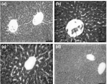
Figure 1. (a) Hematoxylin and eosin staining. Low dose deltamethrin group, liver x40. (b) Intermediate dose deltamethrin group, liver x40. (c) High dose deltamethrin group, liver x40. (d) Control group, liver x20. (Scale bar x20= 250 μm, x40=125 μm). Arrows indicate picnotic nucleus. (a) (c) (b) (d)
RESULTS
Histological resultsGroup 1 (low dose DLM): During the low dose DLM group’s liver histology, vena centralis’ were found dilated in comparison to the control group. Sinusoid structures accompanying to Remark cordons and Disse intervals were observed to be expanded when compared to the control group. In hepatic triangle structure; A. Hepaticus interlobularis, V. interlobularis and Ductus biliferi structures showed dilatation (Table 1, Figure 1a, 2a, 3a).
Group 2 (intermediate dose DLM): At the intermediate dose DLM group’s liver histology, vena centralis’ were found to be dilated more when compared to the control and low dose
DLM group. Disse intervals were observed to be expanded more when compared to the control and low dose DLM group. Pyknotic hepatocyte nuclei have been observed in some areas (Table 1, Figure 1b, 2b, 3b).
Group 3 (high dose DLM): At the high dose DLM group’s liver histology, vena centralis’ were found to be significantly dilated when compared to the other groups. Disse intervals were observed to be significantly expanded when compared to the other groups. Many more pyknotic hepatocyte nuclei and degenerated hepatocytes have been observed when compared to the intermediate dose DLM group. Dissolution of glycogen granules located in the hepatocyte were also observed (Table 1, Figure 1c, 2c, 3c).
Table 1. Histopathological changes of deltamethrin on liver
Liver area Histopathological Group 1 Group 2 Group 3 Group 4 findings (low dose) (intermediate dose) (high dose) (control) Paranchyma
Intracytoplasmic edema + ++ +++ -Nuclear hypertrophy + ++ +++ N Dilatation of vena centralis + ++ +++ N Fibronectin immunostaining ++ + + +++ Stroma
Sinusoidal dilatation + ++ +++ N Dilatation of portal triad + ++ +++ N
Group 4 (control group): During the control group’s liver histology, histological structure of the normal animals has been observed. Vena centralis that is settled at the center of classical liver lobule has been observed in the normal histological structure of the organ covered with tight connective tissue, Glisson capsule. Remark cordons that are leaving vena centralis radially are made of hepatocyte cells formed in a fascicular style. Sinusoids located at the hepatocytes’ laterals were in the normal structure. In portal hepatic triangle structure, A. Hepaticus interlobularis, V. interlobularis and Ductus biliferi structures were observed at their normal structural forms. Sinusoidal structures in hepatocytes basolateral parts were also normal sized (Table 1, Figure 1d, 2d, 3d).
Biochemical results
Plasma alanine transaminase (ALT) level for group 1 is 50.4±7.13 IU/L, for group 2 is
68.2±8.4 IU/L, for group 3 is 81.4±11.9 IU/L, for group 4 is 21.7±2.15 IU/L. Plasma ALT levels increased as the dose of DLM increased. The increment was statistically significant (Table 2).
Plasma MDA level for group 1 is 0.37±0.08 μM, for group 2 is 0.48±0.13 μM, for group 3 is 0.61±0.03 μM, for group 4 is 0.16±0.02 μM. Plasma MDA levels increased dose-dependent at the DLM exposed rats. The increment was statistically significant (Table 2).
Plasma GSH level for group 1 is 6.92±1.29 μM, for group 2 is 5.20±1.18 μM, for group 3 is 3.30±0.97 μM, for group 4 is 12.8±1.62 μM. Plasma GSH levels decreased as the dose of DLM increased. The decrement was statistically significant (Table 2).
Liver MDA level for group 1 is 7.15±0.78 nmol/g tissue, for group 2 is 10.90±1.34 nmol/g tissue, for group 3 is 18.35±1.62 nmol/g tissue, for group 4 is 3.79±0.54 nmol/g tissue. Liver MDA levels increased as the dose of DLM increased. The increment was statistically significant (Table 2).
Liver GSH level for group 1 is 2.78±0.58 nmol/μgr protein, for group 2 is 1.81±0.46 nmol/μgr protein, for group 3 is 1.23±0.4 nmol/μgr protein, for group 4 is 4.33±0.93 nmol/μgr protein. Liver GSH levels decreased as the dose of DLM increased. The decrement was statistically significant (Table 2).
DISCUSSION
Deltamethrin is one of the most preferred insecticides globally because of the high efficiency level in low doses and low toxicity profile.[27] However, in some studies, DLM toxicity has been reported.[28] The liver has a critical role in insecticide elimination and detoxification, so this process may result in serious liver damage.[29] Table 2. Biochemical results
Group 1 Group 2 Group 3 Group 4 (Low dose) (Intermediate dose) (High dose) (Control) Alanine transaminase (IU/L) 50.4±7.13* 68.2±8.4*† 81.4±11.9*†‡ 21.7±2.15 Plasma measurement of malondialdehyde (μM) 0.37±0.08* 0.48±0.13**† 0.61±0.03**†‡ 0.16±0.02 Plasma glutathione (μM) 6.92±1.29* 5.20±1.18*† 3.30±0.97**†‡ 12.8±1.62 Measurement of malondialdehyde (liver) (nmol/g tissue) 7.15±0.78* 10.90±1.34**§ 18.35±1.62**†¶ 3.79±0.54 Glutathione (liver) (nmol/μgr protein) 2.78±0.58† 1.81±0.46*† 1.23± 0.4*†‡ 4.33±0.93
* p<0.05 low, intermediate, high dose versus normal; ** p<0.000 high or intermediate dose versus normal; † p>0.05 intermediate dose versus low dose; † p>0.05 high dose versus intermediate dose; † p>0.05 low dose versus normal; ‡ p<0.05 high dose versus low dose; § p<0.05 intermediate dose versus low dose; ¶ p<0.001 high dose versus low dose.
(a)
(c)
(b)
(d)
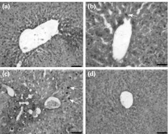
Figure 3. (a) Anti-fibronectin immunohistochemical staining. Low dose deltamethrin group, liver x40. (b) Intermediate deltamethrin group, liver x40. (c) High dose deltamethrin group, liver x40. (d) Control group, liver x20. (Scale bar x20= 250 μm, x40=125 μm). Arrows indicate picnotic nucleus.
Oxidative stress is formed through imbalance between antioxidant processes and free oxygen radicals. It causes instability and apoptotic cell death in cell physiology.[30] DLM causes damage with tissue lipid peroxidases induction and over production of ROS in mammals and other organisms.[31]
Deltamethrin exposure causes an obvious increase in MDA levels. As well known, MDA level is an oxidative stress biomarker. It is the last product of lipid peroxidation and a good indicator of lipid peroxidation level.[32] Lipid peroxidation is a generally used marker in oxidative stress.[33] Under the stress circumstances such as DLM, biologic molecules release oxidant molecules which result in an irreversible oxidation reaction and thus cellular dysfunction develops.[34]
In this study, after ingestion of DLM for 30 days, plasma and tissue MDA levels increased statistically significant amount. In most of the studies, DLM administration duration was determined as 30 days.[35,36] Plasma and tissue MDA levels were significantly increased when compared to the control group. This increment was more obvious between the control group and high dose and intermediate dose groups. A recent study states the “possible mechanism” of the damage by DLM at liver and bone marrow is oxidative stress.[37] This study shows that insecticide toxicity causes oxidative stress and oxidant or free radicals have a major role so plasma MDA and GSH level measurements can be used for insecticide toxicity evaluation. Although plasma MDA and GSH level measurements are not specific to a particular disease or toxicity, they can be used for monitorization of DLM toxicity rather than diagnosis.
Alanine transaminase is a cytosolic enzyme that is found in the cytoplasm. Alanine transaminase serum levels may increase because of the increment of hepatic expression or increment of production with hepatic parenchymal or bile duct damage.[38] Alanine transaminase is a biomarker commonly used for detecting liver damage.[39] In this study ALT plasma level increase was dose-dependent.
Also in this study, parenchymal and stromal histopathological changes were examined as a direct indicator of liver damage. Intracytoplasmic
edema, vena centralis dilatation increased with the dose.
Fibronectin is a tissue glycoprotein which is an indicator of the intact tissue. In this study, we can state that fibronectin levels decreased as the amount of DLM exposure increased. This finding shows us the deteriorating effect of DLM on healthy tissue.
Doses used in this study are important for DLM toxicity. The more exposure caused the more toxicity indicators detected. For each dose exposure, toxicity was shown by histopathological methods and by biomarkers. The difference in the histopathological changes and biochemical parameters are in positive correlation with each other and with the different doses given. These facts are compatible with other studies.[40]
The effects of different doses of DLM has been emphasized in the study. Since insecticide usage differs according to seasonal agriculture and weather conditions (photo degradation, volatilization and rains), instead of continuously toxicity, short term or pulse exposure may give us more clues on DLM toxicity.[41] Therefore, different doses were used in this study.
A commercial formulation has been used instead of the technical material of DLM. Since pesticide formulations can contain different types of chemicals apart from active ingredients (solvents, preservatives, emulsifiers etc.), the usage of technical material of DLM would be more reliable.
Conclusion
In this study on liver tissue and blood, we showed that biomarkers and histopathology can both be used for identifying insecticide toxicity. Plasma ALT levels, plasma MDA levels and liver MDA levels increased significantly and proportionately with the rising dose of DLM. Plasma GSH levels and liver GSH levels decreased significantly and proportionately with the rising dose of DLM.
Deltamethrin causes changes in tissue enzymes by forming oxidative stress. Our study showed that liver biomarkers and oxidative stress parameters can be used for potential toxicity monitorization. Additional comprehensive laboratory studies can provide more specific biomarkers for evaluating insecticide related
environmental pollution. Insecticides specific to insects or less toxic methods should be developed.
Declaration of conflicting interests
The authors declared no conflicts of interest with respect to the authorship and/or publication of this article.
Funding
The authors received no financial support for the research and/or authorship of this article.
REFERENCES
1. Hossain MM, Richardson JR. Mechanism of pyrethroid pesticide-induced apoptosis: role of calpain and the ER stress pathway. Toxicol Sci 2011;122:512-25. 2. Bradbury SP, Coats JR. Comparative toxicology of
the pyrethroid insecticides. Rev Environ Contam Toxicol 1989;108:133-77.
3. Prasamthi K, Muralidhara, Rajini PS. Fenvalerate-induced oxidative damage in rat tissues and its attenuation by dietary sesame oil. Food Chem Toxicol 2005;43:299-306.
4. Chandra N, Jain NK, Sondhia S, Srivastava AB. Deltamethrin induced toxicity and ameliorative effect of alpha-tocopherol in broilers. Bull Environ Contam Toxicol 2013;90:673-8.
5. Food and Agriculture Organisation of the United Nations (FAO/ WHO). Pesticide Residues in Food-1998 Evaluations. Part II-Toxicological. Geneva: World Health Organization; 1999.
6. Barlow SM, Sullivan FM, Lines J. Risk assessment of the use of deltamethrin on bednets for the prevention of malaria. Food Chem Toxicol 2001;39:407-22. 7. Jeyaratnam J. Acute pesticide poisoning: a major
global health problem. World Health Stat Q 1990;43:139-44.
8. Garaj-Vrhovac V, Zeljezic D. Evaluation of DNA damage in workers occupationally exposed to pesticides using single-cell gel electrophoresis (SCGE) assay. Pesticide genotoxicity revealed by comet assay. Mutat Res 2000;469:279-85.
9. Arbuckle TE, Sever LE. Pesticide exposures and fetal death: a review of the epidemiologic literature. Crit Rev Toxicol 1998;28:229-70.
10. Bradberry SM, Cage SA, Proudfoot AT, Vale JA. Poisoning due to pyrethroids. Toxicol Rev 2005;24:93-106.
11. Tosun N, Karabay NU, Sayım F. Pecticide usage and their potential adverse impact on living organisim. Anadolu J of Aari 2001;11:113-25.
12. Moser VC, Stewart N, Freeborn DL, Crooks J, MacMillan DK, Hedge JM, et al. Assessment of serum biomarkers in rats after exposure to pesticides of different chemical classes. Toxicol Appl Pharmacol 2015;282:161-74.
13. Crinnion WJ. The CDC fourth national report on human exposure to environmental chemicals: what it tells us about our toxic burden and how it assist environmental medicine physicians. Altern Med Rev 2010;15:101-9.
14. Aydin B. Effects of thiacloprid, deltamethrin and their combination on oxidative stress in lymphoid organs, polymorphonuclear leukocytes and plasma of rats. Pest Biochem Physiol 2011;100:165-71.
15. Pandey S, Parvez S, Sayeed I, Haque R, Bin-Hafeez B, Raisuddin S. Biomarkers of oxidative stress: a comparative study of river Yamuna fish Wallago attu (Bl. & Schn.). Sci Total Environ 2003;309:105-15. 16. Sharma P, Jan M, Singh R. Deltamethrin toxicity. Ind
J Biol Stud Res 2013;2:91-107.
17. McDonagh B, Tyther R, Sheehan D. Redox proteomics in the mussel, Mytilus edulis. Mar Environ Res 2006;62:101-4.
18. Blomhoff R. Dietary antioxidants and cardiovascular disease. Curr Opin Lipidol 2005;16:47-54.
19. Husain R, Malaviya M, Seth PK, Husain R. Effect of deltamethrin on regional brain polyamines and behaviour in young rats. Pharmacol Toxicol 1994;74:211-5.
20. Hoellinger H, Lecorsier A, Sonnier M, Leger C, Do-Cao-Thang, Nguyen-Hoang-Nam. Cytotoxicity, cytogenotoxicity and allergenicity tests on certain pyrethroids. Drug Chem Toxicol 1987;10:291-310. 21. Michelangeli F, Robson MJ, East JM, Lee AG. The
conformation of pyrethroids bound to lipid bilayers. Biochim Biophys Acta 1990;1028:49-57.
22. Rickard J, Brodie ME. Correlation of blood and brain levels of the neurotoxic pyrethroid deltamethrin with the onset of symptoms in rats. Pest Biochem Physiol 1985;23:143-56.
23. Manna S, Bhattacharyya D, Mandal TK, Dey S. Neuropharmacological effects of deltamethrin in rats. J Vet Sci 2006;7:133-6.
24. Demougeot C, Marie C, Beley A. Importance of iron location in iron-induced hydroxyl radical production by brain slices. Life Sci 2000;67:399-410.
25. Bradford MM. A rapid and sensitive method for the quantitation of microgram quantities of protein utilizing the principle of protein-dye binding. Anal Biochem 1976;72:248-54.
26. Ellman GL. Tissue sulfhydryl groups. Arch Biochem Biophys 1959;82:70-7.
27. Amin KA, Hashem KS. Deltamethrin-induced oxidative stress and biochemical changes in tissues and blood of catfish (Clarias gariepinus): antioxidant defense and role of alpha-tocopherol. BMC Vet Res 2012;8:45.
28. Cengiz EI, Kan Y, Kizmaz V, Bahan M, Yanar M. The protective role of vitamin E on the fatty acid composition of phospholipid structure in gill and liver tissues of Oreochromis niloticus exposed to deltamethrin. Ecotoxicol Environ Saf 2012;80:381-5.
29. Mamat SS, Kamarolzaman MF, Yahya F, Mahmood ND, Shahril MS, Jakius KF, et al. Methanol extract of Melastoma malabathricum leaves exerted antioxidant and liver protective activity in rats. BMC Complement Altern Med 2013;13:326.
30. Sun Y. Free radicals, antioxidant enzymes, and carcinogenesis. Free Radic Biol Med 1990;8:583-99. 31. Saxena R, Garg P, Jain DK. In Vitro Anti-oxidant
Effect of Vitamin E on Oxidative Stress Induced due to Pesticides in Rat Erythrocytes. Toxicol Int 2011;18:73-6.
32. You Y, Yoo S, Yoon HG, Park J, Lee YH, Kim S, et al. In vitro and in vivo hepatoprotective effects of the aqueous extract from Taraxacum officinale (dandelion) root against alcohol-induced oxidative stress. Food Chem Toxicol 2010;48:1632-7.
33. Gawel S, Wardas M, Niedworok E, Wardas P. Malondialdehyde (MDA) as a lipid peroxidation marker. Wiad Lek. 2004;57(9-10):453-5. [Article in Polish] 34. Marklund SL. Role of toxic effects of oxygen in
reperfusion damage. J Mol Cell Cardiol 1988;20 Suppl 2:23-30.
35. Yousef MI, Awad TI, Mohamed EH. Deltamethrin-induced oxidative damage and biochemical alterations in rat and its attenuation by Vitamin E. Toxicology
2006;227:240-7.
36. Manna S, Bhattacharyya D, Mandal TK, Das S. Repeated dose toxicity of deltamethrin in rats. Res Pap 2005;37:160-4.
37. Nieradko-Iwanicka B, Borzecki A. Subacute poisoning of mice with deltamethrin produces memory impairment, reduced locomotor activity, liver damage and changes in blood morphology in the mechanism of oxidative stress. Pharmacol Rep 2015;67:535-41.
38. Obogwu MB, Akindele AJ, Adeyemi OO.
Hepatoprotective and in vivo antioxidant activities of the hydroethanolic leaf extract of Mucuna pruriens (Fabaceae) in antitubercular drugs and alcohol models. Chin J Nat Med 2014;12:273-83.
39. Goorden SM, Buffart TE, Bakker A, Buijs MM. Liver disorders in adults: ALT and AST. Ned Tijdschr Geneeskd 2013;157:A6443. [Abstract]
40. Yousef MI, Awad TI, Mohamed EH. Deltamethrin-induced oxidative damage and biochemical alterations in rat and its attenuation by Vitamin E. Toxicology 2006;227:240-7
41. Burgoa B, Wauchope RD. Pesticides in run-off and surface water. In: Roberts TR, Kearney PC, editors. Environmental Behaviour of Agrochemicals. Volume 9, Chichester: Wiley; 1995. p. 221-55.