Ajda Çoker
1Sand Ahmet Arman
2 1 Istanbul Kultur University, Science and LiteratureFaculty, Molecular Biology and Genetics Department, Atakoy Campus, 34156 Istanbul -Turkey
2 Marmara University, Engineering Faculty, Goztepe Campus, Istanbul - Turkey
Abstract
Growth hormone (GH), is expressed from anterior pituitary gland as a 191 amino acid long polypeptide hormone, has essential role on postnatal growth. In addition to longitudinal growth, GH has various effects on carbohydrate, lipid, protein and mineral metabolisms. GH is expressed from GH-N gene which is located within the GH gene cluster consisting three chorionic somatomammotropin (CS) and GH-V genes on chromosome 17q22-24. Secretion of GH is under control of hypothalamic hormones; somatostatin (SRIF) and growth hormone releasing hormone (GHRH). GH shows it’s biological activities by binding to growth hormone receptor (GHR). After dimerization of GHR, JAK2 is associated with GHR and activated, and JAK2 phosphorylates itself, GHR, signal transducer and activator of transcription (STAT) and intracellular proteins. Activated STAT dimers enter into nucleus, bind to promoter of the target
genes such as Spi and activate transcription. Also, RAS/MAPK and PKC signal transduction pathways are known to take role in GH signaling. GH signaling is negatively controlled by SHP via the reduction of intracellular Ca++levels and also
suppressors of cytokine signaling (SOC) with dephosphorilation of JAK2.
Key Words: Growth, growth hormone, growth
hormone receptor, signal transduction.
Introduction
Human growth is the series of processes initiated with the development of fertilized egg to infant and terminated when adult reaches his/her final height. Human growth process can be divided into two stages; prenatal and postnatal growth periods. Prenatal growth is involved in development of fertilized egg during pregnancy by the effects of parent’s genes and various intrauterine environmental factors. Postnatal growth is the growth and development of infant, embark after birth and terminated when the individual reach to it’s final adult height after puberty. During childhood, both thyroid and growth hormone, during puberty testosterone (in boys) and estrogen (in girls) control the postnatal growth and its under the influence of genetic, environmental factors and nutritional habits.
Prenatal growth is under the control of growth hormone-variant (GH-V), chorionic somatomammotro-pin-A (CS-A), and chorionic somatomammotropin -B (CS-B) hormones but growth hormone (GH-N) is involved in the postnatal growth. GH-N is synthesized and secreted by the somatotroph cells in the anterior lobe of the pituitary gland (Isaksson et al., 1985), while CS-A and CS-B are expressed in syncytiotrophoblastic villous epithelium of the placenta (MacLeod et al.,
Human growth hormone
www.advmolbiol.org
SCorrespondence author:
Istanbul Kultur University, Science and Literature Faculty, Molecular Biology and Genetics Department, Atakoy Campus, 34156 Istanbul - Turkey
E-mail: [email protected]
1991). Both GH-V and CS promote fetal growth by altering the maternal protein, carbonhydrate and fat metabolisms. During pregnancy, GH-V gene expression is up-regulated and GH-V suppresses maternal GH-N expression and the level of GH-N in maternal blood stream is decreased. After birth, GH-V expression is suppressed and the baseline GH-N levels are altered to normal range (Frankenne et al., 1988).
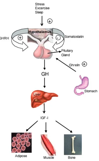
GH synthesis and secretion from the pituitary is under control of two hormones; growth hormone releasing hormone (GHRH) and somatostatin (SRIF). GHRH is a 108 amino acid long polypeptide and it is expressed from a GHRH gene that is located on chromosome 20 (q11.2). GHRH induces the secretion of GH by binding to its receptor, growth hormone releasing hormone receptor (GHRHR). After binding of GHRH to its receptor, cellular Ca++ level is increased and the expression and secretion of GH increases in the pituitary cells (Mayo et al., 1992). SRIF is a cyclic 14- or 28- amino acid residue containing peptide which is expressed from a gene located in 3q28. SRIF inhibits the secretion of GH by decreasing the cellular Ca++ levels (Lussier et al., 1991). Additionally, ghrelin, protein which has a critical role in appetite and food intake, induces the secretion of GH like as GHRH (Tannenbaum et al.,1991). Ghrelin expressed from ghrelin gene located in the 3p25-26 is a 28 amino acid peptide. It is also produced by stomach, intestine, and central nervous system. Ghrelin induces GH secretion by binding to ghrelin receptor and increases the intracellular Ca++ and indirectly stimulates GH secretion via GHRH release (Casanueva et al., 2008).
Growth induction is the major function of GH-N and this function was shown by experiments on hyposectomized animals. The growth chart of hyposectomized animals was halted but the treatment of exogenous GH induced their development (Groesbeck et al.,1987). Somatomedin hypothesis was originated to understand how GH-N promotes growth. In 1957, it was speculated that GH secreted from the pituitary and reaches to the liver and activates sulfation factor which was called somatomedin. And, then somatomedin migrates to the bone tissue to act on bone modeling and leading
longitudinal growth. Cloning and purification of somatomedin demonstrated that this protein shows high homology with insulin and then somatomedin called as insulin like growth factor (IGF); (Green et al., 1985). In 1980’s, somatomedin hypothesis was updated to dual effector hypothesis. In this hypothesis, GH stimulates IGF-I production in liver
Figure 1. Growth hormone secretion and functions: GH secre-tion is negatively controlled by SRIF, positively controlled by GHRH. Also stress, sleep, exercise and ghrelin stimulate growth hormone secretion. After GH secretion to the circulation, it reaches to target organs such as adipose, muscle, bone tis-sues and induces growth. -: negatively, + : positively control.
and migration of IGF-I induces bone growth and modeling. Pituitary derived GH also activates growth both in muscle and bone via stimulating the local IGF-I expression. IGF-In 2000’s, IGF-IGF-IGF-I knock out mice showed 75% of IGF-I level reduction but this couldn’t interrupt the growth and development. Thus GH shows it’s biological activities both directly and indirectly as autocrine/paracrine manner (Le Roith et al., 2001) (Figure 1).
Both in vivo and in vitro experiments demonstrated that GH-N has diverse effects on carbohydrate, lipid, protein, mineral and nitrogen metabolisms (Møller et al., 2009). Also, GH has various effects on the brain and cardiac functions. Additionally, it was shown that GH acts on neuronal development and immune system action (Harvey et al., 2003, Dardenne et al., 2009). The biological functions of GH is well understood via symptoms of GH deficiency or acromegaly patients. GH deficiency in childhood leads short stature but this condition is much more complex in adults. Adult GH deficiency leads sleep disorders, social isolation, bad mood, the decrease in muscle mass and leads abdominal obesity. Adult growth hormone deficiency patients are also under influence of cardiovascular mortality and mobility since GH deficiency leads the decrease in high density lipoprotein (HDL), the increase in low density lipoprotein (LDL) and total cholesterol (TC) levels. These symptoms are taken under control by
GH treatment. Over expression and secretion of GH above its normal range after puberty lead acromegaly. In acromegaly patients, excess GH influences cancer and cardiovascular problems.
Growth hormone gene cluster
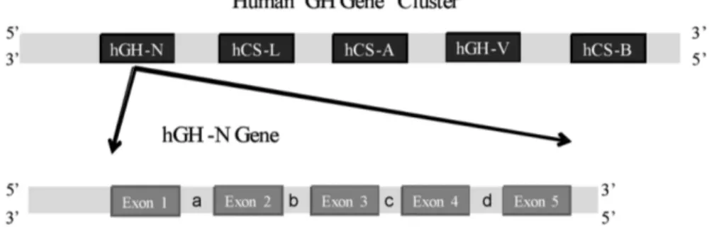
Human GH is expressed from GH-N gene which is located within the GH gene cluster. This gene cluster is composed of 5 closely related members; growth hormone (GH-N), chorionic somatomammotropin L (CS-L), chorionic somatomammotropin A (CS-A), growth hormone V (GH-V), and chorionic somatomammotropin B (CS-B). The genomic organization of GH genes is N, CS-L, CS-A, GH-V, CS-B, respectively. All these genes are located on the long arm of chromosome 17, band q22-24 (Harper et al., 1982). Each gene within the GH gene cluster is composed of 5 exons and 4 introns and CS-A, CS-B, CS-L genes are 2.9 kb, but GH-N and BH-V are 2.6 kb long (Figure 2).
These genes show high homology both in genomic and mRNA nucleotide sequences. The genomic sequence similarity between CS-B/GH-V, CS-A/CS-B and CS-A/GH-N are 91.8%, 98%, 92% respectively. Their transcriptional units have the same orientation and display a high degree of (90%) of sequence similarity. It was suggested that CS and GH genes were evolved over at least 50-60 million years via the duplication of a common ancestral gene
Figure 2. Human growth hormone gene cluster: hGH-N: Human Growth Hormone-N, L: chorionic somatomammotrope L, hCS-A: chorionic somatomammotrope A, hGH-V: Growth hormone V, hCS-B: chorionic somatomammotrope B. GH-N gene’s exons were presented by exon 1, 2, 3, 4, 5 and the introns were presented by a, b, c, d respectively. UTR: Untranslated region.
(Owerbach et al., 1980).
Growth hormone
GH-N gene expresses a 217 amino acid long precursor protein and N-terminal 26 amino acid leader peptide sequence is removed by proteolytic cleavage yielding a mature GH which contains 191 amino acids with a molecular mass of 22 kDa (Niall et al., 1971). Instead of 22 kDa GH, there is also a 20 kDa GH isoform. This isoform is expressed by alternative splicing of GH-N gene (32-46) leading deletion of 15 amino acids. Both 22 kDa and 20 kDa GH are biologically active (Walker et al., 1991). There are also 27, 17 and 5 kDa GH isoforms have been detected both in serum and pituitary extracts by western blotting. 22 kDa GH is the major GH isoform in bloodstream with 75 % percentage, 20% is 20 kDa GH and the rest is the other biologically inactive GH isoforms (Stewart et al.,1992).
Crystal structure of porcine and human GH is determined at 2.8 Aº resolution. It was shown that both of them have four alpha helix and they contain two disulfide bridges. The disulfide linkage in hGH occurs between residues Cys53-Cys165, Cys182-Cys189 (Abdel-Meguid et al., 1987). Helix 1, 2, 3 and 4 are located between residues 9-34, 72-96, 106-128 and 155-184. Among all these amino acids, glycine at 120 position was shown to have critical role in GHR binding. As this glycine was changed to the alanine by site-direct mutagenesis, the mutant GH (GH-G120A) losts its binding affinity to the second GHR. This mutant protein protects its binding affinity to the first GHR but couldn’t lead signal transduction. GH-G120A has been used as a therapeutic drug in acromegaly patients (Goffin et al., 2002).
Growth hormone receptor
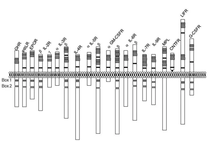
GH shows its biological functions by binding to growth hormone receptor (GHR). GHR is a member of the class I cytokine receptor superfamily that contains interferon, prolactin, granulocyte colony stimulating factor, interleukins and erythropoietin. The features of this family are having extracellular, transmembrane and intracellular domains and conserved the WSXWS (tryptophan, serine, any amino acid, tryptophan, serine) motif at the membrane proximal region of the
extracellular domain. This motif is changed to YGEFS (tyrosine, glycine, glutamic acid, phenylalanin, serine) in GHR (Baumgartner et al., 1994). Intracellular regions of these receptors also share two conserved motifs which are called Box I and Box II regions. Box I is a proline rich region and this region consists of eight conserved amino acids. Box II is composed of hydrophobic and charged amino acids approximately 30 amino acids far from Box I (Kelly et al., 1991) (Figure 3).
GHR is mainly expressed in liver but also it is present on bone, kidney, adipose, muscle, eye, brain, and heart tissues (Mullis et al., 1991, Ohlsson et al., 1998, Tiong et al., 1991, Vikman et al., 1991). In humans, membrane bound GHR are cleaved by proteases; this extracellular GHR circulates in the blood stream as a growth hormone binding protein (GHBP); (Leung et al., 1987)
The human GHR gene is located on chromosome 5p13-p12 and contains 9 coding exons (Barton et al., 1989). The 5’ UTR (Untranslated region) of the GHR gene has multiple exon 1. Exon 2 of GHR encodes the secretory signal peptide, exon 3-7 encodes the extracellular domain, exon 8 encodes the transmembrane domain and exon 9-10 encodes the intracellular domain of the GHR protein. GHR protein is 620 amino acid long and extracellular, transmembrane, and intracellular domains are 246, 24, 350 amino acid long, respectively (Godowski et al., 1989); (Figure 4).
Growth hormone signaling
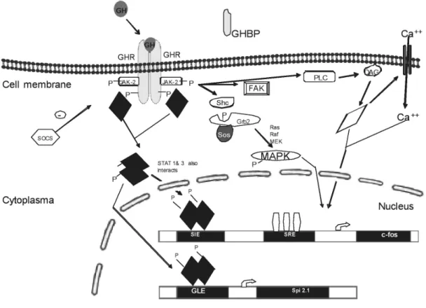
The first step in GH signal transduction is binding of GH to two GHR in order to make GHR:GH:GHR (1:2) complex (Wells et al., 1996). GHR dimerization stimulates the activation of Janus Kinase-2 (JAK-2) which is a member of cytoplasmic tyrosine kinases. JAK-2 is a 993 amino acid long protein with a molecular mass 130 kDa. Once JAK-2 binds to the GHR, JAK-2 is activated and phosphorylates tyrosine residues at the intracellular domain of GHR (Wang et al., 1994). Phosphorylated tyrosine amino acids on GHR have become docking sites for signaling molecules such as signal transducers and activators of transcription (STATs). STAT-1, STAT-3, STAT-5 are the transcription factors in GH signaling. When STAT
Figure 3. Cytokine receptor superfamily: GHR: Growth hormone receptor, PRLR: Prolactin receptor, EPOR: Erythropoietin receptor, IL-2R: Interleukin-2 receptor, IL-3R: Interleukin-3 receptor, IL-4R: Interleukin-4 receptor, IL-5R: Interleukin-5 receptor, GM-SCFR: Granulocyte macrophage colony stimulating factor receptor, IL-6R: Interleukin-6 receptor, IL-7R: Interleukin-7 receptor, IL-9R: Interleukin-9 receptor, MPL: Myeloproliferative leukemia virus receptor, CNTFR: CNTF receptor, LIFR: Leukocyte inhibitory factor receptor, G-CSFR: Granulocyte colony stimulating factor receptor.
: Conserved cysteines, : WSXSW motif, : Box I,: : Box II.
Figure 4. Schematic representation of GHR gene structure: GHR gene has multiple exon 1 at the 5’ UTR region, exon 2 (E2) encodes signal peptide sequence, exons 3-7 (E3-7) encode the extracellular domain, exon 8 (E8) encodes the transmembrane domain and the exon 9 and 10 (E9-10) encode the intracellular region. Introns are represented with a,b,c,d,e,f,g,h,i respectively, UTR: Untranslated region.
proteins bind to GHR, they are phosphorylated by JAK-2 (Meyer et al.,1994, Xu et al., 1996). Either STAT-1:STAT-3 heterodimers or STAT-5:STAT-5 homodimers enter nucleus and activate gene expression. The target genes for GH are IGF-1, c-fos, c-jun and serine protease inhibitor (Spi). GH uses another signaling pathway called MAPK signaling as shown in the figure 5, binding of GH to its receptor leads dimerization of GHR causing association of Shc protein to receptor and JAK2. After tyrosyl phosphorylation, shc protein activates Grb2 (growth factor receptor bound 2) which turn on activates Sos protein (Son of sevenless). Activated Sos protein stimulates the Ras, Raf, MEK and MAPK signaling molecules (Vanderkuur et al., 1997). Another GH signal pathway is protein kinase C (PKC) pathway.
GH induces the formation of 1, 2-diacylglycerol (DAG) which stimulates PKC and expression of the target genes. And it was reported that cytoskeleton organization, chemotaxis and cell migration effects of GH are under control of focal adhesion kinase (FAK) activation by GH (Zhu et al., 1998).
Negative regulation of GH signaling is under control of both suppressor of cytokine signaling (SOCS) and phosphatases including SH-2 domain (SH-P). There are eight known SOCS; SOCS 1, 2, 3, 4, 5, 6, 7 and CIS. SOCS-1 interacts with JAK-2 and inactivates the kinases activity of JAK-2. The negative regulation of SOCS-2 on GH signaling was detected in SOCS-2 knock out mices as a gigantism phenotype. Both SHP1 and SHP2 dephosphorylate JAK-2 and GHR.
Figure 5 . Signal transduction pathways of GH: GH binding with two GHR is the first step in GH signaling. After GHR dimerization, GH signal transduction occurs in JAK/STAT pathway. GH activates target genes such as c-fos using by MAPK, and PKC signal transduction pathways.
In conclusion, GH is a 22 kDa polypeptide hormone which is expressed from anteriory pituitary gland. GH-N mainly controls the postnatal growth and it is essential in longitudinal growth and bone modeling. It has also various roles on carbohydrate, lipid, and protein metabolisms. These biological functions lead GH as an essential hormone in human growth, development and health. GH displays it’s functions by binding GHR and activates the JAK-STAT, MAPK and PKC signaling pathways. And also, GH signaling controlled negatively by SOCS and SHP proteins. The molecular mechanisms of GH in signaling pathways are important for characterization of GH related diseases and also development of drugs to treat patients with GH deficiency and acromegaly.
References
Abdel-Meguid SS, Shieh HS, Smith WW, Dayringer HE, Violand BN and Bentle LA. Three-dimensional structure of a genetically engineered variant of porcine growth hormone. Proceedings of the National Academy of Sciences of the United States of America. 84(18): 6434-6437, 1987.
Barton DE, Foellmer BE, Wood WI and Francke U. Chromosome mapping of the growth hormone receptor gene in man and mouse. Cytogenetics and Cell Genetics. 50(2-3):137-141, 1989.
Baumgartner JW, Wells CA, Chen CM and Waters MJ. The role of the WSXWS equivalent motif in growth hormone receptor function. Journal of Biological Chemistry. 18; 269(46):29094-29101, 1994. Casanueva FF, Camiña JP, Carreira MC, Pazos Y, Varga
JL and Schally AV. Growth hormone-releasing hormone as an agonist of the ghrelin receptor GHS-R1a. Proceedings of the National Academy of Sciences of the United States of America. 23; 105 (51): 20452-20457, 2008.
Dardenne M, Smaniotto S, de Mello-Coelho V, Villa-Verde DM and Savino W. Growth hormone modulates migration of developing T cells. Annals of the New York Academy of Sciences. 1153:1-5, 2009. Frankenne F, Closset J, Gomez F, Scippo ML, Smal J and Hennen G. The physiology of growth hormones (GHs) in pregnant women and partial characterization of the placental GH variant. Journal
of Clinical Endocrinology and Metabolism. 66(6): 1171-1180, 1988.
Godowski PJ, Leung DW, Meacham LR, Galgani JP, Hellmiss R, Keret R, Rotwein PS, Parks JS, Laron Z, and Wood WI. Characterization of the human growth hormone receptor gene and demonstration of a partial gene deletion in two patients with Laron-type dwarfism. Proceedings of the National Academy of Sciences of the United States of America. 86(20):8083-8087, 1989.
Goffin V, Touraine P. and Pegvisomant. (Pharmacia.) Current Opinion in İnvestigational Drugs. 3(5):752-757, 2002.
Green H, Morikawa M and Nixon T. A dual effector theory of growth-hormone action. Differentiation. 29(3): 195-198, 1985.
Groesbeck MD, Parlow AF and Daughaday WH. Stimulation of supranormal growth in prepubertal, adult plateaued, and hypophysectomized female rats by large doses of rat growth hormone: physiological effects and adverse consequences. Endocrinology. 120(5):1963-1975, 1987.
Harper ME, Barrera-Saldaña HA and Saunders GF. Chromosomal localization of the human placental lactogen-growth hormone gene cluster to 17q22-24. American Journal of Human Genetics. 34(2):227-234, 1982.
Harvey S, Kakebeeke M, Murphy AE and Sanders EJ. Growth hormone in the nervous system: autocrine or paracrine roles in retinal function? Canadian Journal of Physiology and Pharmacology. 81(4):371-384, 2003.
Isaksson OG, Edén S and Jansson JO. Mode of action of pituitary growth hormone on target cells.Annual Reviews of Physiology. 47:483-499, 1985.
Kelly PA, Djiane J, Postel-Vinay MC and Edery M. The prolactin/growth hormone receptor family. Endocrine Reviews. 12(3):235-251, 1991.
Le Roith D, Bondy C, Yakar S, Liu JL and Butler A. The somatomedin hypothesis: 2001.Endocrine Reviews. 22(1):53-74, 2001.
Leung DW, Spencer SA, Cachianes G, Hammonds RG, Collins C, Henzel WJ, Barnard R, Waters MJ and Wood WI. Growth hormone receptor and serum binding protein: purification, cloning and expression. Nature. 330(6148):537-543, 1987.
Lussier BT, French MB, Moor BC and Kraicer J. Free intracellular Ca2+concentration and growth hormone (GH) release from purified rat somatotrophs. III. Mechanism of action of GH-releasing factor and somatostatin. Endocrinology. 128(1):592-603, 1991. MacLeod JN, Worsley I, Ray J, Friesen HG, Liebhaber SA and Cooke NE. Human growth hormone-variant is a biologically active somatogen and lactogen. Endocrinology. 128(3): 1298-1302, 1991.
Mayo KE. Molecular cloning and expression of a pituitary-specific receptor for growth hormone-releasing hormone. Molecular Endocrinology. 6 (10): 1734-1744, 1992.
Meyer DJ, Campbell GS, Cochran BH, Argetsinger LS, Larner AC, Finbloom DS, Carter-Su C and Schwartz J. Growth hormone induces a DNA binding factor related to the interferon-stimulated 91-kDa transcription factor. Journal of Biological Chemistry. 18;269(7):4701-4704, 1994.
Møller N Jørgensen JO. Effects of growth hormone on glucose, lipid and protein metabolism in human subjects. Endocrine Reviews. 30(2):152-177, 2009. Mullis PE, Lund T, Patel MS, Brook CG and Brickell PM. Regulation of human growth hormone receptor gene expression by human growth hormone in a human hepatoma cell line. Molecular and Cellular Endocrinology. 76(1-3):125-133, 1991.
Niall HD, Hogan ML, Sauer R, Rosenblum IY and Greenwood FC. Sequences of pituitary and placental lactogenic and growth hormones: evolution from a primordial peptide by gene reduplication. Proceedings of the National Academy of Sciences of the United States of America. 68(4):866-870, 1971. Ohlsson C, Bengtsson BA, Isaksson OG, Andreassen
TT and Slootweg MC. Growth hormone and bone. Endocrine Reviews. 19(1):55-79, 1998.
Owerbach D, Rutter WJ, Martial JA, Baxter JD and Shows TB. Genes for growth hormone, chorionic somatommammotropin, and growth hormones-like gene on chromosome 17 in humans.Science. 209 (4453):289-292, 1980.
Stewart TA, Clift S, Pitts-Meek S, Martin L, Terrell TG, Liggitt D and Oakley H. An evaluation of the functions of the 22-kilodalton (kDa), the 20-kDa, and the N-terminal polypeptide forms of human growth hormone using transgenic mice. Endocrinology.
130(1):405-414, 1992.
Tannenbaum GS. Neuroendocrine control of growth hormone secretion. Acta paediatrica Scandinavica. 372:5-16, 1991.
Tiong TS and Herington AC. Tissue distribution, characterization, and regulation of messenger ribonucleic acid for growth hormone receptor and serum binding protein in the rat. Endocrinology. 129(3):1628-1634, 1991.
Vanderkuur JA, Butch ER, Waters SB, Pessin JE, Guan KL and Carter-Su C. Signaling molecules involved in coupling growth hormone receptor to mitogen-activated protein kinase activation. Endocrinology. 138(10):4301-4307, 1997.
Vikman K, Carlsson B, Billig H and Edén S. Expression and regulation of growth hormone (GH) receptor messenger ribonucleic acid (mRNA) in rat adipose tissue, adipocytes, and adipocyte precursor cells: GH regulation of GH receptor mRNA. Endocrinology. 129(3):1155-1161, 1991.
Walker WH, Fitzpatrick SL, Barrera-Saldaña HA, Resendez-Perez D and Saunders GF. The human placental lactogen genes: structure, function, evolution and transcriptional regulation. Endocrine Reviews. 12(4):316-328, 1991.
Wang X, Xu B, Souza S.C, Kopchick JJ Growth hormone (GH) induces tyrosine-phosphorylated proteins in mouse L cells that express recombinant GH receptors,Proceedings of the National Academy of Sciences of the United States of America. 91(4) 1391-1395 1994.
Wells JA. Binding in the growth hormone receptor complex. Proceedings of the National Academy of Sciences of the United States of America 93(1):1-6, 1996.
Xu BC, Wang X, Darus CJ and Kopchick JJ. Growth hormone promotes the association of transcription factor STAT5 with the growth hormone receptor. J Biol Chem. 271(33):19768-19773, 1996.
Zhu T, Goh EL and Lobie PE. Growth hormone stimulates the tyrosine phosphorylation and association of p125 focal adhesion kinase (FAK) with JAK2. Fak is not required for stat-mediated transcription. J Biol Chem. 273(17):10682-10689, 1998.