World Journal of
Orthopedics
World J Orthop
2019 May 18; 10(5): 219-227
W J O
World Journal of
Orthopedics
Contents
Monthly Volume 10 Number 5 May 18, 2019
ORIGINAL ARTICLE
Basic Study219 Precontoured buttress plate vs reconstruction plate for acetabulum posterior wall fractures: A biomechanical study
Contents
World Journal of Orthopedics
Volume 10 Number 5 May 18, 2019
ABOUT COVER
Editorial Board Member of World Journal of Orthopedics, Antonio Luna, MD,PhD, Assistant Professor, Doctor, MRI Unit, Clínica Las Nieves, Health Time, Jaén, Jaén 23007, Spain
AIMS AND SCOPE
World Journal of Orthopedics (World J Orthop, WJO, online ISSN 2218-5836,DOI: 10.5312 ) is a peer-reviewed open access academic journal that aims to guide clinical practice and improve diagnostic and therapeutic skills of clinicians.
The WJO covers topics concerning arthroscopy, evidence-based medicine, epidemiology, nursing, sports medicine, therapy of bone and spinal diseases, bone trauma, osteoarthropathy, bone tumors and osteoporosis, etc. Priority publication will be given to articles concerning diagnosis and treatment of orthopedic diseases. The following aspects are covered: Clinical diagnosis, laboratory diagnosis, differential diagnosis, imaging tests, pathological diagnosis, molecular biological diagnosis, immunological diagnosis, genetic diagnosis, etc.
We encourage authors to submit their manuscripts to WJO.
INDEXING/ABSTRACTING
The WJO is now abstracted and indexed in PubMed, PubMed Central, Emerging Sources Citation Index (Web of Science), Scopus, China National Knowledge Infrastructure (CNKI), China Science and Technology Journal Database (CSTJ), and Superstar Journals Database.RESPONSIBLE EDITORS
FOR THIS ISSUE
Responsible Electronic Editor: Yun-Xiaojian Wu Proofing Editorial Office Director: Jin-Lei WangNAME OF JOURNAL
World Journal of Orthopedics
ISSN ISSN 2218-5836 (online) LAUNCH DATE November 18, 2010 FREQUENCY Monthly EDITORS-IN-CHIEF Bao-Gan Peng
EDITORIAL BOARD MEMBERS
http://www.wjgnet.com/2218-5836/editorialboard.htm
EDITORIAL OFFICE
Jin-Lei Wang, Director
PUBLICATION DATE
May 18, 2019
COPYRIGHT
© 2019 Baishideng Publishing Group Inc
INSTRUCTIONS TO AUTHORS
https://www.wjgnet.com/bpg/gerinfo/204
GUIDELINES FOR ETHICS DOCUMENTS
https://www.wjgnet.com/bpg/GerInfo/287
GUIDELINES FOR NON-NATIVE SPEAKERS OF ENGLISH
https://www.wjgnet.com/bpg/gerinfo/240
PUBLICATION MISCONDUCT
https://www.wjgnet.com/bpg/gerinfo/208
ARTICLE PROCESSING CHARGE
https://www.wjgnet.com/bpg/gerinfo/242
STEPS FOR SUBMITTING MANUSCRIPTS
https://www.wjgnet.com/bpg/GerInfo/239
ONLINE SUBMISSION
https://www.f6publishing.com
© 2019 Baishideng Publishing Group Inc. All rights reserved. 7041 Koll Center Parkway, Suite 160, Pleasanton, CA 94566, USA E-mail: [email protected] https://www.wjgnet.com
W J O
World Journal of
Orthopedics
Submit a Manuscript: https://www.f6publishing.com World J Orthop 2019 May 18; 10(5): 219-227
DOI: 10.5312/wjo.v10.i5.219 ISSN 2218-5836 (online)
ORIGINAL ARTICLE
Basic Study
Precontoured buttress plate vs reconstruction plate for acetabulum
posterior wall fractures: A biomechanical study
Güray Altun, Gürsel Saka, Teyfik Demir, Fatma Kübra Erbay Elibol, Mehmet Orçun Polat
ORCID number: Güray Altun (0000-0002-0669-8070); Gürsel Saka (0000-0002-9474-112X); Fatma Kübra Erbay Elibol (0000-0002-4117-1098); Teyfik Demir (0000-0001-6352-8302); Mehmet Orçun Polat
(0000-0002-2431-6769).
Author contributions: Altun G and Saka G analyzed the data and prepared the pelvis models prior to laboratory study; Demir T and Elibol FKE performed the biomechanical preparation and tests with the models, as well as the statistical evaluation of the results; Altun G, Polat MO and Saka G wrote the manuscript. Institutional review board statement: This study was exempt from IRB approval, due to its nature as a biomechanical study. Institutional animal care and use committee statement: This study was exempt from IACUC approval, due to its nature as a biomechanical study.
Conflict-of-interest statement: The authors declare no conflicts of interest related to this study or its publication.
Data sharing statement: There are no additional data.
Open-Access: This article is an open-access article which was selected by an in-house editor and fully peer-reviewed by external reviewers. It is distributed in accordance with the Creative Commons Attribution Non Commercial (CC BY-NC 4.0) license, which permits others to
Güray Altun, Department of Orthopaedics and Traumatology, University of Health Sciences Umraniye Training and Research Hospital, Ümraniye, Istanbul 34764, Turkey
Gürsel Saka, Department of Orthopaedics and Traumatology, Intercontinental Hisar Hospital, Ümraniye, Istanbul 34768, Turkey
Teyfik Demir, Fatma Kübra Erbay Elibol, Department of Mechanical Engineering, TOBB University of Economics and Technology, Çankaya, Ankara 06560, Turkey
Mehmet Orçun Polat, Department of Industrial Engineering, Kadir Has University, Department of Industrial Engineering, Fatih, Istanbul 34083, Turkey
Corresponding author: Güray Altun, MD, Doctor, Surgeon, Orthopaedic and Traumatology Surgeon, Department of Orthopaedics and Traumatology, University of Health Sciences Umraniye Training and Research Hospital, Adem Yavuz Str., Ümraniye, Istanbul 34764, Turkey. [email protected]
Telephone: +90-505-7456667
Abstract
BACKGROUNDThe purpose of open reduction and internal fixation of acetabulum posterior wall fractures is to restore anatomical structure and stability of the hip joint, in order to start weight bearing as soon as possible and prevent hip arthrosis; restoration of the anatomy should preserve function of the joint as well. Although “special shaped precontoured plates” have been developed in recent years for surgical treatment of this region, studies comparing the traditional plates with the newly designed precontoured plates are lacking.
AIM
To evaluate the biomechanical properties of precontoured anatomic buttress and conventional curved reconstruction plates (CCRPs) for posterior wall acetabulum fracture treatment.
METHODS
Twelve pelvis models were created for testing plate treatment of fracture in the posterior wall of the acetabulum. These 12 pelvis models were used to create 24 hemipelvis models (experimental) by cutting from the sagittal plane and passing over the center of gravity, after which the posterior wall acetabular fractures (of similar type and size) were created. In these experimental models, the right acetabulum was fixed with a 5-hole CCRP, while the left was fixed with a precontoured anatomic buttress plate (PABP). Samples were placed through the
distribute, remix, adapt, build upon this work non-commercially, and license their derivative works on different terms, provided the original work is properly cited and the use is non-commercial. See: http://creativecommons.org/licen ses/by-nc/4.0/
Manuscript source: Unsolicited manuscript
Received: January 21, 2019 Peer-review started: January 22, 2019
First decision: April 11, 2019 Revised: April 30, 2019 Accepted: May 14, 2019 Article in press: May 14, 2019 Published online: May 18, 2019 P-Reviewer: Liu J
S-Editor: Dou Y L-Editor: A E-Editor: Wu YXJ
test device and were subjected to static load testing, with a constant testing velocity of 2 mm/min until the load reached 2.3 kN or the acetabular fixation failed. Dynamic tests were also performed with sinusoidal wave load, with a maximal load of 2.3 kN and a load ratio of 0.1.
RESULTS
The average stiffness values were 460.83 ± 95.47 N/mm for the PABP and 291.99 ± 118.58 N/mm for the 5-hole CCRP. The precontoured anatomic acetabulum buttress plates had significantly higher rigidity than the CCRPs (P = 0.022). There was a statistically significant difference between the unloaded and 2.3 kN-loaded values of AL (posterosuperior fracture line vertical to the ground surface) and CL (posteroinferior fracture line vertical to the ground surface) parameters for both the PABPs and the 5-hole CCRPs (P = 0.036 and P = 0.045, respectively). According to the static tests, the amount of total displacement was significantly less in the PABPs than in the CCRPs. Comparative analysis of the displacement in the BL (posterior wall fracture line horizontal to the ground) parameter yielded no statistically significant differences between the PABP and the 5-hole CCRPs (P = 0.261).
CONCLUSION
PABP provides more stable fixation in acetabulum posterior wall fractures than 5-hole CCRP, allowing for proximal or distal fracture line screw application without reshaping.
Key words: Acetabular fracture; Anatomical plate; Precontoured plate; Posterior wall fracture; Biomechanical study
©The Author(s) 2019. Published by Baishideng Publishing Group Inc. All rights reserved. Core tip: The results of this biomechanical study demonstrated that application of precontoured anatomic acetabular buttress plates in posterior wall fractures of the acetabulum provide more stable fixation than 5-hole conventional curved reconstruction plates. For the precontoured anatomic acetabular buttress plate, its features of anatomic contour, low profile, avoidance of joint penetration by locking screws, and allowance of placement of more screws through both the proximal and distal parts of a fracture pattern as needed represent marked advantages over the traditional plate.
Citation: Altun G, Saka G, Demir T, Elibol FKE, Polat MO. Precontoured buttress plate vs reconstruction plate for acetabulum posterior wall fractures: A biomechanical study. World J
Orthop 2019; 10(5): 219-227
URL: https://www.wjgnet.com/2218-5836/full/v10/i5/219.htm
DOI: https://dx.doi.org/10.5312/wjo.v10.i5.219
INTRODUCTION
Almost one-fourth to one-third of all fractures of the acetabulum are posterior wall fractures[1]. Furthermore, most of these posterior wall fractures are comminuted and associated with an impaction injury of the articular surface. The Kocher-Langenbeck approach is appropriate for reduction and fixation of posterior acetabular wall fractures, and the traditional fixation methods generally involve reconstruction plates and/or screws.
Over the past decade, few surgical approaches and techniques to repair these fractures have been advanced, and most patients have had good clinical outcomes after anatomic reduction and rigid internal fixation[1-3]. Even nowadays, experienced
surgeons report post-operative rates of osteoarthritis of 20%–60% following acetabular fracture fixation[1,4]. Because of the complex structure of the posterior acetabulum, providing anatomical alignment intra-operatively is sometimes difficult for surgeons. Use of conventional curved reconstruction plates (CCRPs) cause prolongation in the duration of surgery since intra-operative reshaping is required for adaptation to the posterior acetabular surface; moreover, ideal anatomical adaptation cannot always be provided.
Application of two reconstruction plates to obtain better fixation via the buttress effect creates additional risk due to the greater amount of screws used for penetration of the articular surface, which may lead to the development of osteoarthritis[5,6]. This
conundrum makes the precontoured anatomic buttress plates (PABPs) attractive to surgeons, in spite of the fact that little information is available in the literature about the mechanical properties of these anatomical buttress plates.
The purpose of this study was, therefore, to evaluate the stiffness and rigidity of a PABP fixation model for fixation of posterior wall acetabular fractures and to compare with a CCRP model.
MATERIALS AND METHODS
Twelve pelvis models were created, each with fractures made in the same manner through the posterior wall of the acetabulum, to serve as the biomechanical test material for this study. The simulated fracture line of the posterior wall was designed in a similar way and rendered on the basis of the work of Olson et al[7]. Two different types of fixation materials (CCRP and PABP) were used for fixation of the fractures created in these experimental models. Each of the fixated pelvis models were then embedded in a polyurethane block and an alignment tool was used to position the pelvis in an anatomical standing position. Hemipelvis models were then created by cutting each of the pelvis models from the sagittal plane and passing over the center of gravity.
Group 1: CCRP construction group
The right hemipelvis models were fixed by application of a 5-hole, 3.5-mm CCRP. In each model, the fracture fragment was fixed with a screw that was applied through the third hole of the plate, after which one non-locking screw and three locking screws were applied to the proximal and distal parts of the fracture line to complete the reconstruction. For experimental purpose, each hole of the plates on these models was numbered and all of the screws applied were of the same length and diameter (Figure 1A-B).
Group 2: PABP construction group
The left hemipelvis models were fixed by application of a PABP. In each model, the fracture fragment was fixed with two non-locking screws that were applied to the superior and inferior parts respectively, then four locking screws were applied to the proximal and distal parts of the fracture fragment. For experimental purpose, each hole of the plates on these models was numbered and all of the screws applied were of the same length and diameter (Figure 1A-B).
Biomechanical testing
The embedding of the pelvis model in a polyurethane block allowed for us to simulate actual loading conditions and to apply force in the right direction. The embedded samples were placed into the Instron 3300 Universal Testing System (Instron, High Wycombe, United Kingdom) and subjected to axial load according to the anatomical reference position (Figure 2A-B). The samples were then subjected to static loading test, with 2 mm/min constant velocity until load reached 2.3 kN; this maximum load was selected from ISO 7206-4 as the maximum load for a single primer stem, and this value was accepted as the highest load. Load vs displacement values were recorded during the static tests. A high-resolution camera was used, with automatic shot to capture sample data during loading (per-second images taken continuously).
Some parameters determined on the photographs were measured by using the computer-assisted program before and after the 2.3 kN load, and then assessed by statistical comparative analyses (Figure 3). Dynamic tests were performed with sinusoidal wave load, with a maximal load of 2.3 kN and a load ratio of 0.1. In the case of a sample not breaking in the test, it was then subjected to 10 Hz for up to 1000 cycles. Photographs of the samples were taken two times, for measurements of before and after sample loading.
For static tests performed with a 5-hole 3.5-mm CCRP and a PABP, averages were taken for the two groups and a load-shifting distribution graph was generated (Figure 4). Rigidity was calculated for the two different plate types, according to the load-displacement distribution graph.
Statistical analysis
The SPSS statistical software package for Windows (version 11.5; SPSS Inc., Chicago, IL, United States) was used for the statistical analyses. The Shapiro-Wilk test was used to determine if the distributions of continuous variables were normal. The
Figure 1
Figure 1 The plates used to fix the fracture pattern in the experimental model system. A: A 16-hole
precontoured buttress plate with six bicortical screws; B: A 5-hole conventional curved reconstruction plate with five bicortical screws. The screws were fixed throughout the posterior acetabular wall.
parametric Mann-Whitney U test was used to determine which group differed from the other groups significantly according to P value, with the threshold for significance set at 0.05.
RESULTS
The average stiffness values were determined to be 460.83 ± 95.47 N/mm for the PABP group and 291.99 ± 118.58 N/mm for the CCRP group. Rigidity of the PABPs was found to be significantly higher than that of the CCRPs (P = 0.022) (Table 1). There were statistically significant differences between the unloaded and 2.3 kN-loaded values of AL (posterosuperior fracture line vertical to the ground surface) and CL (posteroinferior fracture line vertical to the ground surface) parameters (Figure 3) for both the PABP group and CCRP group (P= 0.036 and P = 0.045, respectively). According to the static tests, the amount of total displacement was significantly less in the PABP group than in the CCRP group (Table 2). Statistical comparison of displacement in the BL (posterior wall fracture line horizontal to the ground) parameter (Figure 3) yielded no statistically significance difference between the PABP and CCRP groups (P = 0.261).
DISCUSSION
The purpose of open reduction and internal fixation of acetabulum posterior wall fractures is to restore anatomical structure and stability of the hip joint, in order to start weight bearing as soon as possible and prevent hip arthrosis; restoration of the anatomy should also preserve function of the joint. However, post-traumatic osteoarthrosis of the hip joint has been reported in association with as many as 20% of fractures of the acetabulum posterior wall following treatment with open reduction and internal fixation[1,8,9].
Abnormal contact stress caused by unsatisfactory joint fixation is one of the crucial factors underlying development of post-traumatic arthritis. Because of the risk of arthritis and the high complication rates of surgical fixation, the option of total hip arthroplasty is preferred for acetabular fractures in older patients as an alternative to surgery[10]. CCRPs and PABPs, with or without inter-fragmentary screws, are the most common fixation methods practiced as treatment for posterior acetabular wall fractures. Biomechanical studies have indicated that the inter-fragmentary lag screws confer additional strength to the CCRP fixation[11,12].
Technological advances in implant technology over the recent past years have included the development of “special shaped precontoured plates” for surgical treatment of this region. However, only a few studies have been published that compare the biomechanical stability of different fixation methods[1,13-15]. Application of
reconstruction plates following primary fixation with inter-fragmentary lag screw(s) is the treatment method used most commonly and safely. Liu et al[16] previously demonstrated that anatomical plates have higher accuracy than conventional ones, which is helpful for fracture reduction and reducing the operation’s difficulty. In our study, the experimental model in which fixation was applied with PABP and
Figure 2
Figure 2 Synthetic hemipelvis models mounted in a polyurethane block for proper positioning when axial load is applied through the polyurethane block. A: A 16-hole precontoured anatomic buttress plate; B: A 5-hole
conventional curved reconstruction plate.
fragmentary screws provided a rigid and reliable fixation, with average 1 mm displacement at an axial load of 2.3 kN.
A paramount issue related to fixation methods is the direction of the locking screws, which is directly associated with the direction of the screw holes. Although we encountered joint penetration in some screws during application of screws into the CCRP for two of the hemipelvis models in our study, joint penetration was not encountered in any screws during the application of the PABP in the hemipelvis samples. Due to design features of the plate itself, locking screws in a PABP have such advantages as inhibiting joint penetration and providing a safer fixation in the proximal and distal parts of a fracture fragment, with use of a greater amount of screws being possible as needed. In addition, the multiple-screw choice for the PABP allows for a greater number of compression screws to be placed into the acetabular fragment, through the plate. There is no need to place any extra screw beyond the plate. Due to this feature, the PABP can provide a safer fixation than the CCRP.
Application of a buttress influences the posterior wall of the acetabulum with a reconstruction plate, and inter-fragmentary screw is the most commonly used fixation method for surgical treatment of posterior wall fractures of the acetabulum. The PABP fixated models in our study had an average displacement of less than 1 mm with a load of 2.3 kN in static and dynamic tests. This feature will both impact comminution favorably and prevent development of degenerative arthrosis due to near-complete joint adaptation. The consequences of malreduction of joint congruency and insufficiency of surgical fixation are the reasons for poor results of acetabular fracture surgery[1,5]. Thus, the choice of fixation method applied in this condition and
knowledge of the biomechanical properties of the methods are crucial.
Among the biomechanical studies in the literature to date, Sawaguchi et al[17] reported on fixation of the anterior column with a plate or lag screw and of the posterior column with one of three different plates; no differences were found between the various modalities. Mehin et al[18] suggested that the locking plate is as
strong as the conventional plate plus inter-fragmentary lag screw for fixing transverse acetabular fractures. Simonian et al[19] evaluated the stability of different types of fixation methods for the T-type acetabular fracture and found that the differences in displacements were not statistically significant. Goulet et al[20] reported in 1994 that when concentric comminuted and transverse comminuted posterior wall fractures of the acetabulum were simulated separately, the stiffness of a reconstruction plate and screws was significantly higher than that achieved with screws alone. Still other studies have mentioned that, for treatment of posterior wall fractures, fixation with combined fixation materials was more reliable and stable due to the single reconstruction plates[21,22]. In our study, displacement of the single PABP was lower
and rigidity was higher than for the CCRP.
It is important that our experimental-based study be discussed in the light of its limitations and strengths. Firstly, our biomechanical study was not performed with Sawbones® models or cadaveric specimens. Although, these pelvic models do not represent the heterogeneity of real bone, the homogeneity of the synthetic models (between each other) provides an advantage over the real specimens for more direct comparison without the potential confounding factors, such as age- or sex-related differences. Secondly, the range of patterns of acetabulum posterior wall fractures is wide but the instability of a fracture and the intra-articular component of a fragment remain the major features of the posterior wall fracture type. As such, we focused on
Figure 3
Figure 3 Parameters measured during the test procedure. AL: Posterosuperior fracture line vertical to the ground
surface; BL: Posterior wall fracture line horizontal to the ground; CL: Posteroinferior fracture line vertical to the ground surface.
an instable and intra-articular fracture pattern for the creation of our simulated fracture figure. Finally, the screw numbers applied to the two groups of plates were not equal; the CCRP group has 5 screws/model and the PABP group had 6 screws/model. Yet this limitation of the study also underlies the PABP feature that increases the stability of the fracture pattern.
In conclusion, the results of this experimental study showed that application of PABP for posterior wall fractures of the acetabulum provides a more stable fixation than CCRP. The features of anatomic contour, low profile, avoidance of joint penetration of locking screws, and allowance for a greater amount of screws through the proximal and distal parts of the fracture pattern as needed are the marked advantages of PABPs. Although biomechanical and experimental data have shown this, further clinical studies are needed to support the findings.
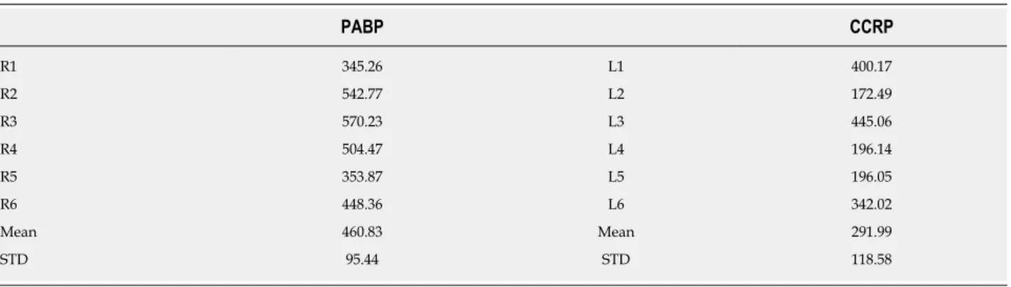
Table 1 Stiffness values for the precontoured anatomical buttress plate and conventional curved reconstruction plate groups PABP CCRP R1 345.26 L1 400.17 R2 542.77 L2 172.49 R3 570.23 L3 445.06 R4 504.47 L4 196.14 R5 353.87 L5 196.05 R6 448.36 L6 342.02 Mean 460.83 Mean 291.99 STD 95.44 STD 118.58
Data are presented in N/mm. CCRP: Conventional curved reconstruction plate; L: Left hemipelvis; PABP: Precontoured anatomical buttress plate; R: Right hemipelvis; STD: Standard deviation.
Table 2 Amount of load-displacement values for the conventional curved reconstruction plate and precontoured anatomical buttress plate groups after static loading
Plate Amount of load-displacement values
CCRP L1 L2 L3 L4 L5 L6 Mean STD AL 0.22 0.46 0.09 0.05 0.12 0.08 0.17 0.16 BL 0.15 0.92 0.21 0.42 0.59 0.09 0.40 0.32 CL 0.41 0.16 0.11 0.35 0.12 0.09 0.21 0.14 PABP R1 R2 R3 R4 R5 R6 Mean STD AL 0.00 0.16 0.05 0.04 0.05 0.04 0.06 0.05 BL 0.33 0.01 0.01 0.17 0.31 0.27 0.18 0.12 CL 0.03 0.05 0.08 0.01 0.41 0.08 0.11 0.15
Data are presented in mm. AL: Posterosuperior fracture line vertical to the ground surface; BL: Posterior wall fracture line horizontal to the ground; CL: Posteroinferior fracture line vertical to the ground surface; CCRP: Conventional curved reconstruction plate; L: Left hemipelvis; PABP: Precontoured anatomical buttress plate; R: Right hemipelvis; STD: Standard deviation.
Figure 4
Figure 4 Comparison of distribution graphs for the conventional curved reconstruction plate and precontoured anatomical buttress plate. The slope of the
linear part of the graph represents stiffness. L: Left hemipelvis model fixated with precontoured anatomical buttress plate; R: Right hemipelvis model fixated with conventional curved reconstruction plate.
ARTICLE HIGHLIGHTS
Research background
The purpose of open reduction and internal fixation of acetabulum posterior wall fractures is to restore anatomical structure and stability of the hip joint, in order to start weight bearing as soon as possible and prevent hip arthrosis; restoration of the anatomy should preserve function of the joint as well.
Research motivation
Although “special shaped precontoured plates” have been developed in recent years for surgical treatment of this region, studies comparing the traditional plates with the newly designed precontoured plates are lacking.
Research objectives
The main objective of this study was to evaluate the biomechanical properties of precontoured anatomic buttress and conventional curved reconstruction plates (CCRPs) for posterior wall acetabulum fracture treatment, particularly to determine if one provides more stable fixation.
Research methods
Hemipelvis models (experimental; divided at the sagittal plane and passing over the center of gravity) with similar posterior wall acetabular fractures were fixed with a 5-hole CCRP (right pelvis model) or a precontoured anatomic buttress plate (left pelvis model). These fixated hemipelvis samples were subjected to static load testing (constant testing velocity of 2 mm/min, applied until the load reached 2.3 kN or the acetabular fixation failed) and dynamic testing (sinusoidal wave load, with a maximal load of 2.3 kN and a load ratio of 0.1).
Research results
Rigidity of the precontoured anatomic acetabulum buttress plates was significantly higher than that of the CCRPs (P = 0.022).
Research conclusions
The findings of this study support the use of anatomic buttress plates for posterior acetabular wall fractures’ surgical treatment. In this surgical treatment, the CCRP is insufficient for safe stability because of its need for anatomic adaptation, lower screw number allowance, and lack of a buttress effect. The precontoured anatomic acetabulum buttress plate is safer, according to its matching the anatomy of the posterior acetabular region and ability to use more screws. In practice, surgeons usually use two reconstruction plates to achieve stable fixation, doubling the surgical time; the use of a single precontoured anatomic acetabulum buttress plate will lessen the surgical time.
Research perspectives
For posterior acetabular wall fractures, only fixing the fragment may not always be the best solution. Because the pelvic girdle is exposed to substantial force, according to its anatomic location, stable fixation with multiple screws is needed. Future research efforts must be made from the perspective of clinical practice, to provide biomechanical data from real-life experiences. We also caution that, in clinical practice, this type of fracture does not usually occur in isolation, and is generally accompanied by fracture in other parts of the pelvic girdle (i.e., sacroiliac), and for best results each single fracture pattern must be considered.
REFERENCES
1 Matta JM. Fractures of the acetabulum: accuracy of reduction and clinical results in patients managed
operatively within three weeks after the injury. J Bone Joint Surg Am 1996; 78: 1632-1645 [PMID:
8934477 DOI: 10.2106/00004623-199611000-00002]
2 Ebraheim NA, Patil V, Liu J, Sanford CG, Haman SP. Reconstruction of comminuted posterior wall
fractures using the buttress technique: a review of 32 fractures. Int Orthop 2007; 31: 671-675 [PMID:
16977451 DOI: 10.1007/s00264-006-0246-0]
3 Kumar A, Shah NA, Kershaw SA, Clayson AD. Operative management of acetabular fractures. A review
of 73 fractures. Injury 2005; 36: 605-612 [PMID: 15826618 DOI: 10.1016/j.injury.2004.11.022] 4 Kreder HJ, Rozen N, Borkhoff CM, Laflamme YG, McKee MD, Schemitsch EH, Stephen DJ.
Determinants of functional outcome after simple and complex acetabular fractures involving the posterior wall. J Bone Joint Surg Br 2006; 88: 776-782 [PMID: 16720773 DOI: 10.1302/0301-620X.88B6.17342] 5 Russell GV, Nork SE, Chip Routt ML. Perioperative complications associated with operative treatment of
acetabular fractures. J Trauma 2001; 51: 1098-1103 [PMID: 11740260 DOI:
10.1097/00005373-200112000-00014]
6 Ebraheim NA, Savolaine ER, Hoeflinger MJ, Jackson WT. Radiological diagnosis of screw penetration of
the hip joint in acetabular fracture reconstruction. J Orthop Trauma 1989; 3: 196-201 [PMID: 2809819
DOI: 10.1097/00005131-198909000-00003]
7 Olson SA, Bay BK, Chapman MW, Sharkey NA. Biomechanical consequences of fracture and repair of
the posterior wall of the acetabulum. J Bone Joint Surg Am 1995; 77: 1184-1192 [PMID: 7642663 DOI:
10.2106/00004623-199508000-00007]
8 Baumgaertner MR. Fractures of the posterior wall of the acetabulum. J Am Acad Orthop Surg 1999; 7:
54-65 [PMID: 9916185 DOI: 10.5435/00124635-199901000-00006]
9 Letournel E, Judet R. Fractures of the acetabulum. Posterior wall fractures. 2nd ed. New York:
Springer-Verlag 1993; 67-88 [DOI: 10.1007/978-3-642-75435-7_6]
10 Tissingh EK, Johnson A, Queally JM, Carrothers AD. Fix and replace: An emerging paradigm for treating
acetabular fractures in older patients. World J Orthop 2017; 8: 218-220 [PMID: 28361014 DOI:
10.5312/wjo.v8.i3.218]
11 Chang JK, Gill SS, Zura RD, Krause WR, Wang GJ. Comparative strength of three methods of fixation of
transverse acetabular fractures. Clin Orthop Relat Res 2001; 433-441 [PMID: 11716419 DOI:
10.1097/00003086-200111000-00057]
12 Shazar N, Brumback RJ, Novak VP, Belkoff SM. Biomechanical evaluation of transverse acetabular
fracture fixation. Clin Orthop Relat Res 1998; 215-222 [PMID: 9678050 DOI:
10.1097/00003086-199807000-00025]
13 Su K, Liu S, Wu T, Yin Y, Zhang R, Li S, Zhang Y. Posterior column acetabular fracture fixation using a
W-shaped angular plate: A biomechanical analysis. PLoS One 2017; 12: e0187886 [PMID: 29155842
DOI: 10.1371/journal.pone.0187886]
14 Zhang Y, Tang Y, Wang P, Zhao X, Xu S, Zhang C. Biomechanical comparison of different stabilization
constructs for unstable posterior wall fractures of acetabulum. A cadaveric study. PLoS One 2013; 8: e82993 [PMID: 24391733 DOI: 10.1371/journal.pone.0082993]
15 Fulkerson E, Egol KA, Kubiak EN, Liporace F, Kummer FJ, Koval KJ. Fixation of diaphyseal fractures
with a segmental defect: a biomechanical comparison of locked and conventional plating techniques. J
Trauma 2006; 60: 830-835 [PMID: 16612304 DOI: 10.1097/01.ta.0000195462.53525.0c] 16 Liu Q, Zhang K, Zhuang Y, Tao K, Fu Y. A morphological study of anatomical plates for acetabular
posterior column. Int J Comput Assist Radiol Surg 2014; 9: 725-731 [PMID: 24135851 DOI:
10.1007/s11548-013-0951-2]
17 Sawaguchi T, Brown TD, Rubash HE, Mears DC. Stability of acetabular fractures after internal fixation.
A cadaveric study. Acta Orthop Scand 1984; 55: 601-605 [PMID: 6524325 DOI:
10.3109/17453678408992404]
18 Mehin R, Jones B, Zhu Q, Broekhuyse H. A biomechanical study of conventional acetabular internal
fracture fixation versus locking plate fixation. Can J Surg 2009; 52: 221-228 [PMID: 19503667] 19 Simonian PT, Routt ML, Harrington RM, Tencer AF. The acetabular T-type fracture. A biomechanical
evaluation of internal fixation. Clin Orthop Relat Res 1995; 234-240 [PMID: 7634640 DOI:
10.1097/00003086-199505000-00030]
20 Goulet JA, Rouleau JP, Mason DJ, Goldstein SA. Comminuted fractures of the posterior wall of the
acetabulum. A biomechanical evaluation of fixation methods. J Bone Joint Surg Am 1994; 76: 1457-1463 [PMID: 7929492 DOI: 10.2106/00004623-199410000-00004]
21 Liu XM, Pan CW, Wang GD, Cai XH, Chen L, Meng CF, Huang JC. Finite element analysis of the
stability of combined plate internal fixation in posterior wall fractures of acetabulum. Int J Clin Exp Med 2015; 8: 13393-13397 [PMID: 26550272]
22 Wu X. A biomechanical comparison of different fixation techniques for fractures of the acetabular
posterior wall. Int Orthop 2018; 42: 673-679 [PMID: 29349502 DOI: 10.1007/s00264-017-3728-3]