Case Report
Desmoplastic fibroma of the jaw bones: A series of twenty-two cases
Devrim Kahraman
a,⇑, Berkem Karakoyunlu
b, Ulker Karagece
c, Umit Ertas
d, Omer Gunhan
aa
TOBB ETU School of Medicine, Department of Pathology, Besßtepe, Yasßam Cd. No:5, 06560 Yenimahalle, Ankara, Turkey
bTOBB ETU School of Medicine, Besßtepe, Yasßam Cd. No:5, 06560 Yenimahalle, Ankara, Turkey c
Private Goren Pathology Laboratory, Cumhuriyet, 1/64-65-66, Sakarya Cd., 06420 Çankaya, Ankara, Turkey
d
Department of Oral and Maxillofacial Surgery, Faculty of Dentistry, Ataturk University, 25240 Yakutiye, Erzurum, Turkey
a r t i c l e i n f o
Article history: Received 17 July 2020 Revised 30 September 2020 Accepted 2 October 2020 Available online 21 October 2020 Keywords: Desmoplastic fibroma Jaw bones Nestin Beta-catenin Neural crest Fibromatosis
a b s t r a c t
Desmoplastic fibroma (DF) is an intraosseous counterpart of desmoid-type soft tissue fibromatosis. It is most frequently seen in the jawbones. The clinical and radiological features of the present cases were nonspecific. The accumulation of beta-catenin in the nuclei of neoplastic cells which is a diagnostic fea-ture of desmoid-type soft tissue fibromatosis could not be detectED in the present DF series. The aim of this study is to report a series of 22 cases of DF involving either mandible or maxilla. A retrospective eval-uation of desmoplastic fibroma and beta-catenin, smooth muscle actin, nestin, cyclin D1 immunostain-ing’s patterns.
Most of the DF cases expressed only cytoplasmic beta-catenin immunostainings. We suggest that nuclear beta-catenin staining may not be used as a corroborating the diagnosis of DF. Immunohistochemical staining difference of jaw bone desmoplastic fibromas from other soft tissue and bone lesions may be related to the origination of jaw bone from The neural crest. Strong nestin and cyclin D1 positivity in our series supported this. A combined clinical, radiological, and histopatholog-ical analysis of the DF cases is essential in the diagnosis and management.
Ó 2020 Published by Elsevier GmbH. This is an open access article under the CC BY-NC-ND license (http:// creativecommons.org/licenses/by-nc-nd/4.0/).
1. Introduction
The word ‘‘fibromatoses” is a generic term used to describe a group of reactive/neoplastic proliferation of fibroblastic/myofi-broblastic cells with rather similar microscopic features but diverse biological behavior [1]. Although the term fibromatosis should preferably be applied to neoplastic ones, it is still in use for various reactive, neoplastic, developmental, and hereditary lesions[1].
Desmoid-type soft tissue fibromatosis, also called a desmoid tumor or aggressive fibromatosis, is neoplastic and it can arise from the soft tissues in almost all parts of the body. It is character-ized by infiltrative growth and a tendency to recur locally. These can be divided into two major groups such as superficial (fascial) and deep (musculoaponeurotic) fibromatoses [1]. Some of these have been labeled according to their localizations such as mesen-teric, palmar, and plantar fibromatosis. Fibromatosis occurs mostly sporadically. Multiple ones are usually accompanied by Gardner syndrome or Tuberous sclerosis[1,2].
Desmoplastic fibroma (DF) is considered as an intraosseous counterpart of desmoid-type soft tissue fibromatosis but is rela-tively rare. They are histologically almost identical to soft tissue fibromatoses. Although the DF can be seen in any bone, the poste-rior mandible is the most common location [3]. It is usually a slowly growing painless lesion. Extragnathic ones usually involve the metaphyseal region of the long bones[3,4]. Data on this lesion is limited to case reports and small series[2,3,5,6]. Radiographi-cally the DF may be seen as a multilocular or unilocular ill-defined radiolucency without mineralization. Thinned and expanded cortex or destruction of the involved bone without a sclerotic border is a common feature[7–9]. There may be an exten-sion into the surrounding soft tissues. Histologically, DF contains spindle-shaped fibroblastic and/or myofibroblastic cells without atypia within a collagenous background with focal myxoid changes.
2. Calculation
The spindle cells of DF do not show any specific diagnostic immunohistochemical staining. Loss of beta-catenin regulation and accumulation of beta-catenin in the nuclei of fibroblastic and/or myofibroblastic cells is a diagnostic feature of desmoid-https://doi.org/10.1016/j.jbo.2020.100333
2212-1374/Ó 2020 Published by Elsevier GmbH.
This is an open access article under the CC BY-NC-ND license (http://creativecommons.org/licenses/by-nc-nd/4.0/).
⇑Corresponding author at: TOBB ETU, Tıp Fakultesi Hastanesi, Yasßam caddesi No: 5 PK: 06510, Ankara, Turkey.
E-mail addresses:[email protected](D. Kahraman),uertas@atauni. edu.tr(U. Ertas),[email protected](O. Gunhan).
Contents lists available atScienceDirect
Journal of Bone Oncology
type soft tissue fibromatosis. However, this immunohistochemical feature is less consistent in DF. It is suggested that, unlike desmoid-type soft tissue fibromatosis, the beta-catenin pathway may not have an essential role in the tumorigenesis of DF[8].
On the other hand, craniofacial bone development is different from long bones and that it develops from the neural crest[10– 13]. Clinical and radiologic appearances of the jaw bone DF are nonspecific and they can mimic several other common lesions including odontogenic tumors. There is a need larger series for a better understanding of the pathogenesis, correct diagnosis, and treatment planning. In this retrospective study, we aimed to report a series of twenty-two cases of DF involving the jawbones. We have also focused on their immunohistology to assess their rela-tionship with desmoid-type soft tissue fibromatosis.
3. Material and methods
Twenty-two desmoplastic fibroma cases involving the jaw-bones diagnosed between 2009 and 2018 were collected retrospec-tively from the archives of private Gören Pathology Laboratory, Ankara, Turkey. Oral and maxillofacial cases form a large part of the tissues evaluated in this laboratory. All hematoxylin and eosin-stained slides and paraffin blocks were retrieved from the files. Patient records were reviewed to extract the information on demographics. They were analyzed by age, gender, tumor location, size, and recurrences if any. The available clinical and radiographic findings were presented inTable 1. The follow-ups of the patients were made on the records of recurrences. The authors re-evaluated the histologic slides. Representative tissue blocks of twenty cases were selected for immunohistochemical studies.
Since nuclear immunostaining for beta-catenin constitutes a marker of the Wnt pathway and is used to diagnose desmoid-type soft tissue fibromatosis, two different monoclonal antibodies against beta-catenin (RB-9035, dilution 1/500, Thermo Fisher Sci-entific, Kalamazoo, MI, USA, and Beta-Catenin 14 mouse mono-clonal antibody, 760-4242, Cell Marque, Sigma-Aldrich, USA) were used for immunostainings on our cases. Desmoid-type soft tissue fibromatosis was used as a positive control.
Labeling with smooth muscle actin Ab-1(MS-113, clone 1A4, dilution 1/800, Thermo Fisher Scientific, Fremont, CA, USA) was used for myofibroblastic differentiation. Vessels within the lesions were used as a positive control for smooth muscle actin stainings. Nestin is a class VI intermediate filament protein; it is known as a specific marker of neural or neural crest stem cells[10,13,27–29]. Nestin, which tends to be more strongly expressed in mandibular bone morphogenetic stem cells (R). The presence of neural crest-associated nestin positivity in desmoplastic fibromas of the jaws was detected (Nestin, RMab, clone EP287, Bio SB, CA, USA). Glomerular nestin staining of kidney tissue was a positive control. Cyclin D1 is a transcriptional target ofb catenin and plays an essential role in the expansion of skeletal precursors[11,12]. Cyclin D1 positivity in the present series was also detected (Cyclin D1/ Bcl-1(SP4) Thermo Scientific, CA, USA). Section of tonsillar tissue was taken as a positive control for cyclin D1.
Immunoreactivity for beta-catenin, smooth muscle actin, nes-tin, and cyclin D1 were graded semiquantitatively as negative ( ), mild (+), and strong (++).
Ki-67 (RM-9106, clone SP6, dilution 1/200, Thermo Fisher Sci-entific, Fremont, CA, USA), a nuclear marker for determination of the growth fraction were analyzed immunohistochemically and positive cells were manually counted on the hot spots at the 400X magnification. The mean percentage of positive cells was recorded. The Ki-67 index of the cases was then labeled accord-ingly as low (<1%) and high (>=2%). Streptavidin-peroxidase tech-nique was used for immunostainings. Immunohistological results are shown inTable 2.
4. Results
As seen inTable 1, the postero-lateral mandible (11 cases) was the most frequently affected site in this series. Others were found in the anterior mandible (7 cases) and the postero-lateral region of maxillae (4 cases). Most of the patients were female (15 female, 7 male) and the age range was between 4 and 66. The largest DF was six centimeters in diameter and it was located in the anterolateral area of the mandible. Asymptomatic swelling, ameloblastoma,
Table 1
Main clinicopathologic data of the present series.
Number Age-gender Location of tumor Radiological description Provisional clinical
diagnosis
1 16F Mandibula left corpus Radiolucent lesion 1 cm in diameter Radiolucent destructive
lesion
2 66F Mandibula anterior Expansive radiolucent lesion Asymptomatic swelling
3 50F Mandibula corpus Expansive radiolucent lesion, 2 cm in diameter Ameloblastoma
4 17M Maxilla, left lateral Expansive well demarcated radiolucent lesion, 3 cm in diameter Soft tissue lesion
5 -F Mandibula, left corpus Radiolucent lesion Giant cell granuloma
6 32M Mandibula corpus Radiolucent lesion well demarcated 4 cm in diameter Solid lesion 7 8F Mandibula, bilateral,
antero-lateral
Large, multilocular, expansive radiolucent lesion Cherubism 8 46F Mandibula right corpus Multilocular, destructive 2 cm in diametere, radiolucent lesion Ameloblastoma
9 22F Mandibula left corpus Radiolucent, lobular 2 cm in diameter lesion –
10 65F Mandibula left corpus Radiolucent, 1 cm in diameter Malignancy
11 42F Maxilla, right posterior mandibula
– Gardner syndrome
12 27M Maxilla left lateral Radiolucent, poorly demarcated 3 cm in diameter lesion with root resorption
– 13 12F Mandibula anterior Multilocular large radiolucent lesion with tooth luxation –
14 52M Maxilla, posterior Asymptomatic radiolucent lesion –
15 9M Mandibula right corpus Radiolucent lesion, 2 cm in diameter Ameloblastoma myxoma
16 25F Mandibula right posterior Radiolucent solid lesion Soft tissue lesion
17 -F Mandibula right posterior Radiolucent lesion 2 cm in diameter Swelling
18 58F Mandibula anterio-lateral Radiolucent lesion, 1 cm in diameter –
19 25F Mandibula Radiolucent lesion, poorly demarcated 6 cm in diameter Ossifying fibroma
20 50F Mandibula lateral Expansive lesion, 1,5 cm in diameter –
21 4M Mandibula anterior Radiolucent destructive lesion Swelling
22 F Mandibula anterior Large radiolucent lesion with cystic change Asymptomatic swelling 2
myxoma, giant cell granuloma, solid soft tissue lesion, malignant tumor, and ossifying fibroma were frequent provisional clinical diagnoses. A panoramic radiograph was available in some cases and it showed a destructive, ill-defined, unilocular, or multilocular radiolucent lesion (Figs. 1and2). There was no discernable perios-teal reaction and/or thickening. Cortical expansion and some perfo-ration were noted. In a single case (case 22) a cystic lesion was seen in the anterior mandible. Displacement of tooth and tooth germs were seen in one child (Fig. 2). The majority of patients suf-fered from local swelling. Four of them were asymptomatic. There was no pathologic fracture. There was no history of chemotherapy or radiotherapy in any of our cases. Complete surgical excision was the usual treatment. No recurrence was found in our files.
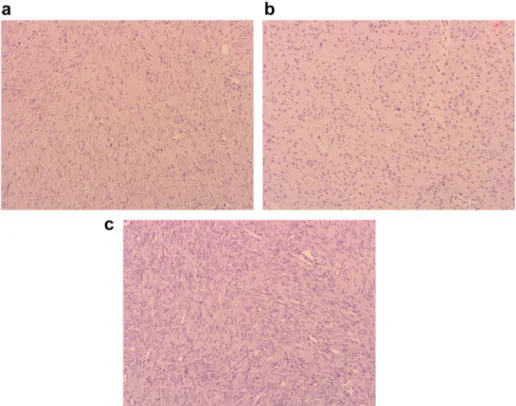
The histopathologic appearances in our series were quite simi-lar. The unencapsulated, infiltrative tumors were composed of irregular fascicles of spindle, uniform fibroblastic/myofibroblastic cells with low to variable cellularity (Figs. 3and4). The cytoplas-mic membrane of the cells was ill-defined and the surrounding matrix was collagenized, variably hyalinized, focally myxoid con-nective tissue (Fig. 4a,b,c). There were no hard tissue formations within the lesions. Mitosis is exceptional and none were atypical. There was no necrosis.
Table 2shows the results of the immunohistochemical
stain-ings of the present cases. Briefly, there was no nuclear reactivity with beta-catenin immunostaining in the present series. Four cases
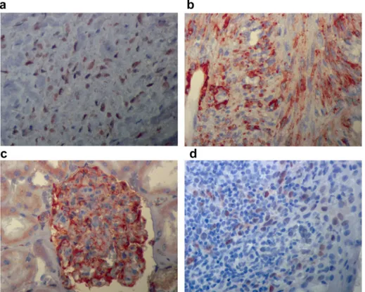
showed mild and ten cases showed strong cytoplasmic beta-catenin immunostainings (Fig. 5a). There was no staining in six cases. Control immuno stainings showed inFig. 6shown (Fig. 6a, b,c,d).
Table 2
Immunoreactivity for smooth muscle actin, beta-catenin and Ki-67.
Patient Smooth muscle actin-cytoplasmic Beta-catenin-cytoplasmic Beta-catenin-nuclear Ki-67(%) Nestin
1 ++ <1 + 2 ++ + <1 3 ++ <1 + 4 ++ ++ <1 5 ++ ++ <2 6 ++ <1 7 + <1 8 ++ <1 9 ++ ++ <1 + 10 + <1 11 ++ <1 + 12 ++ <1 + 13 ++ ++ <2 14 ++ ++ <2 + 15 ++ <1 + 16 ++ ++ <1 17 ++ + <1 + 18 <1 19 ++ <1 20 <1
: negative, + : mild, ++ : strong
Fig. 1. Large multilocular radiolucency and expansion in the left angulus mandible caused by desmoplastic fibroma (case 9).
Fig. 2. The pre-operative panoramic radiograph of a desmoplastic fibroma of the mandible in a child (case 21) shows a radiolucent area in the anterior region causing teeth displacement.
Most of the tumor cells (12/20) showed strong cytoplasmic reactivity for smooth muscle actin. There was no cytoplasmic smooth muscle actin reactivity in eight cases. Histologically, no dif-ference was found between smooth muscle actin positive and neg-ative cases.
There was diffuse, moderate cytoplasmic immunopositivity in neoplastic cells with nestin antibodies (Fig. 5b) in the present cases. Eight cases showed nuclear cyclin D1 positivity (Fig. 5c), others were negative. All control tissues showed positive immunostainings as expected.
Fig. 4. Histologic appearance showing fascicles of spindle fibroblastic proliferation of desmoplastic fibroma (a). Some cases contained plump fibroblasts and more collagenized matrix (b) and others were more cellular (c). (HEX200).
Fig. 5. The neoplastic spindle cells of desmoplastic fibromas showed cytoplasmic immunoreactivity for beta-catenin and nestin (a, b) without any nuclear staining. Some cases showed nuclear cyclin D1 reactivity (c), Streptavidin-peroxidase techniqueX400).
Ki-67 activity index was low (<1%) in most of the cases and reached to a high level (>=2) only in three cases. There was no sig-nificant clinical or histological difference between low and high Ki-67 activity cases.
5. Discussion
The first case of jaw bone DF was reported by Griffith and Irby in 1965 in the mandible of an 8-year-old girl[14]. In 2015, Woods et al reviewed 152 gnathic DFs which had been reported in the English language literature[2]. A female predilection of 56% was noted [2]. Most of the cases were below the age of 30. The mandibular (84%) posterior location was favored [2]. Gnathic lesions were usually painless, slow-growing, asymptomatic swel-lings[2,3]. Facial asymmetry, tooth displacement, root divergence, pain, trismus, and infections were the other symptoms [2,3,15]. Radiologic features of DF affecting the jaws were not unique and they overlapped with many other lesions [2]. Likewise, the age range, tumor location, and radiologic findings of our cases were not significantly different from those reported in earlier studies of DF of the jawbones. The wide range of provisional clinical diag-nosis reflects the overall non-specific clinical and radiologic appearances of DF in this region.
The nuclear and cytoplasmic beta-catenin expression is a typi-cal finding in nearly all of the desmoid-type soft tissue fibromato-sis[16–18]. On the other hand, reports of such genomic alterations in DF are exceptional[8,19,20]. In 2005, Hauben et al suggested that in contrast to desmoid-type soft tissue fibromatosis, the beta-catenin mutation was not essential in diagnosis[8]. Geneti-cally, there seems to be no evidence to consider DF as the intra-osseous counterpart of desmoid-type soft tissue fibromatosis
[8,19,20]. A literature review showed only eight gnathic intraoss-eous DF’s with nuclear beta-catenin reactivity [2,8,9,21–23]. In
the present case series, we did not detect any cases with nuclear catenin expression. On the other hand, cytoplasmic beta-catenin staining of varying intensity was found in most of all our cases (14/20).
Normally, beta-catenin is a cytoplasmic membraneous protein. Activated Wnt signaling causes accumulation of beta-catenin in the cytoplasm then translocates to the nucleus to regulate cell pro-liferation and differentiation. Neoplastic overexpression of Wnt signaling may contribute to an increased level of cytoplasmic and nuclear beta-catenin expression[24]. DF is composed of mature fibroblasts and myofibroblasts as in desmoid-type soft tissue fibro-matosis. Fibroblasts are versatile cells and may show differentia-tion into the other connective tissue cells. Transidifferentia-tions of fibroblasts to myofibroblasts can occur along with the increased activity of beta-catenin[25]. Myofibroblastic differentiation in DF may be less prominent or lacking compared to that of the desmoid-type soft tissue fibromatosis. Our findings support the view that the cellular components of jaw bone lesions are immunologically different. We also believe that nuclear beta-catenin staining should not be a prerequisite for the definitive diagnosis of DF.
Hauben et al reported 7/13 cases of DF were immunoreactive for one or more muscle-specific markers in more than %10 of the cells[8]. In their literature review, Wood et al reported that 77% of the DF cases showed labeling with smooth muscle actin[2]. Sim-ilarly, most of our cases (12/20) expressed cytoplasmic SMA posi-tivity in varying degrees.
Skeletal site-specific functional differences of cells may also affect the neoplasias. Therefore, bone lesions may display unique histological changes in the jawbones. Craniofacial bone develop-ment is different from long bones and that it develops from neural crest [27,13,10,28,29]. This difference may also be effective in immunostaining results. Absence of nuclear beta-catenin
Fig. 6. Control immunostainings: Desmoid-type soft tissue fibromatosis used as control tissue and showed strong nuclear beta-catenin immunopositivity (a). There was strong cytoplasmic positivity for smooth muscle actin in a case of desmoplastic fibroma. Vessels within the lesion were positive control (b). Control positive nestin immunostaining of kidney section seen predominantly in glomeruli(c). Tonsil tissue served control tissue of cyclin D1 immunostaining. Suprabasal squamous epithelial cells, scattered lymphocytes, and endothelial cells showed strong nuclear immunostaining (d). (Streptavidin-peroxidase techniqueX400).
immunoreactivity and the diffuse cytoplasmic positivity of neural crest-associated nestin and in some cases cyclin D1 stainings also may support this idea.
Proliferation markers such as Ki-67 are used as a surrogate mar-ker for evaluating the tumor’s aggressiveness. A low rate of cell turnover expected in DF as in desmoid-type soft tissue fibromato-sis [2]. Our Ki-67 staining results are in agreement with those reported in earlier studies.
The clinical and radiologic appearance of DF may mimic many other jaw bone lesions including ameloblastoma, odontogenic fibroma, odontogenic cysts, aneurysmal bone cyst, chondromyxoid fibroma, central hemangioma, eosinophilic granuloma, osteomyelitis, cemento-ossifying fibroma, giant cell granuloma and rarely primary malignant lesions such as the low-grade fibrosarcoma and osteosarcoma. Averna et al suggested that the gold standard for the diagnosis of DF is the histologic appearance
[26].
The histologic differential diagnosis of DF includes myofibroma, odontogenic fibroma, cemento-ossifying fibroma, and fibrous dys-plasia. The absence of nuclear pleomorphism, very low mitotic activity, lack of traces of odontogenic epithelium, and lack of cementum or bone components should help differentiate DF from other jaw bone lesions histologically[26].
Myofibroma is a solitary circumscribed, unencapsulated nodu-lar lesion of the soft tissues and bone of the head and neck area
[1]. Myofibroma consists of short fascicles and whorls of spindle myofibroblastic cells with characterized haemangiopericytoma-tous pattern. These clinical and pathological features are not the expected findings of DF. The odontogenic fibroma is made up of plump, oval to spindle cells within a collagenous matrix with irreg-ular strands of immature odontogenic epithelial cells. The odonto-genic epithelium is not a component of DF. Cemento-ossifying fibroma presents with radiologically well-defined margins and is composed of vascularised fibro cellular connective tissue with cementoid and osseous components distributed throughout the lesion. Fibrous dysplasia is an ill-defined lesion that contains cellu-lar fibrous connective tissue stroma with irregucellu-lar trabeculae of woven bone blending into the surrounding normal bone. Hard tis-sue formations such as cementum and bone are findings that exclude the diagnosis of DF. However, peripheral parts of a DF may contain adjacent reactive bone which can lead to a misdiagno-sis of the benign fibro-osseous lesion. Said-Al-Naief et al suggested that a generous diagnostic biopsy should be taken from the center of the lesion rather than the periphery in order to avoid misinter-preting the presence of reactive bone at the periphery as osteoid, which may, in turn, lead to a misdiagnosis of the benign fibro-osseous lesion or even osteosarcoma [3]. Khatib and Porgel sug-gested that DFs need to be included in the differential diagnosis for aggressive, radiolucent mandibular lesions, particularly in chil-dren[6]. Immunohistochemical studies are of limited utility in the differentiation of DF from lesions with a morphologically similar fibroblastic/myofibroblastic component[3].
Neither the location nor histology of DFs permits accurate pre-diction of their potentially aggressive clinical course[6]. Optimum treatment strategies for DF of bone are total excision or resection to minimize the risk of recurrence[3]. Treatment with curettage should be avoided since the lack of sharp demarcation can make total removal impossible. Khatib and Pogrel reported that patients who were treated with resection or wide excision showed no recurrence[6]. In other series, the recurrence rate in those treated with simple excision or enucleation was 20% to 40%, as compared to 70% in those treated with curettage alone[3]. Malignant change is very uncommon in desmoplastic fibroma and it is usually in the form of fibrosarcoma characterized with a high rate of recurrence, hypercellularity, increased mitotic activity and, nuclear atypia[15]. In 2011 Min et al reported 20 cases of primary desmoid-type
fibro-matosis arising from the oral and maxillofacial region and three of them were intraosseous[15]. They found a malignant change in six cases, one of which was intraosseous [15]. The records of our patients did not reveal any recurrence or malignant transforma-tions. Other therapeutic interventions like radiotherapy and chemotherapy were suggested in controlling recurrent and aggres-sive tumors especially when complete tumor resection is impossi-ble or avoiding a mutilating operation is a concern[5,6,15]. Further series of DFs of the jawbones may improve our understanding of this lesion allowing us better treatment methods.
This research did not receive any specific grant from funding agencies in the public, commercial, or not-for-profit sectors. CRediT authorship contribution statement
Devrim Kahraman: Investigation, Software, Visualization, Writing - original draft, Writing - review & editing. Berkem Kara-koyunlu: Data curation. Ulker Karagece: Resources. Umit Ertas: Data curation, Formal analysis. Omer Gunhan: Conceptualization, Methodology, Project administration, Supervision, Validation, Writing - original draft, Writing - review & editing.
Declaration of Competing Interest
The authors declare that they have no known competing finan-cial interests or personal relationships that could have appeared to influence the work reported in this paper.
References
[1]J.R. Goldblum, A.L. Folpe, S.W. Weiss, Enzinger, and Weiss’s soft tissue tumors, 6th.ed., Elsevier Saunders, Philadelphia, 2016, p. 239, 292, 297.
[2] T.R. Woods, D.M. Cohen, M.N. Islam, Y. Rawal, I. Bhattacharyya, Desmoplastic fibroma of the mandible: A series of three cases and review of the literature, Head Neck Pathol. 9 (2015) 196–204, https://doi.org/10.1007/s12105-014-0561-5.
[3] N. Said-Al-Naief, R. Fernandes, P. Louis, W. Bell, G.P. Siegal, Desmoplastic fibroma of the jaw: a case report and review of the literature, Oral Surg. Oral Med. Oral Pathol. Oral Radiol. Endod. 101 (2006) 82–94, https://doi.org/ 10.1016/j.tripleo.2005.03.034.
[4] C.Y. Inwards, K.K. Unni, J.W. Beabout, F.H. Sim, Desmoplastic fibroma of bone, Cancer 68 (9) (1991) 1978–1983, https://doi.org/10.1002/1097-0142 (19911101)68:9<1978::aid-cncr2820680922>3.0.co;2-h.
[5] L. Seper, H. Bürger, J. Vormoor, U. Joos, J. Kleinheinz, Aggressive fibromatosis involving the mandible. Case report and review of the literature, Oral Surg. Oral Med. Oral Pathol. Oral Endod. 99 (2005) 30–38,https://doi.org/10.1016/j. tripleo.2004.03.026.
[6] B. Khatib, M.A. Pogrel, Desmoplastic fibroma of the mandible in young children—a case series, Int. J. Oral Maxillofac. Surg. 46 (2017) 173–180,https:// doi.org/10.1016/j.ijom.2016.09.018, Epub 2016 Nov 3.
[7] A. Ikeshima, T. Utsunomiya, Case report of intra-osseous fibroma: a study on odontogenic and desmoplastic fibromas with a review of the literature, J. Oral Sci. 47 (2005) 149–157,https://doi.org/10.2334/josnusd.47.149.
[8] E.I. Hauben, G. Jundt, A. Cleaton-Jansen, A. Yavas, H.M. Kroon, E.V. Marck, P.C. W. Hogendoorn, Desmoplastic fibroma of bone: an immunohistochemical study including catenin expression and mutational analysis for beta-catenin, Human Pathol 36 (2005) 1025–1030, https://doi.org/10.1016/j. humpath.2005.07.004.
[9] U. Flucke, H. Coleman, Who classification of head and neck tumors, in: A. El Naggar, J.K.C. Chan, J.R. Grandis, T. Takata, P.J. Slootweg (Eds.), Lyon, 4th ed., 2017, p. 250,https://doi.org/10.1007/s00428-018-2320-6.
[10] S. Onishi, Y. Baba, F. Yokoi, K. Ide, M. Ohyama, K. Nishifuji, Progenitor cells expressing nestin, a neural crest stem cell marker, differentiate into outer root sheath keratinocytes, Vet Dermatol. 30 (5) (2019),https://doi.org/10.1111/ vde.12771, 365-e107.
[11] A.J. Mirando, T. Maruyama, J. Fu, H.I. Yu, W. Hsu, Beta-catenin/cyclin D1 mediated development of suture mesenchyme in calvarial morphogenesis, BMC Dev. Biol. 10 (2010) 116,https://doi.org/10.1186/1471-213X-10-116. [12] D. Graf, Z. Malik, S. Hayano, Y. Mishina, The common mechanism in
development and disease: BMP signaling in craniofacial development, Cytokine Growth Factor Rev. 27 (2016) 129–139, https://doi.org/10.1016/ j.cytogfr.2015.11.004.
[13] B. Lloyd, B.C. Tee, C. Headley, H. Emam, S. Mallery, Z. Sun, Similarities and differences between porcine mandibular and limb bone marrow mesenchymal stem cells, Arch. Oral Biol. 77 (2017) 1–11, https://doi.org/10.1016/j. archoralbio.2017.01.012.
[14] J.G. Griffith, W.B. Irby, Desmoplastic fibroma report of a rare tumor of the oral structures, Oral Surg Oral Med. Oral Pathol. 20 (1965) 269–275,https://doi. org/10.1186/1746-160X-5-25.
[15] R. Min, Z. Zun, W. Lizheng, D. Minjun, L. Shengwen, Y. Wenjun, Z. Chenping, Oral and maxillofacial desmoid-type fibromatosis in an eastern Chinese population: a report of 20 cases, Oral Surg. Oral Med. Oral Pathol. Oral Endod. 111 (2011) 340–345,https://doi.org/10.1016/j.tripleo.2010.10.019. [16] S. Tejpar, F. Noller, C. Li, et al., Predominance of beta-catenin mutations and
beta-catenin dysregulation in sporadic aggressive fibromatosis (desmoid tumor), Oncogene 18 (1999) 6615–6620, https://doi.org/10.1038/sj. onc.1203041.
[17] T. Saito, Y. Oda, K. Tanaka, et al., Beta-catenin nuclear expression correlates with cyclin D1 overexpression in sporadic desmoid tumors, J. Pathol. 195 (2001) 222–228,https://doi.org/10.1016/j.humpath.2006.01.017.
[18] T. Saito, Y. Oda, M. Nakamori, S. Tamiya, H. Yamamoto, R. Yokoyama, Y. Iwamoto, M. Tsuneyoshi, Possible association between higher beta-catenin mRNA expression and mutated beta-catenin in sporadic desmoid tumors: Real-time semiquantitative assay by TaqMan polymerase chain reaction, Lab. Invest. 82 (2002) 97–103,https://doi.org/10.1016/j.humpath.2008.05.005. [19] E. Hauben, A.M. Cleton-Jansen. Desmoplastic fibroma of bone. In WHO
Classification of Tumors of Soft Tissue and Bone. Edited by Fletcher CDM, Bridge JA, Hogendoorn PCW, Mertens F. IARC Lyon, 2013. p. 298. https://hdl. handle.net/2066/132068.
[20] T. Okubo, T. Saito, T. Takagi, Y. Suehara, K. Kaneko, Desmoplastic fibroma of the rib with cystic change: a case report and literature review, Skeletal Radiol 43 (2014) 703–708,https://doi.org/10.1007/s00256-013-1772-7.
[21] A.M. Azola, C.T. Wartmann, M.K. Fischer, B.T. Ambro, K.D. Pereira, et al., Desmoplastic fibroma arising from the anterior maxillary sinüs in a child, Arch. Otolaryngol. Head Neck Surg. 138 (2012) 859–862, https://doi.org/ 10.4322/acr.2019.091.
[22] L.Y. Taher, M. Saleem, S. Velagapudi, A. Dadado, Fibromatosis arising in association with neuromuscular hamartoma of the mandible, Head Neck Pathol. 7 (2013) 280–284,https://doi.org/10.1007/s12105-012-0418-8. [23] A.N. Arya, B. Saravan, K. Subalakshmi, R. Appadurai, I. Ponniah, Aggressive
fibromatosis of the mandible in a two-month-old infant, J Maxillofac Oral Surg. 14 (2015) S235–S239,https://doi.org/10.1007/s12663-012-0460-9. [24]J.H. Sellin, S. Umar, J. Xiao, A.P. Morris, Increased b-catenin expression and
nuclear translocation accompany cellular hyperproliferation in vivo, Cancer Res. 61 (2001) 2899–2906.
[25] L. Xu, W. Cui, W. Zhou, D. Li, L. Li, P. Zhao, X. Mo, Z. Zhang, J. Gao, Activation of Wnt/beta-catenin signaling is required for TGF-beta/Smad2/3 signaling during myofibroblast proliferation, J. Cell Mol. Med. 21 (2017) 1545–1554,https:// doi.org/10.1111/jcmm.
[26] R. Averna, M. De Flippo, S. Ferrari, E. Bacchini, C. Rossi, Desmoplastic fibroma of the mandible, Acta Biomed 82 (2011) 77–81, https://doi.org/10.1007/s12105-014-0561-5.
[27] J. Bugueno, W. Li, P. Salat, L. Qin, S.O. Akintoye, Canine mesenchymal stem cell bone regenerative capacity is regulated by site-specific multilineage differentiation, Oral Surg. Oral Med. Oral Pathol. Oral Radiol. 123 (2) (2017) 163–172,https://doi.org/10.1016/j.oooo.2016.09.011.
[28] J. Isern, A. Garcia-Garcia, A.M. Martin, L. Arranz, D. Martin-Perez, C. Torroia, F. Sachez-Cabo, S. Mendez-Ferrer, The neural crest is a source of mesenchymal stem cells with specialized hematopoietic stem cell niche function, Elife 25 (3) (2014) e03696,https://doi.org/10.7554/eLife.03696.
[29] M.T. Zeuner, N.N. Didenko, D. Humphries, S. Stergiadis, T.M. Morash, K. Patel, W.D. Grimm, D. Widera, Isolation and characterization of neural crest-derived stem cells from adult ovine palatal tissue, Front. Cell Dev. Biol. 11 (6) (2018) 39,https://doi.org/10.3389/fcell.2018.00039.