INTRODUCTION
Folding of chromatin in higher order structures is most likely one of the epigenetic factors conditioning cell type-specific patterns of gene expression which, finally, results in approximately 200 cell types arising from the background of only one genome (Hendrich and Willard, 1995; Strohman, 1997). Polypeptides involved in the cell type-specific structural organization and modeling of the chromatin fibre belong, as expected, to the fraction of insoluble nuclear proteins with strong DNA affinity. In this context, of special interest are those polypeptides that remain associated with genomic DNA, especially with highly repetitive DNA sequences (Neuer-Nitsche et al., 1988; Werner and Neuer-(Neuer-Nitsche, 1989; Pfütz et al., 1992), even after treatment with harsh denaturants (Neuer et al., 1983; Juodka et al., 1991). There is evidence for covalent bonds existing between polypeptides of this fraction and DNA,
because combined and prolonged protease/nuclease treatment of purified genomic DNA releases molecules comprising peptide-tyrosine residues and deoxynucleotides linked by phosphoester bonds (Neuer et al., 1983; Avramova and Tsanev, 1987; Juodka et al., 1991).
C1D is the first of such polypeptides to have been characterized at the sequence level. The cDNA encoding this antigen was first cloned (murine MC1D cDNA, X95591; human HC1D, X95592; 1995) by expression cloning using a monoclonal antibody to polypeptides released from rigorously extracted and nuclease-digested DNA (Nehls et al., 1998). Later, C1D was also found to be associated with the transcriptional repressor RevErb and the nuclear corepressors N-cor and SMRT, which led to the conclusion that it could function as a component of the complex involved in transcriptional repression (Zamir et al., 1997). The involvement of C1D in transcriptional repression would agree Printed in Great Britain © The Company of Biologists Limited 1999
JCS0179
Apoptosis is induced in various tumor cell lines by vector-dependent overexpression of the conserved gene C1D that encodes a DNA-binding and DNA-PK-activating protein. C1D is physiologically expressed in 50 human tissues tested, which points to its basic cellular function. The expression of this gene must be tightly regulated because elevated levels of C1D protein, e.g. those induced by transient vector-dependent expression, result in apoptotic cell death. Cells transfected with C1D-expressing constructs show terminal deoxynucleotidyl transferase-mediated dUTP nick end-labeling of DNA ends. Transfections with constructs in which C1D is expressed in fusion with the (enhanced) green fluorescent protein from A. victoria (EGFP) allow the transfected cells to be identified and the morphological changes induced to be traced. Starting from intense nuclear spots, green fluorescence reflecting C1D expression increases dramatically at 12-24 hours post-transfection. Expression of C1D-EGFP protein is accompanied by
morphological changes typical of apoptotic cell death, e.g. cytoplasmic vacuolation, membrane blebbing and nuclear disintegration. Cell shrinkage and detachment from extracellular matrix are observed in monolayer cultures while suspension cells become progressively flattened. The facility to differentiate between transfected and non-transfected cells reveals that non-non-transfected cells co-cultured with transfected cells also show the morphological changes of apoptosis, which points to a bystander effect.
C1D-dependent apoptosis is not induced in cells with non-functional p53. Accordingly, C1D-induced apoptosis is discussed in relation to its potential to activate DNA-PK, which has been considered to act as an upstream activator of p53.
Key words: C1D gene, DNA-binding protein, Vector-dependent overexpression, DNA-PK, p53, p21Cip1(WAF1), Apoptosis, Bystander effect, Enhanced green fluorescent protein SUMMARY
Induction of apoptosis by overexpression of the DNA-binding and
DNA-PK-activating protein C1D
Karsten Rothbarth1, Eberhard Spiess2, Benediktas Juodka3, Ugur Yavuzer4, Peter Nehls5,*, Hermann Stammer1and Dieter Werner1,‡
1Division Biochemistry of the Cell and 2Biomedical Structure Analysis Group, German Cancer Research Center, D-69120 Heidelberg, Germany
3Department of Biochemistry and Biophysics, Vilnius University, Vilnius, Lithuania 4Bilkent University, Molecular Biology Department, Ankara, Turkey
5Institute of Hygiene and Occupational Medicine, University of Essen, Medical School, D-45122 Essen, Germany *Present address: Am Brunnen 8, D 85551 Kirchheim, Germany
‡Corresponding author (e-mail: werner@dkfz-heidelberg.de) Accepted 20 April; published on WWW 10 June 1999
well with the functional role previously proposed for tightly DNA-bound polypeptides. However, if so, C1D is apparently a multifunctional protein because it can serve as a DNA-end independent activator of the protein kinase DNA-PK (Yavuzer et al., 1998). Since activation of DNA-PK by DNA ends has been reported to be the decisive step in radiation-induced apoptosis (Shieh et al., 1997), it was of interest to study whether C1D induces apoptosis in nonirradiated cells.
MATERIALS AND METHODS
Expression constructs
The cDNA sections encoding C1D proteins (human C1D, HC1D, accession no. X95592; murine C1D, MC1D, accession no. X95591) were PCR-amplified. Restriction sites were added during PCR amplification. For recombinant expression in E. coli, KpnI (5′end) and HindIII (3′ end) sites were added, and the corresponding amplification products were recloned in the pQE30 vector (Qiagen). For expression in prokaryotic cells the same products were first recloned in the pBluescript KS+ vector (Stratagene), then excised by
KpnI/NotI and recloned in the pcDNA3 vector (Invitrogen).
Fusions with the sequence encoding the enhanced green fluorescent protein (EGFP) were prepared by PCR amplification of the EGFP-encoding sequence (Clontech) and either KpnI (5′end)/SalI (3′end) sites or SalI (5′ end)/HindIII sites were added during amplification. The amplification products were recloned in the corresponding sites of the pBluescript KS+ vector, resulting in the plasmid vectors pBlue-(KpnI, SalI)-EGFP-(HindIII) (I), and pBlue (KpnI)-EGFP-SalI (II). Insertion of the KpnI-C1D-SalI amplification product into (I) resulted in pBlue-C1D-(SalI)-EGFP, and insertion of the SalI-C1D-HindIII amplification product into (II) resulted in pBlue-EGFP-(SalI)-C1D. The fused cDNA segments C1D-EGFP and EGFP-C1D were excised from the pBluescript-constructs with suitable restrictases and recloned in eukaryotic expression vectors (pcDNA3, pMEP 4; Invitrogen), pJ3Ω(Morgenstern and Land, 1990) and in the prokaryotic expression vector pQE30 (Qiagen). For controls, the EGFP sequence was excised (KpnI, NotI) from the pBluescript construct II and recloned in the pcDNA3 vector.
Recombinant proteins, mobility shift experiments
C1D proteins and EGFP-tagged C1D proteins were expressed in E.
coli M15. The recombinant proteins were purified by adsorption to
Ni-NTA resin according to the instructions of the producer (Qiagen). The protein was released from the resin by EDTA in 8 M urea. Samples were mixed with DNA as described in the legend of Fig. 1 and diluted with TE 0.1 buffer to achieve a final urea concentration of ≤0.5 M. Samples of such mixtures were subjected to agarose gel electrophoresis.
RNA master blot
The human RNA masterblot (Clontech, PT3004-1) was prehybridized (for 3 hours) and hybridized in a rotating tube (at 68°C) with a solution containing 7% SDS, 1 mM EDTA and 0.5 M sodium phosphate, pH 7.2 (Church and Gilbert, 1984). The HC1D probe was radiolabelled by [α-32P]dCTP and the Pharmacia Ready-to-Go DNA labeling kit. Following hybridization the blot was washed three times with hybridization solution and the stringency wash was performed with 0.1× citrate-buffered saline (68°C, 2 hours). After drying the blot was exposed to Kodak X-Omat film. An image of the film was captured by an electronic camera (Herolab) and relative intensities of the hybridization signals were estimated by the NIH-image software.
Protein blots
Non-transfected and pcDNA3-MC1D-EGFP-transfected EAT cells
(48 hours post-transfection) were collected by centrifugation and lysed in 4 volumes of 2× sample buffer containing 5% β-mercaptoethanol at 95°C, for 3 minutes. Samples containing equal amounts of proteins and prestained marker proteins were subjected to SDS-polyacrylamide (12% w/v) gel electrophoresis. Gels were blotted to nitrocellulose membranes, which were probed with the polyclonal serum against the recombinant MC1D protein (diluted 1:100 in PBS/1% BSA; Nehls et al., 1998). The second antibody was 125I-labeled anti-rabbit Ig (Amersham).
Identical blots were also probed with an anti-p53 antibody (Santa Cruz, sc-6243) and with an antibody to p21Cip1(WAF1) (Santa Cruz, sc-397).
Cells, transfections, microscopy and microphotography Qia-tip (Qiagen)-purified plasmids were used for transfections. Suspension cells (Ehrlich ascites tumor (EAT) and HeLa S3 cells) were transiently transfected with 5-20 µg of plasmid DNA by electroporation using the Biorad Gene Pulser II (cell density 107/ml, electrode distance D=4 mm, 366 V/950 µF). Monolayer cells (LCLC 103H, Huh7, M059J) were transiently transfected with FuGene-6 (Roche Molecular Biochemicals) according to the descriptions of the producer. Briefly, 2.5×10⁵cells were transfected 16-18 hours post-seeding on uncoated glass slips in a 5 cm Petri dish containing 4 ml of the appropriate complete medium by addition of a mixture of 12 µl Fugene-6, 3 µg plasmid DNA and 200 µl serum-free medium. 12-14 hours post-transfection the slides with transfected cells were mounted into a closed POC chamber system (LaCon, Ulm, Germany).
For control purposes a stably transfected and EGFP-expressing EAT cell line was prepared by transfection with pcDNA3-EGFP and G418 selection. In contrast, attempts to achieve a stably transfected EAT cell line expressing C1D-EGFP by transfection of EAT cells with pcDNA3-MC1D-EGFP (G418 selection) and with pMEP 4-MC1D-EGFP (hygromycin selection) were not successful.
Two different Zeiss microscopes for phase contrast and fluorescence mode (FITC-compatible filters) were in use: (1) a Zeiss IM35 equipped with a planachromat 63×objective for phase contrast and fluorescence mode was used for photography of living cells, (2) a Zeiss Axiovert S100 TV equipped with fluar 40×and 63×ojectives for phase contrast and fluorescence mode. The latter microscope was also equipped with a digital electronic camera (Hamamatsu C4742-95), and controlled by a Power MacIntosh computer (Cell Imaging software Openlab, Improvision, Warwick, UK). It was used for capturing fluorescence and bright field images consecutively and for long-term studies of living cells.
Terminal deoxynucleotidyl transferase-mediated dUTP nick end-labeling (TUNEL assay)
EAT cells were transiently transfected by electroporation with 20 µg/107 cells with the pcDNA3 vector and with pcDNA3-MC1D expression construct, respectively. A third culture was supplemented with Etoposide (5 µM) and another culture remained untreated. After 48 hours the cells were collected, washed with PBS, fixed in 4% formaldehyde solution and submitted to the in situ cell death detection assay (Boehringer, Mannheim, Cat. No. 1684795), which was performed according to the manufacturers’ instructions. Fluorescein label incorporated was detected by fluorescence microscopy and images were captured by the electronic camera.
RESULTS
DNA binding characteristics of C1D proteins and C1D-EGFP fusion proteins
The almost identical (Nehls et al., 1998) human and murine C1D proteins (16 kDa) expressed in E. coli (Fig. 1A) are
insoluble in the absence of ionic detergents or chaotropic agents such as urea or guanidine hydrochloride. The recombinant EGFP-tagged proteins show similar physico-chemical characteristics. The C1D and the EGFP-tagged C1D proteins, however, refold on DNA when mixed with DNA in 8 M urea followed by dialysis or dilution resulting in urea concentrations ≤0.5 M. The complexes are electrophoretically stable and result in the DNA mobility shifts shown in Fig. 1B,E. These mobility shifts are not due to a change in DNA conformation because digestion of the complexes with proteinase K releases DNA that migrates like naked DNA (Fig. 1C). C1D is kept in solution at ≤0.5 M urea by supercoiled DNA and by linear DNA. However, the binding affinity to supercoiled DNA is higher than that to linearized DNA. Only the supercoiled DNA is shifted and migrates like the linear DNA if equal amounts of supercoiled and linearized DNA are mixed with a limited amount of C1D
(Fig. 1D). EGFP-tagged C1D shows essentially the same binding and mobility shift characteristics as the non-tagged C1D protein (Fig. 1E). The somewhat stronger shift observed in mixtures with the fusion protein (43 kDa) is consistent with the higher molecular mass of the DNA-fusion protein complexes. Fig. 1 displays only the results performed with the recombinant murine MC1D protein and the recombinant murine MC1D-EGFP fusion protein, but the human HC1D protein and its EGFP-tagged derivative show no significant differences in corresponding assays.
Ubiquitous expression of the C1D gene
The HC1D cDNA probe hybridizes with poly(A)+ RNAs from 50 human tissues, suggesting that the gene is expressed in all human cells, which indicates that the physiological C1D gene product is an essential component in all cells. The human RNA masterblot (Clontech PT3004-1) to which poly(A)+ RNAs from 50 human tissues have been immobilized in separate dots, along with several controls, was hybridized with the radiolabelled HC1D cDNA probe. All dots containing human poly(A)+ RNAs showed positive hybridization signals when exposed to Kodak X-Omat film for periods that did not result in hybridization signals with the negative controls on the same blot (with the exception of
E. coli DNA, which hybridized with the vector sequences
contained in the probe). The relative intensities of the hybridization signals were recorded by means of the NIH-image software. Rather low levels of C1D mRNA were detected in tissues like skeletal muscle, appendix, heart, lung and colon. Fetal tissues showed generally somewhat higher C1D expression levels (lung, liver, kidney; 2- to 3-fold). Expression in several glands (mammary gland, thyroid gland, salivary gland) was still higher (3- to 4-fold) while the highest expression was found in hippocampus and medulla oblongata (4- to 5-fold) (data not shown).
Transient transfection of EAT suspension cells with the control plasmid pcDNA3-EGFP
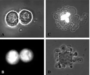
The CMV promoter-based expression construct pcDNA3-EGFP is most suitable for the design of optimal transient transfection conditions because the vector-dependent expression can easily be recorded in living cells by means of the bright EGFP fluorescence. For example, EAT cells show onset of green fluorescence about 12 hours post-electroporation in the presence of the pcDNA3-EGFP control plasmid. Optimal expression is observed at 24-48 hours post-transfection. The green fluorescence reflecting EGFP is distributed in the whole cell and no cytotoxic effects are detectable either in transiently transfected cells or in a stably transfected cell line (Fig. 2A,B). Transient transfection efficiencies are in the order of 20-40%. High plasmid concentrations (20 µg/107cells) result in a higher percentage of transfected cells than do low plasmid concentrations (5-10 µg/107 cells). However, there is no strict linear relationship, and sequential experiments performed under identical conditions, e.g. the same plasmid concentrations, may result in different ratios of non-transfected and genetically manipulated cells. In contrast, parallel assays show rather identical fractions of transfected cells. Consequently, transfections with the control plasmid pcDNA3-EGFP were performed throughout in parallel as an indicator of the efficiency of the transfections
Fig. 1. DNA binding of the recombinant C1D protein. (A) Expression of MC1D protein in E. coli M15 transformed with the pQE30-MC1D construct. The figure shows a Coomassie Blue-stained 12% polyacrylamide gel loaded with the proteins of an E. coli lysate (lane 1) and with MC1D protein (lane 2) isolated by means of Ni-NTA agarose. (B) MC1D-induced DNA mobility-shift on a 1% (w/v) ethidium bromide-stained agarose gel. Purified MC1D protein was folded on a supercoiled plasmid (3 kbp). Lane 1 shows the supercoiled plasmid without MC1D protein. Lanes 2 and 3 show the mobility of this plasmid associated with increasing amounts of recombinant MC1D protein (lane 2, one MC1D molecule per 10 bp; lane 3, two MC1D molecules per 10 bp). (C) Release of the supercoiled plasmid after digestion of the MC1D/plasmid complexes with proteinase K. Lane 1, complexes of the type shown in B, lane 3, were digested with proteinase K and submitted to a 1% (w/v) ethidium bromide-stained agarose gel (lane 2). Mainly supercoiled plasmid is released, which indicates that the mobility shift shown in B is due to a stable DNA-protein interaction and not caused by a change of DNA conformation, e.g. linearization of the plasmid. (D) Preferential binding of MC1D to supercoiled DNA. Samples comprising equal amounts of linearized and supercoiled plasmid DNA (lane 1) were mixed with increasing quantities of MC1D protein and analysed on an agarose gel (lane 2) as described for B. The mobility of the supercoiled plasmid is significantly reduced at a protein-DNA ratio where the migration of the linearized conformation remains unchanged. (E) Mobility shift induced by the MC1D-EGFP fusion protein. The MC1D-EGFP fusion protein was expressed in E. coli and purified as described for the MC1D protein (A). It becomes folded, like the MC1D protein, on the supercoiled plasmid and reduces its mobility, which indicates that the EGFP portion of the fusion protein does not interfere with the DNA-binding capacity of the MC1D protein. Lane 1 shows the supercoiled plasmid and lane 2 shows the mobility-shifted plasmid loaded with MC1D-EGFP protein.
with other plasmids (e.g. pcDNA3-C1D) whose expression cannot be controlled visually.
Transient transfection of EAT suspension cells with the pcDNA3-MC1D expression construct
Electroporation of cell cultures generally results in some mechanically or otherwise damaged cells. Accordingly, it is almost impossible to differentiate between disrupted cells and potentially apoptotic cells. Moreover, several methods commonly used to provide information on apoptosis-related effects potentially induced by vector-dependent C1D overexpression are not applicable because the changes in the 20-40% subset of transfected cells become obscured by the unchanged characteristics of the cells in the larger non-manipulated fraction. In this situation, the terminal deoxynucleotidyl transferase-mediated dUTP (fluorescein) nick end-labeling assay (TUNEL, Boehringer) is the method of choice to detect apoptosis, because this test allows a correlation of the size of the cell fraction containing apoptosis-related DNA strand breaks with the size of the fraction of genetically manipulated cells. 48 hours post-transfection, transiently pcDNA3-C1D-transfected EAT cell cultures contain a subset of cells (10-20%) showing significant incorporation of the fluorescein label (Fig. 3). The size of this fraction correlates well with that of EGFP-positive cells in parallel assays transfected with the control plasmid pcDNA3-EGFP, suggesting that pcDNA3-MC1D-transfected cells are
the positives in the TUNEL assay. As mentioned above the electroporation procedure results in some damaged and obviously non-viable cells. Such cells can also be detected in the cultures transfected as a control with the pcDNA3 vector. In vector controls, however, these physically injured cells show no significant fluorescence label, which points to the significance of the TUNEL assay. Actually, inspection of several hundred cells from vector-transfected assays alone revealed less than 1% of significantly fluorescein-labelled cells. Significant fluorescein label is also detected in cells of Etoposide-treated cultures (5 µM), but the fraction of Etoposide-induced positive cells is significantly smaller (≤5%) than that induced by pcDNA3-C1D transfection. Accordingly, these results can be considered as the first evidence for induction of apoptosis by vector-dependent overexpression of C1D.
Transient transfection of EAT suspension cells with the pcDNA3-MC1D-EGFP expression construct
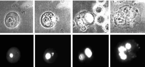
Transfections with EGFP-tagged MC1D expression constructs allow the transfected cells to be identified and the morphological changes induced by vector-dependent overexpression to be traced in single cells. In a first series of experiments suspension cells (EAT and Hela S3) were transfected by electroporation in the presence of pcDNA3-MC1D-EGFP. The results obtained with these two cell lines were rather similar and only EAT cell data are shown. Transfected EAT cells elicite nuclear onset of green fluorescence about 9-12 hours post-transfection. Later stages are characterized by an increasing fluorescent spot, which develops in the cell nucleus. This process is accompanied by morphological changes typical for apoptotic cell death, e.g.
Fig. 2. Morphology of EAT cells stably transfected with pcDNA3-EGFP, transiently transfected with pcDNA3-MC1D-EGFP and cultured in the presence of 5 µM Etoposide, respectively. (A,B) EAT cells stably transfected with the control plasmid pcDNA3-EGFP in bright field (A) and in fluorescence mode (B). The morphology and the viability of the cells indicate that the enhanced green fluorescent protein (EGFP) itself has no cytotoxic effects. (C) The morphology of an EAT cell transiently transfected with the pcDNA3-MC1D-EGFP expression construct (48 hours post-transfection, simultaneous bright field and fluorescence mode). For comparison (D) shows an EAT cell with the typical morphology of apoptotic cells in cultures supplemented with 5 µM Etoposide (only bright field). Zeiss IM35, phase 63×.
Fig. 3. Terminal deoxynucleotidyl transferase-mediated dUTP (fluorescein) nick end-labeling (TUNEL assay) following transfection of EAT cells with pcDNA3-MC1D. EAT cells were transiently transfected with the MC1D expression construct pcDNA3-MC1D. At 48 hours post-transfection the cells were collected, fixed, and submitted to a procedure which adds
fluorescein-labeled dUTP at 3′-OH ends of DNA breaks. The upper row of micrographs shows cells in bright field. The lower row shows the same fields in fluorescence mode (FITC filter set). Images were captured by means of an electronic camera. Improvision Openlab system, Zeiss S100 TV, phase 40×. Bars, 10 µm.
cytoplasmic vacuolation, membrane blebbing and, finally, nuclear disintegration (Fig. 4). The conclusion of apoptotic cell death of transfected cells is supported by comparison of the rather identical morphologies of pcDNA3-MC1D-EGFP-transfected cells (Fig. 2C) with those triggered into apoptosis by 5 µM Etoposide, respectively (Fig. 2D). In contrast to monolayer cells, EAT cells show no shrinkage and become progressively flattened during apoptosis. Cells of a transfected culture go asynchronously through this process, e.g. cells with complete nuclear disintegration can be observed as soon as 24 hours post-transfection while cells showing only intense nuclear fluorescent spots are still found 48 hours post-transfection.
Stably transfected cell lines expressing the non-toxic EGFP can be easily prepared by G418 selection of pcDNA3-EGFP transfected EAT cells (Fig. 2A,B). In contrast, no fluorescent EAT cells survive during G418 selection of pcDNA3-C1D-EGFP-transfected cells, which is a further indication for the cytotoxic effect of over-physiological levels of C1D.
The cytotoxic effects of C1D overexpression are not species-specific. Expression constructs containing the human C1D sequence are as effective in murine cells as in human cells and vice versa (not shown).
Transient transfection with other EGFP-tagged MC1D expression constructs
The relative positions of the C1D and EGFP sequences in the fusion proteins do not change the effectiveness of the constructs because transient transfection with either pcDNA3-MC1D-EGFP or pcDNA3-EGFP-MC1D induces the morphological changes described in Figs 2 and 4. These changes are also observed following transfection with an expression construct driven by the weaker SV40 early promoter (pJ3Ω-MC1D-EGFP), which indicates that a minor increase of C1D expression is sufficient to induce the cytotoxic effects. This conclusion is also supported by unsuccessful attempts to generate stable transfectants using the metalotionein promoter-based pMEP 4-MC1D-EGFP construct. Before hygromycin selection in the absence of metal
ions a fraction of cells shows barely detectable fluorescence, which points to low-level expression of MC1D-EGFP under these conditions. Apparently, the residual transcriptional
Fig. 4. Stages of cell death following transient
transfection of EAT cells with the expression construct pcDNA3-MC1D-EGFP. Living EAT cells transiently transfected and inspected 24-48 hours post-transfection show morphological changes including (from left to right) local nuclear accumulation of the EGFP-tagged MC1D protein, cytoplasmic vacuolization, membrane blebbing and finally nuclear disintegration. The
micrographs in the upper row show the transfected EAT cells in bright field; those in the lower row show the same
cells in fluorescence mode (FITC filter set). It should be noted that cells transfected with other C1D expression constructs (e.g. pcDNA3-EGFP-MC1D, pJ3Ω-MC1D-EGFP, pcDNA3-HC1D-EGFP) show essentially the same morphological changes. Zeiss IM35, phase 63×.
Fig. 5. Estimation of the level of vector-dependent MC1D-EGFP expression, p53 expression and p21Cip1(WAF1) expression in EAT cells. Autoradiography of a 12% (w/v) SDS polyacrylamide gel loaded with total cellular protein from non-transfected EAT cells (lane 1), and from EAT cells transiently transfected with the expression construct pcDNA3-MC1D-EGFP (lanes 2 and 3). Following electroblotting the nitrocellulose membrane was probed with an MC1D-specific antibody and 125I-labeled anti rabbit Ig (Amersham). Transfected cells (lane 2, 5 µg of plasmid/107cells; lane 3, 20 µg of plasmid/107cells) were harvested 48 hours post-transfection. It should be noted that MC1D is present in EAT cells in a monomeric (M) and in dimeric forms (D) (Nehls et al., 1998). Molecular masses refer to a prestained protein marker present on the gel (not shown). The same protein lysates were used for identical blots, which were probed with antibodies specific for p53 and p21Cip1(WAF1) respectively. Only the relevant sections of the latter blots are shown.
activity of the non-induced metalotionein promoter is sufficient to produce low but lethal amounts of the MC1D-EGFP protein. Accordingly, all cells die during hygromycin selection. The transfected cells are killed because they express MC1D-EGFP and the non-transfected cells because they are not hygromycin-resistant.
Quantitation of MC1D-EGFP expression in EAT suspension cells
The intense fluorescence of the EGFP protein is a most
sensitive tool for identifying transfected cells; however, it may be unreliable with respect to the actual quantity of EGFP-tagged C1D, which is lethal for transfected cells. More reliable estimates would be expected from protein blots probed with C1D-specific antibody. However, there is a problem with this technique in that the C1D-EGFP protein present in transfected cells is diluted by protein from the fraction of non-transfected cells. Fig. 5 shows a protein blot loaded with equal amounts of total cellular proteins from non-transfected EAT cells and from EAT cells non-transfected with increasing amounts of the pcDNA3-MC1D-EGFP plasmid. It should be noted that the endogenous C1D protein is present in EAT cells in a monomeric (16 kDa) and in non-disulfide-bonded dimeric (32 kDa) forms (Nehls et al., 1998). The MC1D-specific serum detects the endogenous 16 kDa and 32 kDa proteins on the three lanes while it detects a protein of 43 kDa, which is the molecular mass of the MC1D-EGFP fusion protein, only on the lanes loaded with protein from transfected EAT cells (Fig. 5). The fusion protein signal on the lane loaded with protein from cells transfected with 20 µg/107 cells is almost as intense as the signal(s) of the endogenous C1D. Since the transfection efficiency was about 40% it can be included that a two- to threefold increase of the C1D level induces the cytotoxic effects.
Membrane irritation and morphological changes of non-transfected cells cocultured with pcDNA3-MC1D-EGFP- and pcDNA3-HC1D-EGFP-transfected cells
The EGFP-dependent differentiation between transfected and non-transfected cells allows us to investigate the fate of the population of apparently non-transfected cells cocultured with transfected cells. Microscopical inspection of pcDNA3-MC1D-EGFP-transfected EAT cultures revealed membrane changes of non-fluorescent cells, suggesting the release of cytotoxic factors from transfected cells dying by apoptosis (Fig. 6). These effects are preferentially seen in experiments with high transfection efficiencies (≥20%). Although the cells are kept in full medium during microscopical observation these morphological changes could be due to the non-optimal culture conditions.
This bystander effect is also observed, however, after transfection of human lung cancer-derived LCLC 103H monolayer cells cultured and microscopically inspected under more optimal culture conditions. LCLC 103H cells show, like EAT cells, no significant changes of the cell morphology and cell growth when transfected with the pcDNA3-EGFP control construct. They grow up during 48 hours forming a perfect monolayer (Fig. 7A,A′). In contrast, 48 hours post-transfection with the HC1D-EGFP-expressing construct, the transfected cells and almost all of the apparently non-transfected cells show shrinkage and detachment from the support (Fig. 7B,B′). The transfected LCLC 103H monolayer cells can be long-term cultured and microscopically inspected under almost optimal culture conditions using a temperature-controlled incubation chamber (Pentz and Hörler, 1992). This device allows us to study dynamic processes post-transfection with the pcDNA3-HC1D-EGFP expression construct (Fig. 8). Onset of green fluorescence is observed 8-12 hours post-transfection. This initial green fluorescence, indicating EGFP-tagged HC1D expression, is located, like in EAT suspension
Fig. 6. Morphological changes of non-transfected EAT cells cocultured with transfected EAT cells. A group of EAT cells from a transiently pcDNA3-MC1D-EGFP-transfected culture was observed for 1 hour in full medium under the microscope. Observation started at 36 hours post-transfection. The uppermost image was captured only in fluorescence mode while the other images were captured under simultaneous bright field and fluorescence mode. Only one cell of this group is transfected because it shows bright fluorescence. At the time the observation began (0′) the bystander cells showed a rounded morphology, which is typical for EAT cells. Arrows point to membrane changes of non-transfected cells arising during the observation period (10-60 minutes). Zeiss IM35, phase 6×.
cells, in dense nuclear spots (Fig. 8). Later, as shown at 24 hours, the transfected cells show bright fluorescence, predominantly in cell nuclei. In addition Fig. 8 shows a selected field comprising only one transfected cell. Continued observation of this highly fluorescent cell for another 4 hours shows retraction of the plasma membrane, cell shrinkage and detachment from the support. Concomitantly the apparently non-transfected bystander cells show similar morphological changes which, in this case, cannot be attributed to non-optimal culture conditions. Finally, at 48 hours post-transfection this bystander effect leads to about 50% of detached and spherical-shaped cells, while only about 10% of the cells appear to be transfected and exhibit significant green fluorescence (not shown). The cytotoxic factors inducing this bystander effect are unknown. However, they appear to possess rather short half-life times because the cytotoxic effects could not be induced in non-transfected cultures by conditioned medium taken from transfected EAT or LCLC 103H cultures.
Exploration of the mechanism involved
The putative mechanism for C1D-induced apoptosis predicts DNA-end independent activation of DNA-PK with the consequence of p53 phosphorylation and activation of its downstream effectors. This assumption is based on the fact that C1D interacts in vivo with DNA-PK and that DNA-PK is activated by C1D in a DNA end-independent manner (Yavuzer et al., 1998). Accordingly, DNA-PK deficient cells, e.g. M059J cells (Lees-Miller et al., 1995), should escape C1D-induced apoptosis. However, this approach meets with technical problems. Regular M059J cell cultures contain a
significant fraction of dying and detached cells. This fraction increases post-transfection with any plasmid DNA, e.g. with the control plasmid pcDNA3-EGFP. Accordingly it is difficult to differentiate between C1D-dependent effects and the effects induced by the transfection procedure. Viable and C1D-EGFP transfected cells can be found at 12-24 hours post-transfection; however, this fraction is low and neither proves nor disproves the insensitivity of M059J cells to C1D-induced apoptosis.
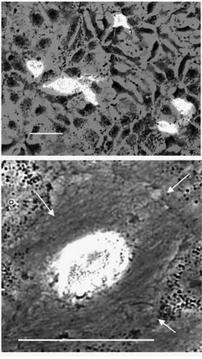
In contrast, the downstream steps of the proposed mechanism are supported by additional experimental evidence. Wild-type p53 appears to be required for C1D-induced apoptosis in cells where p53 cannot be replaced by functionally related proteins. Huh7 cells are known to possess mutated and non-functional p53, and p53 function is not replaced in these cells by functionally similar proteins (Muller et al., 1997). Following transfection with the C1D-EGFP expression construct these cells grow up and form perfect monolayers (Fig. 9). At higher magnification the C1D-EGFP-expressing cells appear to be well spread and without morphological changes that are typical for apoptosis, e.g. they do not show membrane retraction, detachment and plasma vacuolization.
The p53 level itself remains unchanged in C1D-EGFP-transfected cells (Fig. 5B), which is to be expected because p53 activation is due to phosphorylation rather than increased expression (Shieh et al., 1997). In contrast, expression of the p53 downstream effector p21Cip1(WAF1) is significantly increased in C1D-EGFP transfected cells (Fig. 5C). It should be noted that the induction of p21Cip1(WAF1) expression is somewhat underestimated by
Fig. 7. LCLC 103H cells transfected with the control construct pcDNA3-EGFP and the HC1D-EGFP-expressing construct pcDNA3-HC1D-EGFP. The cells of an LCLC 103H culture transiently transfected with the control plasmid pcDNA3-EGFP show, 48 hours post-transfection, perfect cell morphology. The cells grow up during this period normally and form a dense monolayer (A, bright field; A′, fluorescence mode, FITC filter set). Two cells of this group are transfected and express significant amounts of EGFP, which has apparently no toxic effect. In contrast, LCLC 103H cells transfected with the expression construct pcDNA3-HC1D-EGFP show, 48 hours post-transfection, disturbed morphology, especially shrinkage and detachment (B, bright field; B′, fluorescence mode, FITC filter set). It should be noted that all cells in this field show disturbed morphology whether they are transfected or not. Improvision Openlab system, Zeiss Axiovert S100 TV, phase 40×.
the western blot shown in Fig. 5 because only about 40% of the cells are transfected.
DISCUSSION
Apoptosis is induced by many and rather different environmental stimuli, such as treatments with various drugs and death factors like Fas ligand or TNF α, cell injury, irradiation, viral infection and growth factor deprivation. Any of these exogenous factors or treatments is able to activate a genetic program that culminates in apoptotic cell death. A large number of gene products that are required for, favour or inhibit apoptosis induced by exogenous factors have been identified in lower and higher eukaryotes. Moreover, intracellular death signals play a major role during development; e.g. it has been reported that in organisms such as the nematode C. elegans large numbers of cells are eliminated by programmed cell death during development (Sulston and Horvitz, 1977; Ellis et al., 1991), which indicates that cells can activate their own suicide programs. However, cellular genes which are able to initiate the programmed cell death, e.g. by transient overexpression, are rather rare. The IL-1β-converting enzyme (ICE), which is a mammalian homolog of the C. elegans cell death gene ced-3, has been reported to induce apoptosis in fibroblasts (Miura et al., 1993). Other examples include the Drosophila proteins HID and GRIM,
whose transient overexpression induce apoptosis in SF-21 cells (Vucic et al., 1998). Overexpression of a serine/threonine kinase, designated ZIP kinase, has been reported to induce the morphological changes of apoptosis in NIH 3T3 cells (Kawai et al., 1998).
In this paper we present evidence for the induction of programmed cell death of various tumor cells by transient overexpression of the conserved 16 kDa DNA-binding protein C1D. Several criteria suggest that the C1D-induced cell death occurs by apoptosis rather than by necrosis. The fraction of terminal transferase-labeled cells corresponds with the fraction of C1D-transfected cells and the morphological changes induced by overexpression of EGFP-tagged C1D are typical for apoptotic cell death. Surprisingly, the endogenous C1D gene is ubiquitously expressed. Apparently, physiological levels of this protein are harmless and most probably essential for the cell. However, in the tumor cells tested, additional quantities of the gene product are not tolerated, with the consequence of cell death. These additional quantities inducing the cytotoxic effects are rather low and it should be noted that the bright fluorescence of the EGFP-tagged C1D protein overestimates the true amount of the vector-expressed C1D protein. Blots loaded with protein from transfected EAT cell cultures show that the C1D level is increased at most two- to threefold in transfected cells. Since the C1D expression levels of different tissues cover a broader range (one- to fivefold) it must be concluded that the
Fig. 8. Long-term microscopical observation of LCLC 103H cells transfected with pcDNA3-HC1D-EGFP. Transiently transfected LCLC 103H cells cultured in a temperature-controlled incubation chamber (Pentz and Hörler, 1992) show nuclear onset of green fluorescence 8-12 hours transfection (8 hours). 24 hours post-transfection the transfected cells show bright and predominantly nuclear fluorescence (24 hours). During a further 4 hour observation period the transfected cell shows shrinkage (24+4 h; retraction of the plasma membrane, arrows). Concomitantly the apparently non-transfected cells of this group show similar
morphological changes. They become progressively spherical-shaped and detached from the support. Openlab Improvision system, Zeiss S100 TV, phase 40×.
expression level of the endogenous gene is cell type-specifically regulated and that different cell types require and/or tolerate different levels of C1D protein.
The induction of apoptosis by overexpression of C1D paralleled by increasing levels of p21Cip1(WAF1), and missing apoptosis in cells with non-functional p53, correlate well with previously published data on the specific interaction of the C1D protein with DNA-PK. The latter enzyme is known to be activated by DNA strand breaks, e.g. by those generated by γ-irradiation (Anderson and Lees-Miller, 1992; Jackson, 1997; Smith et al., 1998). The DNA end-activated enzyme phosphorylates p53 at SER 15 and SER 37, resulting in p53 activation and in p53-dependent radiation-induced apoptosis (Shieh et al., 1997). This pathway also appears to be inducible by overexpressed C1D in the absence of DNA ends. It has been shown that C1D directs the activation of DNA-PK in a manner that does not require DNA termini. Moreover, DNA-PK activated by C1D is able to
phosphorylate p53 (Yavuzer et al., 1998). Accordingly, C1D-induced apoptosis can be consistently explained by the assumption that overexpression of C1D mimics or substitutes DNA strand breaks, resulting in an initial DNA end-independent activation of DNA-PK, which results in p53 activation and finally in apoptosis.
While the induction of apoptosis in C1D-overexpressing cells can be consistently explained, there is at present no such explanation for the morphological changes of the bystander cells, which are apparently non-transfected with the constructs expressing EGFP-labeled C1D. Unknown factors released by the transfected and dying cells must be postulated. However, it cannot be ruled out unequivocally that the non-fluorescent cells are indeed transfected and express EGFP-tagged protein in amounts that cannot be detected by fluorescence microscopy but which are sufficient to induce the morphological changes.
It has been suggested that survival of tumor cells in all or in part is due to their inability to activate a suicide program (Donehower et al., 1992). In this paper it is shown that vector-dependent overexpression of C1D can override this failure of tumor cells in vitro, provided that p53 function is retained. The in vivo application of this procedure would meet the basic problems of gene therapy, consisting of insufficient transfection rates of the target cells by appropriate vector systems. However, all cells of an organism, including tumor cells, do contain the C1D gene. Thus it should be possible to substitute the vector-dependent overexpression of C1D by stimulation of the endogenous C1D promoter. Accordingly, the identification of the promoter which regulates the physiological C1D expression and the search for factors or procedures which could systemically activate the C1D promoter seems a promising task.
One of us (B. J.) is grateful for a guest scientist fellowship provided by the German Cancer Research Center, Heidelberg. The authors thank P. Jeggo (University of Falmer, UK) for supplying M059J cells, J. Turner (Cross Cancer Institute, Edmonton, Canada) for permission to use these cells, and C.-H. Schroeder (German Cancer Research Center) for giving us Huh7 cells.
REFERENCES
Anderson, C. W. and Lees-Miller, S. P. (1992). The nuclear serine/threonine
protein kinase DNA-PK. Crit. Rev. Euk. Gene Exp. 2, 283-314.
Avramova, Z. and Tsanev, R. (1987). Stable DNA protein complexes in
eukaryotic chromatin. J. Mol. Biol. 196, 437-440.
Church, G. M. and Gilbert, W. (1984). Genomic sequencing. Proc. Natl. Acad. Sci. USA 81, 1991-1995.
Donehower, L. A., Harvey, M., Slagle, B. L., McArthur, M. J., Montgomery, C. A., Butel, J. S. and Bradley, A. (1992). Mice deficient
for p53 are developmentally normal but susceptible to spontaneous tumors. Nature 356, 215-221.
Ellis, R. E., Yuan, J. and Horvitz, H. R. (1991). Mechanisms and functions
of cell death. Ann. Rev. Cell Biol. 4, 663-689.
Hendrich, B. D. and Willard, H. F. (1995). Epigenetic regulation of gene
expression: the effect of altered chromatin structure from yeast to mammals. Hum. Mol. Genet. 4, 1765-1777.
Jackson, S. P. (1997). DNA-dependent protein kinase. Int. J. Biochem. Cell. Biol. 29, 935-938.
Juodka, B., Pfütz, M. and Werner, D. (1991). Chemical and enzymatic
analysis of covalent bonds between peptides and chromosomal DNA. Nucleic Acids Res. 19, 6391-6398.
Fig. 9. pcDNA3-HC1D-EGFP transfection of cells (Huh7) with non-functional p53. Transiently transfected Huh7 cells are growing normally and they form a monolayer at about 48 hours post-transfection (upper image, overview). Inspection of individual cells of such cultures at higher magnification shows that the morphology of the transfected cells is unchanged when compared with non-transfected cells. The non-transfected cells remain well spread and attached to the support (lower image). The images represent merged layers captured sequentially in phase contrast and fluorescence mode. Openlab Improvision software, Zeiss S100. Bars, 50 µm.
Kawai, T., Matsumoto, M., Takeda, K., Sanjo, H. and Akira, S. (1998).
ZIP kinase, a novel serine/threonine kinase which mediates apoptosis. Mol. Cell. Biol. 18, 1642-1651.
Lees-Miller, S. P., Godbout, R., Chan, D. W., Weinfeld, M., Day, R. S., Barron, G.M. and Allalunis-Turner, J. (1995). The absence of p350
subunit of DNA-activated protein kinase from a radiosensitive human cell line. Science 267, 1183-1185.
Miura, M., Zhu, H., Rotello, R., Hartwig, E. A. and Yuan, J. (1993).
Induction of apoptosis in fibroblasts by IL-1β-converting enzyme, a mammmalian homolog of the C. elegans cell death gene ced-3. Cell 75, 653-660.
Morgenstern, J. P. and Land, H. (1990). A series of mammalian expression
vectors and characterisation of a reporter gene in stably and transiently transfected cells. Nucleic Acids Res. 18, 1068.
Muller, M., Strand, S., Hug, H., Heinemann, E. M., Walczak, H., Hofmann, W. J., Stremmel, W., Krammer, P. H. and Galle, P. R. (1997)
Drug-induced apoptosis in hepatoma cells is mediated by the CD5 (Apo-1/Fas) receptor/ligand system and involves wild-type p53. J. Clin. Invest.
99, 403-413.
Nehls, P., Keck, T., Greferath, R., Spiess, E., Glaser, T., Rothbarth, K., Stammer, H. and Werner, D. (1998). cDNA cloning, recombinant
expression and characterization of polypeptides with exceptional DNA affinity. Nucleic Acids Res. 26, 1160-1166.
Neuer, B., Plagens, U. and Werner, D. (1983). Phosphodiester bonds
between polypeptides and chromosomal DNA. J. Mol. Biol. 164, 213-235.
Neuer-Nitsche, B., Lu, X. and Werner, D. (1988). Functional role of a highly
repetitive DNA sequence in anchorage of the mouse genome. Nucleic Acids Res. 16, 8351-8360.
Pentz, S. and Hörler, H. (1992). A variable cell culture chamber for open and
closed cultivation, perfusion and high microscopic resolution of living cells. J. Microsc. 167, 97-103.
Pfütz, M., Gileadi, O. and Werner, D. (1992). Identification of human
satellite DNA sequences associated with chemically resistant nonhistone polypeptide adducts. Chromosoma (Berlin) 101, 609-617.
Shieh, S.-Y., Ikeda, M., Taya, Y. and Prives, C. (1997). DNA
damage-induced phosphorylation of p53 alleviates inhibition by MDM2. Cell 91, 325-334.
Smith, G. C. M, Dicheva, N., Lakin, N. D. and Jackson, S. P. (1998). The
DNA-dependent protein kinase and related proteins. Biochem. Soc. Symp.
64, 87-100.
Strohman, R. C. (1997). The coming Kuhnian revolution in biology. Nature Biotechnol. 15, 194-200.
Sulston, J. and Horvitz, H. R. (1977). Postembryonic cell lineages of the
nematode C. elegans. Dev. Biol. 82, 110-156.
Werner, D. and Neuer-Nitsche, B. (1989). Site-specific location of covalent
DNA-polypeptide in the chicken genome. Nucleic Acids Res. 17, 6005-6015.
Yavuzer, U., Smith, G. C. M., Bliss, T., Werner, D. and Jackson, P. (1998).
DNA end-independent activation of DNA-PK is mediated via association with the DNA-binding protein C1D. Genes Dev. 12, 2188-2199.
Vucic, D., Kaiser, W. J. and Miller, L. K. (1998). Inhibitor of apoptosis
proteins physically interact with and block apoptosis induced by Drosophila proteins HID and GRIM. Mol. Cell. Biol. 18, 3300-3309.
Zamir, I., Dawson, J., Lavinsky, R. M., Glass, C. K., Rosenfeld, M. G. and Lazar, M. A. (1997). Cloning and characterization of a corepressor and
potential component of the nuclear hormone receptor repression complex. Proc. Natl. Acad. Sci. USA 94, 14400-14405.


