Research Article
Prognostic Significance of Tumor Tissue NeuGcGM3 Ganglioside
Expression in Patients Receiving Racotumomab Immunotherapy
Necdet Uskent ,
1Sule Ayla ,
2Nil Molinas Mandel ,
3Metin Ozkan ,
4Mehmet Teomete,
5Huseyin Baloglu,
6Cenk Aydıncer,
1Hale Yergok,
3Ender Dogan ,
4Barkın Berk ,
7and Aziz Yazar
81Department of Medical Oncology, Anadolu Health Center, Istanbul, Kocaeli, Turkey
2Department of Histology and Embryology, School of Medicine, Istanbul Medipol University, Istanbul, Turkey 3Department of Medical Oncology, School of Medicine, Koc University, Istanbul, Turkey
4Department of Medical Oncology, School of Medicine, Erciyes University, Kayseri, Turkey 5Department of Medical Oncology, Altunizade Acibadem Hospital, Istanbul, Turkey 6Department of Pathology, Anadolu Health Science Center, Istanbul, Kocaeli, Turkey
7Department of Pharmaceutical Chemistry, School of Pharmacy, Istanbul Medipol University, Istanbul, Turkey 8Department of Internal Diseases, School of Medicine, Acibadem University, Istanbul, Turkey
Correspondence should be addressed to Necdet Uskent; [email protected]
Received 8 January 2020; Revised 1 May 2020; Accepted 19 May 2020; Published 29 June 2020 Academic Editor: Hakan Buyukhatipoglu
Copyright © 2020 Necdet Uskent et al. This is an open access article distributed under the Creative Commons Attribution License, which permits unrestricted use, distribution, and reproduction in any medium, provided the original work is properly cited. Expression of N-glycolyl GM3 (NeuGcGM3) ganglioside was detected in the tumor specimens of patients who were on Racotumomab anti-idiotype vaccine maintenance treatment, and prognostic significance as a biomarker was investigated. No statistically significant association was observed in the multivariate analysis between overall survival and tissue NeuGcGM3 IHC levels. Although numerically there was a difference favoring less intense IHC for better prognosis, this did not reach statistical power. However, there was a strong correlation between Racotumomab doses and overall survival (OS). Mean OS of the patient with more than 10 Racotumomab application was significantly longer than the patient who had less than 10 injections (70.7 months vs. 31.1 months, p < 0.001). We propose that, regardless of staining intensity, the presence of NeuGcGM3 in patient tissues might be an indicator of benefit in Racotumomab treatment.
1. Introduction
Numerous molecules have been considered as targets for cancer immunotherapy because of their levels of expression on tumor cells and their ability of immunogenicity. Among them, a ganglioside GM3 (Neu5Gc) which presents on the outer surface of the plasma membrane of vertebrate cells attracted most attention as a tumor specific antigen, a target for cancer immunotherapy.
NeuGc is not normally expressed in human cells due to the lack of the enzyme cytidine monophospho-N-ace-tylneuraminic acid hydroxylase, responsible for its synthesis. Aberrant accumulation of NeuGc has been shown in many human malignant cell membranes. In particular, the
aberrant expression of the ganglioside NeuGcGM3 in certain human tumors, like lung cancer, breast cancer, neuroblas-toma (Scursoni et al. (Clin Dev Immunol, 2011)), pancreatic cancer, and gastrointestinal tumors, made this molecule an attractive target for immunotherapy. The most common sialic acids in humans are N-acetyl (NeuAc) and N-glycolyl (NeuGc) neuraminic acids(Figure 1) [1–6].
There are numerous hypotheses explaining the existence of NeuGcGM3 in human malignancies, from changing di-etary habits to the modified metabolism of malignant cells as a result of genetic mutations and due to the hypoxic con-ditions of the tumor [7].
Yin et al. showed in their study that hypoxic condition resulted in overexpression of sialic acid transporter sialin
Volume 2020, Article ID 1360431, 7 pages https://doi.org/10.1155/2020/1360431
and induced cancer-associated gangliosides in human cancer cells [8].
Given its tumor-associated expression, GM3 (Neu5Gc) targeting antibodies have been developed, including the mouse 14F7 monoclonal antibody and its humanized variant 14F7hT [9–11].
Anti-idiotypic antibodies functionally resemble the an-tigen itself in their own idiotype and Racotumomab is this kind of monoclonal antibody, which mimics NeuGcGM3 and triggers an immune response in various tumors [12–15]. The production of Racotumomab anti-idiotype mono-clonal antibody involves the immunization of Balb/c mice with NeuGcGM3 ganglioside, followed by isolation of IgM monoclonal antibody (Ab1). This Ab1 antibody was again used in mice to generate an IgG1 monoclonal antibody (Ab2), named as Racotumomab which has a high affinity towards Ab1 idiotype. Racotumomab by mimicking the ganglioside NeuGcGM3 induces a specific cellular and humoral immune response against the actual antigen (Figure 2).
Segatori et al. demonstrated that anti-NeuGcGM3 Abs induced by Racotumomab vaccination are able to mediate an antigen-specific antibody-dependent cellular cytotoxicity (ADCC) response against tumor cells in NSCLC patients [16].
Patients vaccinated with Racotumomab produce anti-bodies against NeuGcGM3. Hern´andez et al. demonstrated that immunized patients’ sera had cytotoxic effects on tumor cells in the L1210 culture expressing NeuGcGM3 [17].
Racotumomab is currently registered in Cuba and Argentina in non-small-cell lung cancer (NSCLC) as a
second-line treatment. In a multicenter double-blind ran-domized, placebo controlled Phase II/III trial involving 176 patients with advanced NSCLC, Alfonso et al. demonstrated an overall survival benefit, almost doubled in one year (22.5% vs. 40.2%) and tripled in 2 years (6.7% vs. 18.4%) favoring the study group [18].
The prognostic role of NeuGcGM3 expression in NSCLC is controversial. Blanco et al. assessed the relationship be-tween NeuGcGM3 expression intensity and overall survival of advanced NSCLC patients using 14F7 Mab and concluded that NeuGcGM3 expression was associated with higher proliferation index and poor OS of patients. In their study, five-year survival probability in NSCLC patients with higher levels of NeuGcGM3 expression was 3 to 4 times lower than those with reduced levels [3].
In another study, a group from Japan confirmed the high expression of NeuGc gangliosides in NSCLC using the monoclonal antibody GMR8, which is specific to ganglio-sides with NeuGc alpha 2,3Gal-terminal structures and demonstrated for the first time that an inverse relationship exists between NeuGcGM3 expression levels and progres-sion-free survival [19].
There are also other studies claiming that higher tissue NeuGcGM3 levels represent a more aggressive phenotype, like EGFR and EGF expressions [19, 20].
1.1. Aim of the Study. Although scarce reports in the
liter-ature suggested that high tissue NeuGcGM3 levels might represent a more aggressive tumor, prognostic implication of this was not known in the patients who are on
anti-Ceramide OH O O O HN H H H H H H H H H H H H H H H OH OH OH OH OH OH CH2OH CH2CONH Glucose O O O O Galactose COOH HCOH HCOH H2COH GM3(Neu5Gc) ganglioside N-glycoylneuraminic acid (a) Ceramide OH O HN H H H H H H H H H H H H H H H OH OH OH OH OH CH2OH CH2OH CH3CONH Glucose O O O O O O Galactose COOH HCOH HCOH H2COH GM3(Neu5Ac) ganglioside N-acetylneuraminic acid (b)
NeuGcGM3 treatments. In this study, we investigated the prognostic significance of tissue NeuGcGM3 expression level in the patients who are on switch maintenance treat-ment with Racotumomab.
2. Materials and Methods
This study was accredited by Noninvasive Clinical Studies Ethical Committee of Istanbul Medipol University, regis-tration number 10840098-604.01.01-E.44203. Expression of NeuGcGM3 was detected in the tumor specimens of 25 patients, using 14F7 monoclonal antibody (14F7 Mab) by immunohistochemistry (IHC) test and graded as 0, 1+, 2+, or 3+ according to the intensity of the stain. IHC staining was also performed in 10 benign tissue specimens as con-trols. 14F7 monoclonal antibody was obtained from mice that were immunized with purified gangliosides and sup-plied by the manufacturer’s institute, Center of Molecular Immunology (CIM), Havana, Cuba.
Detection of the ganglioside in the tumor tissue was done by IHC with primer antibody: 14F7 Mab 20 mcgr/ml after using biotinylated link universal and a streptavidin HRP commercial kit. Afterwards, the enzymatic activity was vi-sualized with a diaminobenzidine (DAB). All samples were counterstained with Mayer’s Hematoxylin. Immunoreac-tivity on the sample was evidenced as a brown staining. 5-microns thick sections of tissue (paraffin-embedded block) were used to observe the expression of NeuGcGM3.
IHC using 14F7 Mab was evaluated according to the percentage of positive cells (0–100 percent) and graded as 0,
1+ (weak), 2+ (moderate), or 3+ (strong) according to the intensity of the reaction. The score was calculated for each specimen by multiplication of the intensity of reaction and the grade of positive cells. Classification of the score was slightly modified from the method used by Blanco et al. as follows: 0 (total score 0); IHC 1+ (total score 1–100); IHC 2+ (total score 101–150); and IHC 3+ (total score > 150) [3, 15].
2.1. Racotumomab Administration. The route of
adminis-tration was intradermal; each dose was divided into four different anatomic sites in the medial surfaces of arms, thighs, or abdomen. The vaccination program consisted of two phases. In the induction phase, 5 doses were admin-istered 2 weeks apart, followed by the maintenance phase, when 10 more doses were administered monthly.
3. Results
All 48 patients were on Racotumomab maintenance treat-ment following chemotherapy who had at least partial re-mission. Out of a total of 48 patients, 42 had lung cancer (40 NSCLC, 2 SCLC), 6 had miscellaneous cancers (breast, pancreas, etc.) of 40 non-small-cell lung cancer (NSCLC) patients, 12 patients were in stage III, and 28 were in stage IV at the time of the diagnoses. 19 adenocarcinomas and 21 squamous cell carcinomas were identified. None of the patients had sensitive mutations like EGFR, ALK, and ROS-1. There were also 2 patients with small-cell lung cancer (SCLC). Male/female ratio was 3.36 (37/11) and median age was 63 (31–85) (Table 1).
Racotumomab is an anti-idiotype monoclonal antibody that mimics tumor neoantigens, such as the ganglioside NeuGcGM3
+
Adjuvant (aluminum hydroxide)
Immunotherapeutic product for cancer treatment
Lymphocyte Cancer
cell
Neoantigen NeuGc ganglioside
The product administered intradermally to a patient induces a strong specific cellular and humoral response against the NeuGc
ganglioside that induces apoptosis of tumor cells
NeuGcGM3 IHC was performed in 25 out of 48 patients whose paraffin blocks were available and adequate for testing. 9 patients had strong 3+, 12 had moderate 2+, and 4 patients had weak 1+ NeuGcGM3 staining intensity. None of the benign tissues, as controls, showed positivity. Positive IHC staining of NeuGcGM3 from a patient’s tumor tissue with NSCLC and a control staining of a benign tissue using 14F7 Mab are depicted in Figure 3.
Mean overall survival (OS) time for subsequent IHC groups was 88.1 months for IHC 1+, 51.6 months for IHC 2+, and 51.7 months for IHC 3+. Mean survival of the patients who died was 22.6 months for IHC 3+ and 27.9 months for IHC +2, whereas in living patients, mean OS was 51.1 months in IHC 3+, compared to 57.8 months and 74.3 months in IHC 2+ and IHC 1+ patients, consecutively.
In the multivariate Cox regression model, all the vari-ables in Table 2 were included in the model to identify the independent predictors of survival. The analysis showed that the total number of Racotumomab cycles used during the course of treatment was statistically associated with better overall survival rates (HR � 0.750, 95% CI 0.601–0.936,
p �0.011), as was also found in the univariate analysis in Figure 4. That is, after adjusting for age, gender, IHC, and treatment protocol, important reduction in mortality rate was attributed to each dose of Racotumomab.
The total number of Racotumomab cycles used in the course of treatment was categorized into two groups as ≤10 (n � 23) and >10 (n � 25), and it was found that more than 10 cycles significantly were associated with better overall sur-vival (p < 0.001) (Figure 4).
When IHC groups were considered, the patients with IHC scores of 2 have 2.6 times higher risk of death compared to the patients with IHC scores of 1. Also, the patients with IHC scores of 3 have 1.3 times higher risk of death compared to the patients with IHC scores of 1. Due to the small sample size, the findings regarding the IHC groups were not found to be statistically significant (Table 2).
Univariate (Table 3) and multivariate (Table 2) survival analyses were conducted to compare overall survival (OS) rates. Survival curves were generated with the Kaplan–Meier method and log-rank test was used to compare survival curves as the univariate survival analysis. Multivariate analysis was performed with Cox proportional hazards regression to identify the independent risk factors of OS.
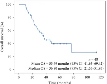
Median follow-up time was 34.2 months, ranging from 10.1 to 113.7 months. After 5 years of follow-up, 27/48 (56.25%) of patients died and 11/48 (22.9%) were alive more than 5 years after diagnosis (Figure 5).
The overall survival rate throughout the follow-up period was found to be higher in the patients with IHC score of 1, with mean overall survival of 88.1 months (95% CI: 43.9–132.2). When the patients with IHC scores of 2 and 3 were considered, the mean of overall survival time was approximately the same (51.7 months (95% CI: 35.5–67.9) vs. 51.6 months (95% CI: 38.14–64.97), respectively). However, the sample size was limited to draw any statisti-cally significant difference between IHC groups (Figure 6).
4. Discussion
Pilco-Jeneto and colleagues assessed the expression of NeuGcGM3 in fifty paraffin embedded specimens from patients diagnosed with sarcomas and investigated the re-lation of this expression with clinicopathological features and overall survival of patients. They concluded that ex-pression of NeuGcGM3 was a significantly poor prognostic factor for overall survival [19].
Blanco et al. investigated the relationship between the NeuGcGM3 expression, Epidermal Growth Factor Receptor (EGFR), and Epidermal Growth Factor (EGF) with overall survival of patients with advanced NSCLC. Higher NeuGcGM3 expression was associated with a poorer OS of the patients. Moreover, the double and triple positivity of tumors with EGFR and EGF identified more aggressive biological behavior in both univariate (p: 0.020) and mul-tivariate analyses (p: 0.010) [3].
In their work, Blanco et al. evaluated the expression of NeuGcGM3 using 14F7 Mab and proposed an IHC score called H-score. The authors classified the staining intensity as 0 (0), 1 (1 to 100), 2 (101–200), and 3 (>200). However, subsequently, they divided H-scores into 2 group as low expression (scores < 150) and high expression (scores > 150), while in our study we decided to use a slightly different scoring system like 0 (0), 1 (1–100), 2 (101–150), and 3 (>150). This modification was proposed by our Pathology Department due to the scarcity of that >200 staining.
NeuGcGM3 is capable to bind to the extracellular do-main of the epidermal growth factor receptor (EGFR). This binding may provoke EGFR system activation mediated by
Total patients (n) NSCLC adeno NSCLC sq cell SCLC Miscel.
Number of patients 48 19 21 2 6 Female 11 6 2 1 2 Male 37 13 19 1 4 Mean age 63.0 62.4 60.2 51.6 58.1 Stage — IV 28∗ 13 15 2 6 IIIB 10∗ 5 5 — — IIIA 2∗ 1 1 — —
NSCLC: non-small-cell lung cancer; adeno: adenocarcinoma; sq cell: squamous cell; Miscel.: miscellaneous.∗Patients with NSCLC (adeno and sq cell) in
the growth factor ligand. Activation of epidermal growth factor in the presence of overexpressed NeuGcGM3 con-stitutes a more aggressive immunophenotype [3]. In our oncoming studies, we are planning to include tissue EGFR levels and serum EGF levels in addition to tissue NeuGcGM3 expression, in order to better understand the value of anti-EGF plus anti-NeuGcGM3 combination strategies.
To the best of our knowledge, there were no studies in the literature, assessing the predictive and prognostic value of NeuGcGM3 expression in the tumor tissues who are on anti-NeuGcGM3 antibody treatment. In our study, we demon-strated a strong relation with Racotumomab dose cycles and overall survival of the patients. Mean survival of the patient who received more than 10 intradermal injections was significantly higher than the patient who received less than 10 injections (70.7 months vs. 31.1 months, p < 0.001).
In the multivariate Cox regression model, all the vari-ables were included in the model to identify the independent predictors of survival (Table 2). The analysis showed that the total number of Racotumomab cycles used during the course of treatment was statistically associated with better overall
survival rates (HR � 0.750, 95% CI 0.601–0.936, p � 0.011). This finding was also confirmed in the univariate analysis; that is, after adjusting for age, gender, IHC, and treatment protocol, the 25% reduction in mortality rate was attributed to the cumulative dose of Racotumomab (log-rank p value <0.001).
These findings may be well comparable with HER-2 expression in breast cancer. HER-2 was considered a bad prognostic biomarker, a messenger for progressive disease before anti-HER2 strategies had been invented. However, today, best outcomes are achieved in this subtype.
The presence of NeuGcGM3 antigen is favorable for the treatment with Racotumomab since its antitumor activity is mediated by an immune response directed against the NeuGcGM3 antigen. Thus, we may argue that regardless of the intensity of the staining, the presence of NeuGcGM3 at the tissues of patients is an indicator of benefit in Racotu-momab treatment.
Our study did not show any significant association between tissue antigen staining intensity and overall
(a) (b)
Figure 3: Positive and negative IHC staining results for NeuGcGM3. (a) Positive IHC 2+ staining for GM3, X10. (b) Negative IHC result for GM3, X10.
Table 2: Multivariate analysis of overall survival with Cox pro-portional hazards regression analysis.
Variables HR (95% CI) p value Age 0.940 (0.869–1.016) 0.119 Gender, male (0.290–52.896)3.920 0.304 IHC +1 Reference +2 2.615 (0.156–43.928) 0.504 +3 1.311 (0.052–33.028) 0.869 Chemotherapy + Racotumomab treatment 0.298 (0.038–2.316) 0.247 Total Racotumomab (0.601–0.936)0.750 0.011 IHC: immunohistochemistry; HR: hazard ratio; CI: confidence interval. Bold p value indicates statistically significant association.
Total racotumomab > 10 (mean OS = 70.7 months, n = 25) Total racotumomab ≤ 10 (mean OS = 31.1 months, n = 23) 0 20 40 60 80 100 O verall sur vi val (%) 20 40 60 80 100 120 0 Time (months) Log-rank p-value < 0.001
Figure 4: Kaplan–Meier survival estimates by the number of total Racotumomab cycles used during the course of treatment (n � 48).
survival, probably due to the small number of patients that were analyzed. However, this may also be the result of compensatory effect of Racotumomab, balancing aggressive biological phenotype with anti-NeuGcGM3 effect.
significance of serum anti-NeuGcGM3 antibodies and cy-totoxicity tests in cell lines using hyperimmune patient’s sera of the patients who are on Racotumomab treatment. In that study, we concluded that cytotoxicity test was a good tool as a biomarker of response to Racotumomab immunotherapy, also related to the better prognosis of the patients with positive tests. However, in the same study, tissue NeuGcGM3 levels did not give us valuable information as to the prediction of response or prognosis although the number of the patients was limited [21].
5. Conclusion
No statistically significant association was observed in the multivariate analysis between overall survival and tissue NeuGcM3 IHC level. Although numerically there was a difference favoring less intense IHC for better prognosis, this did not reach statistical power.
However, looking at the overall survival curve of our patients, we concluded that, regardless of the intensity of staining, the presence of NeuGcGM3 at the tissues of pa-tients is an indicator of benefit in Racotumomab treatment. We believe although high expression of NeuGcGM3 of tumor tissues is generally considered as a poor prognostic marker, anti-NeuGcGM3 treatment with Racotumomab overrides this phenomenon.
Data Availability
The data provided in this publication will be available from the corresponding author upon request.
Conflicts of Interest
The authors declare that they have no conflicts of interest regarding the publication of this paper.
Acknowledgments
The authors would like to thank Assistant Prof. Dr. Oya Kalaycıo˘glu for her help with biostatistics. The authors are also thankful to Biologist Arzu Baykaldı for her outstanding effort in the pursue of coordination between centers.
References
[1] M. Labrada, D. Dornvignit, G. Hevia et al., “GM3 (Neu5Cc) ganglioside: an evolution fixed neoantigen for cancer im-munotherapy,” Seminars in Oncology, vol. 45, no. 1-2, pp. 41–51, 2018.
[2] A. M. Scursoni, L. Galluzzo, S. Camarero et al., “Detection of
N-glycolyl GM3 ganglioside in neuroectodermal tumors by
immunohistochemistry: an attractive vaccine target for ag-gressive pediatric cancer,” Clinical and Developmental
Im-munology, vol. 2011, Article ID 245181, 6 pages, 2011.
[3] R. Blanco, E. Dom´ınguez, O. Morales et al., “Prognostic significance of N-glycol GM3 ganglioside expression in non-small cell lung carsinoma patients: new evidences,” Pathology
Research International, vol. 2015, Article ID 132326, 12 pages,
2015. and dead.
Alive (n � 20) Dead (n � 28) Age, years (mean ± SS) 63.1 ± 11.5 63.8 ± 11.1 Gender Male 14 23 Female 6 5 IHC +3 6 3 +2 5 7 +1 3 1 Not available 6 17
Total Racotumomab (mean ± SS) 13.9 ± 5.2 9.6 ± 4.0
Fisher’s exact test and independent samples t-test were used for categorical and continuous variables, respectively.
n = 48 0 20 40 60 80 100 O ve rall s ur vi val (%) 20 40 60 80 100 120 0 Time (months)
Mean OS = 55.69 months (95% CI: 41.95–69.42) Median OS = 36.80 months (95% CI: 21.65–51.95)
Figure 5: Overall survival curve for all patients (n � 48).
Log-rank p-value = 0.469
IHC = 2 (mean OS = 51.7 months, n = 12)
IHC = 1 (mean OS = 88.1 months, n = 4)
IHC = 3 (mean OS = 51.6 months, n = 9) 0 20 40 60 80 100 O ve rall s u rv iv al (%) 20 40 60 80 100 120 0 Time (months)
[4] R. Blanco, “N-glycolyl GM3 ganglioside as a relevant tumor antigen in humans,” Journal of Molecular Biomarkers &
Diagnosis, vol. 7, no. 6, 2016.
[5] A. Carr, A. Mullet, Z. Mazorra et al., “A mouse IgG1 monoclonal antibody specific for N-glycolyl GM3 ganglioside recognized breast and melanoma tumors,” Hybridoma, vol. 19, no. 3, pp. 241–247, 2000.
[6] R. Blanco, E. Rengifo, M. Cedeño, C. E. Rengifo, D. F. Alonso, and A. Carr, “Immunoreactivity of the 14F7 mab raised against N-glycolyl GM3 ganglioside in epithelial malignant tumors from digestive system,” ISRN Gastroenterology, vol. 2011, Article ID 645641, 8 pages, 2011.
[7] P. Tangvoranuntakul, P. Gagneux, S. Diaz et al., “Human uptake and incorporation of an immunogenic nonhuman dietary sialic acid,” Proceedings of the National Academy of
Sciences, vol. 100, no. 21, pp. 12045–12050, 2003.
[8] J. Yin, A. Hashimoto, M. Izawa et al., “Hypoxic culture in-duces expression of sialin, a sialic acid transporter, and cancer-associated gangliosides containing non-human sialic acid on human cancer cells,” Cancer Research, vol. 66, no. 6, pp. 2937–2945, 2006.
[9] R. Blanco, Y. Quintena, and D. Blanco, “Tissue reactivity of the 14F7 mab raised against N-glycolyl GM3 ganglioside in tumors of neuroectodermal, mesodermal and epithelial ori-gin,” Journal of Biomarkers, vol. 2013, Article ID 602417, 9 pages, 2013.
[10] D. Dorvignit, K. F. Boligan, E. Relova-Hern´andez et al., “Antitumor effects of GM3 ganglioside-specific humanized antibody 14F7hT against Cmah-transfected cancer cells,”
Scientific Reports, vol. 9, no. 1, 2019.
[11] D. Dorvignit, L. Garc´ıa-Mart´ınez, A. Rossin et al., “Antitumor and cytotoxic properties of a humanized antibody specific for the GM3 (Neu5Gc) ganglioside,” Immunobiology, vol. 220, no. 12, pp. 1343–1350, 2015.
[12] M. Alfonso, A. D´ıaz, A. M. Hern´andez et al., “An anti-idiotype vaccine elicits a specific response to N-glycolyl sialic acid residues of glycoconjugates in melanoma patients,” The
Journal of Immunology, vol. 168, no. 5, pp. 2523–2529, 2002.
[13] S. Alfonso, R. M. D´ıaz, A. De La Torre et al., “1E10 anti-idiotype vaccine in non-small cell lung cancer: experience in stage IIIb/IV patients,” Cancer Biology & Therapy, vol. 6, no. 12, pp. 1847–1852, 2007.
[14] R. E. Gomez and M. L. Ardigo, “Anti-idiotype antibodies in cancer treatment: the pharmaceutical industry perspective,”
Frontiers in Oncology, vol. 2, p. 147, 2012.
[15] R. Blanco, C. E. Rengifo, M. Cedeño, M. Fr´ometa, E. Rengifo, and A. Carr, “Immunoreactivity of the 14F7 mab (raised against N-glycolyl GM3 ganglioside) as a positive prognostic factor in non-small-cell lung cancer,” Pathology Research
International, vol. 2012, Article ID 235418, 12 pages, 2012.
[16] V. I. Segatori, H. A. Cuello, C. A. Gulino et al., “Antibody-dependent cell-mediated cytotoxicity induced by active im-munotherapy based on racotumomab in non-small cell lung cancer patients,” Cancer Immunology, Immunotherapy, vol. 67, no. 8, pp. 1285–1296, 2018.
[17] A. M. Hern´andez, N. Rodr´ıguez, J. E. Gonz´alez et al., “Anti-NeuGcGM3 antibodies, actively elicited by idiotypic vacci-nation in nonsmall cell lung cancer patients, induce tumor cell death by an oncosis-like mechanism,” The Journal of
Im-munology, vol. 186, no. 6, pp. 3735–3744, 2011.
[18] S. Alfonso, A. Valdes-Zayas, E. R. Santiesteban et al., “A randomized, multicenter, placebo-controlled clinical trial of racotumomab-alum vaccine as switch maintenance therapy in
advanced non-small cell lung cancer patients,” Clinical Cancer
Research, vol. 20, no. 14, pp. 3660–3671, 2014.
[19] N. Hyashi, H. Chiba, K. Kuronumo et al., “Detection of N-glycosylated gangliosides in non-small cell lung cancer using GMR8 monoclonal antibody,” Cancer Science, vol. 104, no. 1, pp. 43–47, 2013.
[20] D. Pilco-Janeta, M. De la Cruz Puebla, J. Soriano et al., “Aberrant expression of N-glycolyl GM3 ganglioside is as-sociated with the aggressive biological behavior of human sarcomas,” BMC Cancer, vol. 19, no. 1, p. 556, 2019. [21] N. Uskent, N. M. Mandel, Z. Gulbas et al., “Cytolytic tests with
hyperimmune patient sera is a good prognostic tool in racotumomab immunotherapy in advanced non-small cell lung cancer,” Advances in Modern Oncology Research, vol. 3, no. 5, pp. 223–231, 2017.
![Figure 1: Chemical structures of NeuGcGM3 and NeuAcGM3 [1].](https://thumb-eu.123doks.com/thumbv2/9libnet/5455230.105125/2.900.107.795.116.529/figure-chemical-structures-neugcgm-neuacgm.webp)
![Figure 2: Mechanism of action of Racotumomab [14].](https://thumb-eu.123doks.com/thumbv2/9libnet/5455230.105125/3.900.157.749.104.550/figure-mechanism-of-action-of-racotumomab.webp)