Determination of optimal conditions for activity of arginase in kidney
of rabbit
Beytullah ŞENGÜL
1, Mine ERİŞİR
21Palu Directorate of DistrictFood, Agriculture and Livestock, 2University of Fırat, Faculty of Veterinary Medicine, Department of Biochemistry, Elazığ, Turkey.
Summary: Optimum conditions for arginase in kidney tissue may differ from those for arginase of liver. Because optimum
conditions for measuring arginase activity in rabbit kidney tissue were not determined, it was aimed to optimize arginase of rabbit kidney tissue and compare it with arginase of liver. In the present study, the kidneys of six New Zealand rabbits aged 1 year old were used. It was found to be preincubation temperature 53 °C, preincubation period is 15 minutes, incubation period is 10 minutes and optimum pH is 10.1 in NaHCO3-NaOH buffer for arginase in rabbit kidney tissue. Enzyme achieved its highest activity at 1 mM MnCl2 concentration. Km of arginase in rabbit kidney tissue for L-arginine was measured as 12.5 mM. It was determined that Mn+2 ions and preincubation at 53 °C were essential for the activation of the enzyme. As a result, optimal conditions for the kidney arginase were found to be different from those for the liver arginase. Our study indicates that arginases encoded by separate gene loci may have different optimal conditions.
Keywords: Arginase, kidney, optimization, rabbit.
Tavşan böbrek arginaz aktivitesi için optimal şartların belirlenmesi
Özet: Böbrek dokusundaki arginazın optimum şartları karaciğer arginazınınkinden farklı olabilir. Tavşan böbrek dokusundaki
arginazın ölçülmesi için optimum şartlar tespit edilmediğinden böbrek dokusundaki arginazın optimize edilmesi ve karaciğer arginazınınki ile karşılaştırılması amaçlanmıştır. Çalışmada bir yaşında 6 adet Beyaz Yeni Zelanda Tavşanı’nın böbrek dokuları kullanıldı. Tavşan böbrek doku arginazı için preinkübasyon ısısı 53°C, preinkübasyon zamanı 15 dakika, inkübasyon zamanı 10 dakika, en uygun tampon NaHCO3-NaOH tamponu ve optimum pH 10.1 şeklinde tespit edilmiştir. Enzimin aktivitesi, 1 mM MnCl2 konsantrasyonunda en yüksek değere ulaşmıştır. Tavşan böbrek dokusu arginazının L-arginine karşı olan Km’i 12.5 mM olarak bulunmuştur. Enzimin aktive olması için Mn+2 iyonları ve 53 °C’ de preinkübasyon gereklidir. Böbrek arginazı için tayin edilen optimum şartlar karaciğer arginazınınkinden farklı olarak bulundu. Yapılan çalışma, ayrı gen lokusları tarafından kodlanan arginazların farklı optimum şatlara sahip olduğunu göstermektedir.
Anahtar sözcükler: Arginaz, böbrek, optimizasyon, tavşan.
Introduction
Arginase (L-arginine amidino hydrolase, EC 3.5.3.1) is the final enzyme in the urea cycle which is the major pathway for the detoxification of ammonia in mammals that catalyzes the hydrolysis of arginine to urea and L-ornithine. The richest source of arginase is the liver of the ureotelic animals which contains complete enzymes of urea cycle but it is present in other tissues such as kidney which is lack of a complete urea cycle (1, 22). Extrahepatic tissues use arginase for purposes other than urea synthesis. Arginase produces L-ornithine which acts as a biosynthetic precursor for proline, glutamate, and polyamines such as putrescine, spermine and spermidine (16).
Arginase is present in two isoforms as arginase I, i.e. the hepatic isoform, and arginase II, i.e. the extrahepatic
isoform. There are two distinct structural gene loci that encode arginase. The one structural gene locus expresses arginase in the liver, whereas the other expresses arginase in the kidney and many other extrahepatic tissues (15, 21). Tissue arginases, except for liver, are known to give different responses to hormones, some vitamins and minerals (8, 9, 11, 12). Optimum conditions for arginase in kidney tissue may also differ from arginase for liver. Because optimum conditions for measuring arginase activity in rabbit kidney tissue were not determined, it was aimed to optimize arginase of rabbit kidney tissue and compare it with arginase of liver.
Materials and Methods
Six, one-year-old New Zealand rabbits obtained from Fırat University, Faculty of Medicine, Experimental
Research Center were used in the study. Permission of ethics committee regarding the animal experiments was received for this study (Fırat University, Faculty of Medicine, Experimental Animal Research Ethic Committee, Protocol No: 2014/107).
The rabbits were euthanized with carbon dioxide ventilation method. Kidney tissues were taken immediately after euthanasia. After the tissues were excised and weighed, they were homogenized with 10 volumes of distilled water in glass-teflon homogenizer in an ice bath. The homogenates were centrifuged at 20000
g for 15 min at 4 ºC. The supernatants were used for the
arginase assay. The supernatants was pooled to test the optimum assay conditions.
Arginase activity was measured
spectrophotometrically (Shimadzu UV-1800) with the thiosemicarbazide diacetylmonoxime urea (TDMU) method of Geyer and Dabich (13). The principle of arginase activity determination is spectrophotometric measurement of urea produced by hydrolysis of L-arginine by arginase. One unit (U) of enzymatic activity was defined as µmol of the product formed per hour at 37 ºC. The results are presented as U/mg protein. The protein content of tissue samples was analyzed by using the method of Lowry et al. (20). Bovine serum albumin was used as the standard.
Results
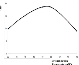
Determination of preincubation temperature: The
samples were preincubated for 10 min at various temperatures (30-70 °C) in the presence Mn+2 ions. Kidney
arginase showed highest activity at 50-55 °C. After this temperature, the enzymatic activity decreased progressively. Therefore, the optimum preincubation temperature for kidney arginase was determined to be 53 °C (Figure 1).
Determination of preincubation period: The samples
were preincubated at various preincubation period (0-30 min) at 53°C preincubation temperature. The enzymatic activity increased approximately 2.5 times within 15-min preincubation at 53°C. Thus, the optimum preincubation period for kidney arginase was determined to be 15 min (Figure 2).
Determination of incubation period: Kidney
arginase activity was linear up to 15 min for incubation at 37 oC. The optimum incubation period for kidney arginase
was accepted to be 10 min (Figure 3).
Influence of manganese concentration: To determine
the influence of Mn+2 ions on arginase activity, Mn+2 ions
at varying concentrations (0-5 mM) were added during preincubation. The highest arginase activity was detected at 1 mM Mn+2 concentration. The arginase activity
increased 5 times in the presence of 1 mM Mn+2
concentration at 53oC (Figure 4).
Figure 1. The effect of preincubation temperature on rabbit kidney arginase.
Şekil 1. Tavşan böbrek arginazı üzerine preinkübasyon sıcaklığının etkisi.
Figure 2. The effect of preincubation period on rabbit kidney arginase.
Şekil 2. Tavşan böbrek arginazı üzerine preinkübasyon zamanının etkisi.
Figure 3. The effect of incubation period on rabbit kidney arginase.
Şekil 3. Tavşan böbrek arginazı üzerine inkübasyon zamanının etkisi.
Influence of pH: To determine optimum pH for
arginase activity, various buffers were used. These buffers were Glycine-NaOH (pH 8.6-10.6), NaHCO3-Na2CO3
(pH 9.2-10.2) and NaHCO3-NaOH (pH 9.7-10.9). The
arginase activity was highest at NaHCO3-NaOH buffer,
pH 10-10.2. The most appropriate buffer system and pH for rabbit kidney arginase was NaHCO3-NaOH buffer and
pH 10.1 (Figure 5).
Km (Michaelis-Menten constant) analysis: The
changes in arginase activity were assessed on various L-arginine concentrations according to Michaelis-Menten kinetic analysis (Figure 6). As shown in figure 6, arginase activity demonstrated a linear increase up to 12.5 mM L-arginine concentrations. At L-L-arginine concentrations higher than 12.5 mM, enzyme activity lost the trend of linear increase and followed a hyperbolic curve. At 45 mM L-arginine concentrations, arginase enzyme activity reached a plateau, and zero order kinetic was noted. The effect of substrate concentration on the activity of kidney arginase was also determined by Lineweaver-Burk plots (Figure 7). In both Michaelis-Menten and Lineweaver-Burk plots, Km value of the enzyme against L-arginine was 12.5 mM.
Figure 4. The effect of Mn+2 ions on rabbit kidney arginase. Şekil 4. Tavşan böbrek arginazı üzerine Mn+2 iyonlarının etkisi.
Figure 5. The effect of pH on rabbit kidney arginase. Şekil 5. Tavşan böbrek arginazı üzerine pH’ın etkisi.
Figure 6. The changes in arginase activity in various L-arginine concentrations.
Şekil 6. Çeşitli L-arginin konsantrasyonlarında arginaz aktivite-sindeki değişiklikler.
Figure 7. Lineweaver-Burk plot of kidney arginase activity measured at various concentrations of L-arginine.
Şekil 7. Çeşitli L-arginin konsantrasyonlarında ölçülen böbrek arginaz aktivitesinin Lineweaver-Burk eğrisi.
Table 1. Arginase activities in kidney of different species. Tablo 1. Farklı türlerin böbreklerindeki arginaz aktiviteleri.
Species Enzyme activities (Unit)
Quail 59.63 ± 13.03 Chicken 20.86 ± 4.53 Rabbit 20.84 ± 3.38 Rat 16.28 ± 2.04 Goat 13.51 ± 1.97 Sheep 4.17 ± 1.19 Cattle 1.22 ± 0.25
The arginase activities in kidneys of different species: After optimum conditions were determined, the
arginase activities in kidneys of the sheep, goat, cattle, rabbit, chicken, quail and rat were also defined (Table 1). Among the studied species, the highest arginase activity was found in kidneys of the quails, the lowest arginase activity was found in kidneys of the cattle and sheep.
Discussion and Conclusion
Determination for optimum conditions of the enzyme in various animal species and tissues is important to detect the most active level of the enzyme during measurements and reveal physiological roles of the enzyme. The arginase activity depends on the concentration of manganese ions (Mn+2), the
pre-incubation temperature and period, and pH.
Arginase measured in various tissues required heat activation with Mn+2 (pre-incubation) before incubation in
order to reach a maximum activity (6, 7, 10). The optimum preincubation temperature for the liver arginases of different species were found between 60-68oC (2, 7, 10,
17). The optimum preincubation temperature for rabbit kidney arginase was detected to be 53oC in the presence of
MnCl2 (Figure 1). These findings indicated that the
arginase from kidney in the presence of Mn+2 was less
resistant to elevated temperature than arginases from liver. The pre-incubation period as well as the preincubation temperature are also important in the arginase activity. The optimum preincubation period for rabbit kidney arginase was determined to be 10 min at 53oC (Figure 2).
This period was similar to the preincubation period of the liver arginase in rabbit (7). On the contrary, Erişir et al. (10) reported that the preincubation for activity of kidney arginase in rats was unnecessary because the preincubation temperature and period has no effect on the activity of kidney arginase in rats. Furthermore, Kadowaki and Nesheim (18) observed a decrease in the activity of the chick kidney enzyme by prior heat treatment (for 5 min at 55°C) with Mn+2. We may suggest that preincubation
conditions varied for the kidney arginases of different species. Likewise, the arginase activity levels in kidney from different species were also different (Table 1). Interestingly, the highest arginase activity was found in kidney of the quails, while the lowest activity was present in kidneys of the sheep and cattle. Results of this study are in agreement with those found by Sepehrimanesh and Aminlari (24) who reported higher arginase activity in kidney of the quails compared to the chicken.
Arginase is a metalloenzyme in which manganese acts as a cofactor, and arginase activity is manganese dependent. It is known that manganese ion stabilizes and/or activates arginases from different tissues (5, 10). At 53oC, the presence of 1 mM Mn+2 concentration increased
the arginase activity 5 times (Figure 4). Results of this study showed that arginase activity in the kidney of rabbit are also Mn+2 dependent and Mn+2 ions are required for
preincubation. Mn+2 ions establish a bridge between
arginase and substrate and, thus, the enzyme becomes active (3). Mn+2 concentration (1 mM) at which arginase
of rabbit kidney tissue show the highest activity was found to be close to the value determined for arginase of rabbit liver tissue (2 mM) (7).
Generally various tissue arginases were shown to have basic pH optimal (9.5–10.5) and to be NaHCO3
-Na2CO3 buffer of the most stable buffer system (4, 5, 7,
10, 17). In the present study, the highest activity of rabbit kidney arginase was determined at pH 10.1. But, the most suitable buffer system was NaHCO3-NaOH buffer which
is different from that for other arginases including liver arginase (Figure 5).
When determining optimum incubation time, it was observed that reaction continued linearly until 15th min.,
and linearity was replaced with a hyperbolic appearance after 15th min. The reason behind why linear increase in
enzyme activity deviated from linearity after 15th min.
may be that ornithine and urea occurring as a result of reaction caused inhibition of the enzyme. Both products (L-ornithine and urea) of the arginase reaction were inhibitors of arginase (14, 23).
The calculated Km value by both Michaelis-Menten and Lineweaver-Burk kinetic analyses was 12.5 mM. The
Km value for rabbit kidney arginase was higher than in the
range of other reported Km values (1-6.8 mM) for liver arginases (2, 4, 7, 10, 17, 19, 23). The fact that Km of kidney arginase was greater than Km of liver arginase points out that kidney arginase has a low affinity against its substrate L-arginine.
There are two distinct structural gene loci that encode arginase in the rat. The one structural gene locus expresses arginase in the liver, while the other one expresses arginase in the brain and kidney (15, 21). The determined optimal conditions for the kidney arginase was found to be different from that of the liver arginase. Our study indicates that arginases encoded by separate gene loci have the different optimal conditions.
Acknowledgement
This article was taken from the master thesis entitled “Tavşan Böbrek Arginaz Aktivitesi için Optimal Şartların Belirlenmesi ” and it was supported by the Fırat University Scientific Research Fund (Project No: VF.15.03).
References
1. Aminlari M, Shahbazkia HR, Esfandiari A (2007): Distribution of arginase in tissues of cat (Felis catus). J Feline Med Surg, 9 (2), 133-139.
2. Benzer F, Temizer Ozan PS (2002): The levels of arginase enzyme activity in sheep with fasciolasis and some biochemical properties of arginase in liver tissue. FÜ Sağ Bil Vet Derg, 16 (2), 217-222.
3. Colombo JP, Konarska L (1984): Arginase. In: Bergmayer GM. (Editor). Methods of Enzymatic Analysis. 3rd Edition, Weinheim: Vertag Chemie, 285-294. 4. Dabir S, Dabir P, Somvanshi B (2005): Purification,
properties and alternate substrate specificities of arginase from two different sources: Vigna catjang cotyledon and buffalo liver. Int J Biol Sci, 1 (3), 114-122.
5. Dabir S, Dabir P, Somvanshi B (2006): The kinetics of inhibition of Vigna catjang cotyledon and buffalo liver arginase by L-proline and branched-chain amino acids. J Enzyme Inhib Med Chem, 21 (6), 727-731.
6. Demir F, Ozan G, Temizer Ozan PS (2015): Ratlara uygulanan kurşun asetatın karaciğer arginazına etkisi ve enzimin bazı kinetik özellikleri. FÜ Sağ Bil Vet Derg, 29 (1), 37-43.
7. Erdem N, Erişir M (2013): Tavşan Karaciğer Arginazının Optimize Edilmesi ve Dokulardaki Dağılımı. FÜ Sağ Bil Vet Derg, 27 (2), 81-86.
8. Erişir M, Aydilek N, Aksakal M (2005): Effect of Vitamin E on Arginase Activity in the Liver and Kidneys of Testesterone Treated and Castrated Rabbits. Acta Vet Brno, 74, 527-531.
9. Erişir M, Beytut E, Ozan TS, et al. (2003): Effects of dietary vitamin E and Selenium on Arginase Activity in the liver, kidney and heart of rats treated with high doses of glucocorticoids. Cell Biochem Funct, 21, 331-335. 10. Erişir M, Erçel E, Yılmaz S, et al. (2005): Evaluation of
optimal conditions for arginase activity in streptozotocin induced diabetic rats. Vet Med-Czech, 50, 69-76. 11. Erişir M, Nazıroğlu M, Taşdemir B, et al. (2006): Effect
of a dietary combined vitamin C and E supplementation on the liver, kidneys and brain arginase activity in non-pregnant and non-pregnant rats with streptozotocin-induced diabetes. Revue Med Vet, 157, 445-449.
12. Erişir M, Tamser M, Taşdemir, et al. (2006): Ovariektomili Ratlara Estradiol 17-β ve Vitamin E Verilmesinin Arginaz Aktivitesi Üzerine Etkisi. Kafkas Üniv Vet Fak Derg, 12 (1), 31-35.
13. Geyer JW, Dabich D (1971): Rapid method for determination of arginase activity in tissue homogenates. Anal Biochem, 39, 412-417.
14. Glass RD, Knox WE (1973): Arginase isozymes of rat mammary gland, liver, and other tissues. J Biol Chem, 248, 5785-5789.
15. Gotoh T, Sonoki T, Nagasaki A, et al. (1996): Molecular cloning of cDNA for nonhepatic mitochondrial arginase (arginase II) and comparison of its induction with nitric oxide synthase in a murine macrophage-like cell line. FEBS Lett, 395, 119-122.
16. Guoyao WU, Morris SM (1998): Arginine metabolism: nitric oxide and beyond. Biochem J, 336, 1-17.
17. Halifeoğlu İ (1993): İnsan Karaciğerinde, Eritrosit Ve Uterus Doku Arginazının Kinetik Özellikleri. Doktora Tezi, Elazığ Fırat Üniversitesi Tıp Fakültesi. Biyokimya Anabilim Dalı.
18. Kadowaki H, Nesheim MC (1978): An assay for arginase in chicken kidney. Comp Biochem Physiol B, 61, 281–285. 19. Kaysen GA, Strecker HJ (1973): Purification and properties of arginase of rat kidney. Biochem J, 133, 779-788.
20. Lowry OH, Rosenbrough NJ, Farr AL, et al. (1951): Protein measurements with the folin phenol reagent. J Biol Chem, 193, 265-275.
21. Miyanaka K, Gotoh T, Nagasaki A, et al. (1998): Immunohistochemical localization of arginase II and other enzymes of arginine metabolism in rat kidney and liver. Histochem J, 30, 741–751.
22. Powers GS, Meister T (1982): Urea synthesis and ammonia metabolism. In: Arias I, Popper H, Schachter D, Shafrits DA, editors: The liver: Biology and Pathobiology. Raven Press. New York, 251-263.
23. Reczkowski RS, Ash DE (1994): Rat liver arginase: kinetic mechanism, alternate substrates, and inhibitors. Arch Biochem Biophys, 312, 31–37.
24. Sepehrimanesh M, Aminlari M (2014): Arginase distribution in tissues of domestic avian species. Comp Clin Pathol, 23 (2), 353-356.
Geliş tarihi: 28.06.2016 / Kabul Tarihi: 24.01.2017
Address for correspondence:
Prof. Dr. Mine ERİŞİR
Fırat University, Faculty of Veterinary Medicine, Department of Biochemistry,
231119 / Elazığ, Turkey e-mail: [email protected]