Emerging Infectious Diseases • www.cdc.gov/eid • Vol. 21, No. 12, December 2015 2213
Selcuk Kilic,1 Dawn N. Birdsell,1 Alper Karagöz, Bekir Çelebi, Zekiye Bakkaloglu, Muzaffer Arikan,
Jason W. Sahl, Cedar Mitchell, Andrew Rivera, Sara Maltinsky, Paul Keim, Duran Üstek,
Rıza Durmaz, David M. Wagner
Francisella tularensis DNA extractions and isolates from the environment and humans were genetically characterized to elucidate environmental sources that cause human tulare-mia in Turkey. Extensive genetic diversity consistent with genotypes from human outbreaks was identified in environ-mental samples and confirmed water as a source of human tularemia in Turkey.
T
ularemia is a disease caused primarily by 2 subspecies of Francisella tularensis: F. tularensis subsp.tularen-sis, which is restricted to North America; and F. tularensis
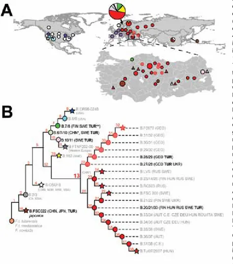
subsp. holarctica, which is found widely throughout the northern hemisphere but is the only subspecies in most of Eurasia (1). Through whole-genome sequencing and canonical single-nucleotide polymorphism (canSNP) ge-notyping, F. tularensis subsp. holarctica has been divided into 4 major genetic groups (B.4, B.6, B.12, and B.16) con-sisting of multiple subgroups (Figure 1) (1–3). Geographic distribution of these subgroups in Europe, Japan, and the USA are well described (1–3).
The phylogeography of F. tularensis in Asia is poorly understood because of undersampling in many regions, but recent studies have revealed new insights. A report has described rich phylogenetic diversity of the bacterium in China (4), including the rare B.16 group (biovar japonica). Previously, B.16 was known only in Japan (1) and Turkey (6). Sweden reportedly has the highest overall phylogenetic diversity among regions worldwide (2).
In Turkey, tularemia cases in humans have increased since 2009 (7), but little is known about environmental sources. Tularemia was first reported in Turkey in 1936 and then was sporadically reported for several decades (7). After improved surveillance, the number of tularemia cases
increased in the 1980s and led to registration of tularemia as a reportable disease in 2004 (7,8). Incidence has contin-ued to increase since then (7), and tularemia is now consid-ered a reemerging zoonotic disease in Turkey.
Patients with oropharyngeal signs and symptoms ac-count for ≈90% of tularemia cases in Turkey (8), and cases emerge seasonally from August–March (7). Seasonality of incidence of cases is presumably associated with consump-tion of contaminated water (9), but confirming sources is difficult. Reports of confirmation of F. tularensis from wa-ter samples by PCR (10) or culture (6) are rare, and defini-tive studies that link water to tularemia in humans are lack-ing. How water sources become seasonally contaminated is also unknown, but contamination could be caused by ro-dents. Recently, F. tularensis was confirmed by PCR from 2 mice captured in Thrace (11), but in Turkey, confirmation has not been obtained from ticks or mosquitoes, which are known vectors of F. tularensis (1,4).
Genetic characterization of clinical samples from tu-laremia outbreaks in Turkey in 2011 showed that multi-ple phylogenetic groups cause disease in multimulti-ple regions across Turkey (5); however, no environmental samples were assessed in that study. We report our findings from genetically characterized samples positive for F. tularensis from environmental and human sources located in multiple active tularemia areas in Turkey. Our results provide new insights into F. tularensis transmission from environmental sources to humans.
The Study
To examine environmental reservoirs that could be possible sources for human infections, during 2010–2013, we sam-pled water sources and rodent populations from suspected sites where transmission of F. tularensis infection could oc-cur in Turkey. To survey and compare phylogenetic diversi-ty of environmental samples and clinical samples, we exam-ined 33 clinical samples of mostly oropharyngeal tularemia cases from approximately the same sites where environmen-tal samples were collected. DNA was extracted (DNeasy Blood & Tissue Kit, QIAGEN GmbH, Hilden, Germany) from 6 water, 1 rodent spleen, and 33 human samples (on-line Technical Appendix Table 1, http://wwwnc.cdc.gov/ EID/article/21/12/15-0634-Techapp.pdf).
The extractions were confirmed F. tularensis–positive by using PCR and targeting the tul4 gene (12). Analysis
Water as Source of Francisella tularensis
Infection in Humans, Turkey
Author affiliations: Public Health Institution of Turkey, Ankara, Turkey (S. Kilic, A. Karagöz, B. Çelebi, Z. Bakkaloglu, R. Durmaz); Northern Arizona University, Flagstaff, Arizona, USA (D.N. Birdsell, J.W. Sahl, C. Mitchell, A. Rivera, S. Maltinsky, P. Keim,
D.M. Wagner); Istanbul University, Istanbul, Turkey (M. Arikan); Medipol University, Istanbul (D. Üstek)
by using 21 published canSNP assays, as previously de-scribed (5), assigned these samples to 3 major phylogenetic groups and distinct subgroups: B.16 (n = 11); B.6 (2 sub-groups: B.6/7/10, n = 1; and B.10/11, n = 6); and B.13 (2 subgroups: B.27, n = 5; and B.20/21/33, n = 17) (Figure 1; online Technical Appendix Table 1). Of the subgroups, 3 were previously unknown in Turkey: B.6/7/10, B.10/11, and B.16. The 7 environmental samples collected included most of the known phylogenetic diversity in Turkey and represented the 3 major groups: B16, B6 (B.6/7/10 and B.10/11), and B.13 (the group previously known to be in Turkey). Of the subgroups identified, all but B.6/7/10 were also found in the human samples.
To determine detailed associations between environ-mental and human clinical samples, we examined the genet-ic diversity among these samples by using multilocus vari-able number of tandem repeats analysis (MLVA) (13). All samples contained a single MLVA genotype (online Tech-nical Appendix Figure, panels A–C); no mixed allele calls were observed at any of the examined variable number of
tandem-repeats loci. Three different environmental samples (F0922, F0910, and F0916) had canSNP and MLVA geno-types that were identical to those of clinical samples (online Technical Appendix Table 1). In 2 instances (F0910 and F0916), the environmental sample and its respective geneti-cally identical clinical sample(s) were recovered from dif-ferent geographic regions, resulting in identical genotypes being found in different localities and suggesting that close genotypes are dispersed widely in Turkey. One environmen-tal sample (F0922) had genetic, geographic, and temporal data (online Technical Appendix Figure, panel A) concor-dant with data from human samples. This water sample shared identical canSNP and MLVA genotypes with 5 clini-cal samples recovered 2 weeks previously at the same loclini-cal- local-ity, strongly suggesting that the human cases are linked with this infected water source.
The genetic characterization of F. tularensis from en-vironmental sources provides insights into transmission of tularemia from the environment to humans, but little is known about how water is contaminated. The seasonal
Figure 1. Phylogeography of Francisella tularensis subsp. holarctica. A) Global
distribution of known phylogenetic groups determined on the basis of previous studies (2–4); enlarged map of Turkey shows locations of phylogenetic groups identified among the 40 samples positive for F. tularensis examined in this and previous studies (5). Circle size indicates number of samples (small circles, 1–3; medium circles, 4–6; large circles, 7–9). Colors of circles (human samples) and triangles (environmental samples) represent the phylogenetic subgroups to which these samples were assigned (see panel B). Subgroup B.16 (biovar japonica) is represented by the dot inside the brown circles and triangles. B) Phylogenetic tree for F. tularensis subsp.
holarctica constructed on the basis of current
canonical single-nucleotide polymorphism genotyping. Red numbers indicate nomenclature of canonical single-nucleotide polymorphism groups. Terminal subgroups representing sequenced strains are shown as stars, and intervening nodes representing collapsed branches are indicated by circles. Countries of origin for samples assigned to relevant phylogenetic groups are as follows: AUT, Austria; CE, central Europe, unknown country; CHN, China; CZE, Czech Republic; DEU, Germany; FIN, Finland; GEO, Georgia; HUN, Hungary; ITA, Italy; NOR Norway; ROU, Romania; RUS, Russia; SWE, Sweden; TUR, Turkey; UKR, Ukraine; USA, United States. CHN* indicates approximate phylogenetic placement because of a lack of resolved information on single-nucleotide polymorphisms (4). TUR** indicates identification from a previous study (5).
DISPATCHES
Emerging Infectious Diseases • www.cdc.gov/eid • Vol. 21, No. 12, December 2015 2215 Francisella tularensis Infection in Humans, Turkey
nature of human outbreaks suggests that water sources are not constant reservoirs but rather are contaminated by an-other source. Rodents were identified as reservoirs (21% tularemia positive) in Bulgaria, where mainly oropharyn-geal tularemia is endemic (14). We found a rodent sample (F0910) with canSNP and MLVA genotypes identical to an oropharyngeal clinical sample (F0898) (online Technical Appendix Table 1), a finding consistent with water contam-ination that originates from animal sources. However, the converse is also possible: animals could become infected by contaminated water.
Analysis of the 7 environmental F. tularensis subsp.
holarctica samples from Turkey revealed extensive
phylo-genetic diversity that represents most known major groups in the world. Three of the 4 major F. tularensis subsp.
hol-arctica phylogenetic groups (B.4, B.6, B.12, and B.16) are
found in Turkey, including the highly basal B.16 group (biovar japonica) (Figure 1). This finding indicates that no single phylogenetic type is dominant in Turkey, unlike in Western Europe (3). Diversity was also represented in the clinical samples, suggesting that all major groups have similar capacities to cause disease, as other studies have suggested (15).
To gain insights into the evolutionary origin of the B.16 group, we examined the phylogenetic relationships among 3 published B.16 strains: 1 from Turkey (PHIT-FT049) (6) and 2 from Japan (FSC021 and FSC022) (GenBank acces-sion nos. CP007148.1, SRX147922, and DS264138.1, re-spectively; Figure 2). We generated a global core-genome SNP phylogeny (online Technical Appendix) for these 3 B.16 strains and 5 strains from other groups (online Tech-nical Appendix Table 2). As expected, PHIT-FT049 clus-ters with the Japanese B.16 strains from Japan and shares 448 putative SNPs; however, it is also distinct from the 2 strains from Japan, which together share 640 putative SNPs
(Figure 2). The distinctiveness of the B.16 strain from Tur-key strongly suggests that it has an evolutionary history dif-ferent from that of the Japanese strains. The MLVA phy-logeny of B.16 strains (online Technical Appendix Table 1) reveals greater diversity among the 8 strains from Japan than among the 8 strains from Turkey. These data show that the B.16 strains from Turkey and Japan are highly dis-tinct, and the greater diversity in strains from Japan sup-ports the possibility that the place of ancestral origin of the B.16 group is Asia.
Conclusions
Phylogenetically diverse strains of F. tularensis subsp.
holarctica are environmentally established in Turkey and
cause human disease. The strains in Turkey now include many phylogenetic groups previously found only in Scan-dinavia or Asia.
Acknowledgments
We thank Charles Williamson, Katy Califf, Bridget Barker, and Heidie Hornstra-O’Neill for assistance with the manuscript. This study was funded by the US Department of Homeland Security, Science and Technology Directorate, Award NBCH2070001, and by the Cowden Endowment in Microbiology at Northern Arizona University.
Dr. Kilic is a professor and a principal investigator of F. tularensis at the Public Health Institution of Turkey, National Tularemia Ref-erence Laboratory, Ankara, Turkey. His research interests include the evolution, epidemiology, and control of bacterial zoonoses.
References
1. Vogler AJ, Birdsell D, Price LB, Bowers JR, Beckstrom- Sternberg SM, Auerbach RK, et al. Phylogeography of Francisella
tularensis: global expansion of a highly fit clone. J Bacteriol.
2009;191:2474–84. http://dx.doi.org/10.1128/JB.01786-08
Figure 2. Maximum-parsimony
phylogeny constructed by using 10,443 putative single-nucleotide polymorphisms discovered from whole-genome sequences of 8
Francisella tularensis strains.
Gray shading indicates the B.16 (biovar japonica) strain from Turkey (PHIT_FT049). Detailed methods are described in the online Technical Appendix (http://wwwnc.cdc.gov/EID/ articles/21/12/15-0634-Techapp.pdf). Reference strains were retrieved from GenBank (online Technical Appendix Table 2). Countries of origin are indicated as follows: FRA, France; JPN, Japan; RUS, Russia; SWE, Sweden; TUR, Turkey; USA, United States. Scale bar indicates single-nucleotide polymorphisms.
2. Karlsson E, Svensson K, Lindgren P, Byström M, Sjödin A, Forsman M, et al. The phylogeographic pattern of Francisella
tularensis in Sweden indicates a Scandinavian origin of
Eurosiberian tularaemia. Environ Microbiol. 2013;15:634–45. http://dx.doi.org/10.1111/1462-2920.12052
3. Gyuranecz M, Birdsell DN, Splettstoesser W, Seibold E, Beckstrom-Sternberg SM, Makrai L, et al. Phylogeography of
Francisella tularensis subsp. holarctica, Europe. Emerg Infect Dis.
2012;18:290–3. http://dx.doi.org/10.3201/eid1802.111305 4. Wang Y, Peng Y, Hai R, Xia L, Li H, Zhang Z, et al. Diversity
of Francisella tularensis subsp. holarctica lineages, China. Emerg Infect Dis. 2014;20:1191–4 . http://dx.doi.org/10.3201/ eid2007.130931
5. Özsürekci Y, Birdsell DN, Çelik M, Karadağ-Öncel E, Johansson A, Forsman M, et al. Phylogenetically diverse Francisella tularensis strains cause human tularemia in Turkey. Emerg Infect Dis. 2015;21:173–5. http://dx.doi.org/10.3201/eid2101.141087 6. Kiliç S, Celebi B, Acar B, Atas M. In vitro susceptibility of
isolates of Francisella tularensis from Turkey. Scand J Infect Dis. 2013;45:337–41. http://dx.doi.org/10.3109/00365548.2012.751125 7. Kiliç S. Tularemia: the pathogen and epidemiology [in Turkish].
Turkiye Klinikleri J.E.N.T–Special Topics. 2014;7:52–61. 8. Erdem H, Ozturk-Engin D, Yesilyurt M, Karabay O, Elaldi N,
Celebi G, et al. Evaluation of tularaemia courses: a multicentre study from Turkey. Clin Microbiol Infect. 2014;20:O1042–51. http://dx.doi.org/10.1111/1469-0691.12741
9. Willke A, Meric M, Grunow R, Sayan M, Finke EJ, Splettstößer W, et al. An outbreak of oropharyngeal tularaemia linked to natural spring water. J Med Microbiol. 2009;58:112–6. http://dx.doi.org/ 10.1099/jmm.0.002279-0
10. Ulu Kiliç A, Kiliç S, Sencan I, Cicek Sentürk G, Gürbüz Y, Tütüncü EE, et al. A water-borne tularemia outbreak caused by Francisella tularensis subspecies holarctica in Central Anatolia region [in Turkish]. Mikrobiyol Bul. 2011;45:234–47.
11. Unal Yilmaz G, Gurcan S, Ozkan B, Karadenizli A. Investigation of the presence of Francisella tularensis by culture, serology and molecular methods in mice of Thrace Region, Turkey [in Turkish]. Mikrobiyol Bul. 2014;48:213–22. http://dx.doi.org/ 10.5578/mb.7028
12. Sjöstedt A, Kuoppa K, Johansson T, Sandströom G. The 17 kDa lipoprotein and encoding gene of Francisella tularensis LVS are conserved in strains of Francisella tularensis. Microb Pathog. 13. Vogler AJ, Birdsell D, Wagner DM, Keim P. An optimized,
multiplexed multi-locus variable-number tandem repeat analysis system for genotyping Francisella tularensis. Lett Appl Microbiol. 2009;48:140–4. http://dx.doi.org/10.1111/ j.1472-765X.2008.02484.x
14. Christova I, Gladnishka T. Prevalence of infection with
Francisella tularensis, Borrelia burgdorferi sensu lato and Anaplasma phagocytophilum in rodents from an endemic focus of
tularemia in Bulgaria. Ann Agric Environ Med. 2005;12:149–52. 15. Johansson A, Lärkeryd A, Widerström M, Mörtberg S,
Myrtännäs K, Ohrman C, et al. An outbreak of respiratory tularemia caused by diverse clones of Francisella tularensis. Clin Infect Dis. 2014;59:1546–53. http://dx.doi.org/10.1093/cid/ciu621
Address for correspondence: David M. Wagner, Northern Arizona University, PO Box 4073, Flagstaff, AZ 86011, USA; email: [email protected]
TM
Check out EID’s 20-year-anniversary timeline
and find an array of fascinating seminal moments in the journal’s history.
DISPATCHES
2216 Emerging Infectious Diseases • www.cdc.gov/eid • Vol. 21, No. 12, December 2015 http://wwwnc.cdc.gov/eid/page/20-year-timeline