ORIGINAL ARTICLE
Screening and selection of novel animal probiotics isolated
from bovine chyme
Alper D. Ozkan&Diren Han&Ozgun C. O. Umu&Pinar Angun&
Berna Senturk&Oncay Yasa&Turgay Tekinay
Received: 9 July 2012 / Accepted: 5 December 2012 / Published online: 27 December 2012 # Springer-Verlag Berlin Heidelberg and the University of Milan 2012
Abstract Probiotics, gut-colonizing microorganisms capa-ble of conferring a number of health benefits to their hosts, are highly desirable as animal feed supplements. Members of the Gram-positive genus Bacillus are often utilized as probiotics, since endospores formed by those bacteria render them highly resistant to environmental extremes and there-fore capable of surviving gastrointestinal tract conditions. In this study, 84 distinct bacterial colonies were obtained from bovine chyme and 29 isolates were determined as Bacillus species. These isolates were principally screened for their antimicrobial activity against a group of two Gram-positive and four Gram-negative bacteria, including known human and animal pathogens such as Salmonella enterica, Klebsiella pneumoniae, Pseudomonas aeruginosa, and Staphylococcus aureus. Seven strains displaying strong antimicrobial activity against the test cohort were further evaluated for other prop-erties desirable from animal probiotics, including high spore-forming capacity and adhesiveness, resistance to pH extremes and ability to form biofilms. The isolates were found to resist simulated gastrointestinal conditions and most of the antibiotics tested. In addition, plasmid pres-ence was checked and cytotoxicity tests were performed to evaluate the potential risks of antibiotic resistance transfer and unintended pathogenic effects on host, respectively. We pro-pose that the bacterial isolates are suitable for use as animal probiotics.
Keywords Antimicrobial . Bacillus . Feed supplement . Isolation . Probiotics
Introduction
The widespread and intensive use of antibiotics for thera-peutic purposes has led to a considerable increase in the number of antibiotic-resistant pathogenic strains, resulting in frequent occurrence of serious and hard-to-treat infections in both humans and livestock (Barbosa and Levy 2000; Barbosa et al.2005; Chaiyawan et al.2010). As such, there has been an increasing concern about non-therapeutic uses of antibiotics, ultimately culminating in bans of their use as animal feed additives. The European Parliament and the Council of the European Union have encouraged the devel-opment of alternative products to replace antibiotics as feed supplements for growth promotion (Santini et al.2010; The European Parliament and the Council of the European Union2003). Consequently, there is an ongoing search for replacement products capable of enhancing growth and preventing disease (Chaiyawan et al. 2010; Santini et al.
2010). An effective and safe alternative to antibiotic imple-mentation is the use of probiotics, which protect the animal from pathogens by improving the microbial balance in the gastrointestinal tract to exclude potentially harmful bacteria (Chaiyawan et al.2010; Modesto et al.2009; Patterson and Burkholder2003; Santini et al.2010).
Probiotics are beneficial live microorganisms primarily including various species of bacteria and fungi (Duc et al.
2004; Lutful Kabir2009). Probiotics influence the host organ-isms’ health by maintaining the normal intestinal microbiota, preventing the growth of pathogenic microorganisms, promot-ing digestion and intake of feed, and inducpromot-ing the immune system (Kim et al.2009; Lutful Kabir2009). As such, the use of probiotics on livestock enhances the growth of animals, improves efficiency of feed conversion, and decreases the rate of mortality (Kim et al.2009; Lutful Kabir2009). Probiotic microorganisms should ideally be pathogenic and non-toxic, improve growth of the host animal, and be stable and
A. D. Ozkan
:
D. Han:
O. C. O. Umu:
P. Angun:
B. Senturk:
O. Yasa
:
T. Tekinay (*)Laboratory of Sustainable Technologies, UNAM, Institute of Materials Science and Nanotechnology, Bilkent University, 06800, Ankara, Turkey e-mail: [email protected]
active during processing and storage. In addition, probiotics should be able to survive and continue their metabolic activ-ities in gastrointestinal conditions and produce compounds that inhibit the growth of pathogenic microorganisms (Kim et al.2009; Patterson and Burkholder2003).
A number of bacterial species are currently used as pro-biotics, mostly in genera Bacillus, Bifidobacterium, Enterococcus, Escherichia, Lactobacillus, Lactococcus, and Streptococcus (Patterson and Burkholder 2003). Some yeast species, such as Saccharomyces cerevisiae, are also utilized as probiotics (Lutful Kabir2009).
The most common probiotics used in humans are species of Lactobacillus and Bifidobacterium, while Bacillus, Enterococcus, and Saccharomyces species are frequently used in livestock (Patterson and Burkholder 2003). Bacillus species in particular are advantageous for use as probiotics, since members of this genus are spore-formers displaying high resistance to heat, chemicals, and other stress factors (Cartman et al.2008; Chaiyawan et al. 2010; Nicholson et al. 2000; Setlow 2006., Bacillus spores can survive in harsh pH conditions of the gastric fluids (Cutting
2011) and reach the small intestine relatively unharmed, making them suitable for use as feed supplements. In addi-tion, spores can be kept for a long time in desiccated form with little to no loss of viability, allowing for ease of trans-port (Duc et al. 2004; Mazza 1994). Bacillus subtilis, B. clausii, B. cereus, B. coagulans, and B. licheniformis are the Bacillus species most widely researched and used as animal probiotics (Cutting2011).
In this study, novel bacterial strains were isolated from bovine chyme samples, and Bacillus strains obtained in this manner were characterized and screened for their potential to be used as animal probiotics. Criteria utilized for assess-ing probiotic capacity include survival in gastrointestinal tract (GIT) conditions, lack of toxicity and invasiveness, ability to adhere to the small intestine, and inhibition of pathogenic bacteria in the GIT by competitive exclusion or synthesis of antimicrobial compounds.
Materials and methods
Bacillus isolation and growth conditions
Microorganisms were isolated from chyme samples of eight male cattle between 2 and 4 years of age. The small intestine was removed as a whole and chyme samples were collected by squeezing the intestinal contents into sterile tubes. Isolation of the microorganisms was performed as described by Lalloo et al. (2007) with minor modifications. An amount of 1 g of chyme fluid from each sample was sus-pended in 3 ml of 0.9 % NaCl solution and inoculated in 9 ml of nutrient broth (NB). Samples were incubated at
37 °C for 24 h, followed by incubation at 45 °C for 10 min to initiate spore formation. Then, 50 % (v/v) ethanol was added to a volume of 20 ml and the suspension was incubated at 20 °C for 1 h. Centrifugation was performed at 12,000 g for 30 s, the supernatant was decanted, and pellets were incubated at 105 °C for 5 min. Dry pellets were resuspended in 20 ml of 0.9 % NaCl solution and serially diluted in ten-fold increments.
An amount of 150 μl of each dilution was spread on MYP (Mannitol–Egg Yolk–Polymyxin) agar plates and incubated at 37 °C for 24 h. Bacillus species were differentiated according to their ability to ferment man-nitol or degrade lecithin, while polymyxin was utilized to inhibit the growth of Gram-negative bacteria. Single col-onies with distinct morphologies were collected and transferred on Luria–Bertani (LB) agar plates. Cultures were incubated at 37 °C and 125 rpm until visible colony formation, and stored at 4 °C to maintain viabil-ity. Cultures were maintained at−80 °C in 30 % glycerol for long-term storage.
Antimicrobial activity
Antimicrobial activity assay was adapted from the method described by Saravanakumari and Mani (2010). Overnight incubated cultures of the isolates were horizontally streaked on LB agar plates and incubated at 37 °C for 48 h. After the incubation period, plates were exposed to chloroform vapor for 90 min for the inactivation of active cells. Plates were aerated for 20 min to completely remove residual chloro-form by vaporization and plate covers were subsequently changed (Barbosa et al. 2005). Representative bacteria (Table 1) were then streaked vertically on the horizontally streaked colonies and plates were incubated at 37 °C for 24 h. Inhibition on the bacterial growth line (Table 1) was assessed to evaluate the antimicrobial activity of isolates against each representative strain.
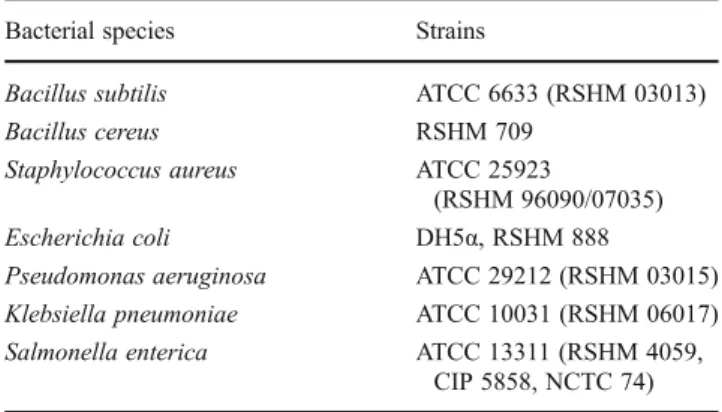
Table 1 Microorganisms used in the present study; all strains were obtained from RSHM (Refik Saydam National Type Culture Collec-tion Laboratory, Ankara, Turkey)
Bacterial species Strains
Bacillus subtilis ATCC 6633 (RSHM 03013)
Bacillus cereus RSHM 709
Staphylococcus aureus ATCC 25923
(RSHM 96090/07035)
Escherichia coli DH5α, RSHM 888
Pseudomonas aeruginosa ATCC 29212 (RSHM 03015)
Klebsiella pneumoniae ATCC 10031 (RSHM 06017)
Salmonella enterica ATCC 13311 (RSHM 4059,
Sporulation efficiency
Sporulation efficiencies of the isolates were measured as described by Barbosa et al. (2005). Isolates were inoculated (0.1 %v/v) in Difco Sporulation Medium (DSM) and incu-bated at 125 rpm and 37 °C for 24 h. After 24 h, serial dilutions were prepared in ten-fold increments and spread on LB agar plates before and after heat exposure at 80 °C for 20 min. Plates were incubated at 37 °C overnight and colony-forming unit (CFU) numbers were counted; colony growth before and after heat exposure was compared to assess the sporulation capacities of the isolates.
Spore formation
Spores were harvested and purified to test the spore tolerance of each isolate to simulated gastric fluids and bile salts. Cultures were grown in 50 ml DSM medium at 37 °C, 250 rpm for 24 h and centrifuged at 1,500 g for 5 min to refresh the medium of spores. Harvested spores were then inoculated in DSM medium and incubated at 37 °C and 250 rpm for 48 h. After the incubation period, spores were centrifuged at 5,000 g for 30 min; spore pellets were washed with 20 ml sterile distilled water and resuspended in 5 ml sterile water. Spores of each isolate were stored at 4 °C for further use.
Resistance to simulated gastric fluids
Vegetative cells of the isolates were tested for survival capac-ity in simulated gastric fluids as described by Barbosa et al. (2005), Hong et al. (2008), and Patel et al. (2009) with minor modifications. Briefly, bacteria grown overnight were inocu-lated in LB broth (pH=2.0, adjusted with 37 % HCl) and incubated at 37 °C and 125 rpm for 30 min. Samples were then serially diluted, spread on LB agar plates and incubated at 37 °C for 24 h. Colonies were counted and compared with those of control cultures grown in unmodified LB broth.
Spore tolerance to simulated gastric fluids was also tested as described by Barbosa et al. (2005), Duc et al. (2004), Hong et al. (2008), and Patel et al. (2009). Approximately 108–109purified spores per ml were resuspended in 0.85 % NaCl solution (pH=2, adjusted with 37 % HCl), incubated and spread on LB agar plates as described for vegetative cells. Spores resuspended in 0.85 % NaCl solution with no pH modification served as controls. Colonies were counted and compared with those of controls to assess the tolerance of spores to simulated gastric fluid conditions.
Bile salt tolerance
Bile salt tolerances of vegetative cells were tested as described by Barbosa et al. (2005). Overnight cultures of isolated
bacteria were inoculated in fresh LB supplemented with 0.2 % bile salts (50 % cholic acid sodium salt, 50 % deoxy-cholic acid sodium salt; Sigma-Aldrich, USA) and incubated at 37 °C and 125 rpm. Bacteria inoculated in fresh LB not containing bile salts were incubated in parallel as controls. Aliquots were taken after 3 h of incubation, serially diluted and plated on LB agar; colonies were counted after incubation at 37 °C for 24 h.
Bile salt tolerances of spores were also assayed as de-scribed by Barbosa et al. (2005). Spores were purified as described by Henriques et al. (1995) and approximately 108–109purified spores per ml were resuspended in an iso-tonic buffer [Bott and Wilson salts: 1.24 % K2HPO4, 0.76 %
H2PO4, 0.1 % trisodium citrate, 0.6 % (NH4)2SO4, pH=6.7)]
containing 0.2 % bile salts. Spores were incubated at 37 °C and 125 rpm, aliquots were taken at 0, 1, and 3 h and colony counts were performed to compare survival efficiencies. Spores resuspended in isotonic buffer not containing bile salts were incubated in parallel as controls.
Biofilm formation
Biofilm formation abilities of isolates were tested as de-scribed by Barbosa et al. (2005) with minor modifications. Instead of Sterlini–Mandelstam medium, a biofilm growth medium consisting of LB medium (pH=7) supplemented with 0.15 mol/l ammonium sulfate, 0.1 mol/l potassium phosphate, 0.034 mol/l sodium citrate, 0.001 mol/l MgSO4
and 0.1 % glucose, (Hamon and Lazazzera 2001) was uti-lized in this study. Isolates were grown in this biofilm growth medium to exponential phase, transferred to 3 ml of the same medium in a culture tube and incubated at 37 °C without agitation to an optical density of∼0.01 at 600 nm. An uninoculated culture tube and a culture tube freshly inoculated with exponential phase cells were utilized as controls. Tubes were rinsed with water and stained with a 1 % crystal violet solution for 15 min. Excess stain was then removed by washing. A ring of staining on the medium suggests that the bacteria are capable of forming biofilm. Antibiotic susceptibility
Antibiotic resistances of the isolates were tested as described by Chaiyawan et al. (2010) and Santini et al. (2010) with modifications. Ampicillin, kanamycin, chloramphenicol, erythromycin, vancomycin, neomycin, ciprofloxacin, streptomycin, and gentamicin were the antibiotics tested. Two-fold serial dilutions of antibiotics were prepared in con-centrations ranging between 1 and 256μg/ml. Next, 160 μl of fresh LB broth, 20μl of appropriate dilutions of antibiotics, and 20μl of overnight grown isolates diluted to 106CFU/ml were added in each well of 96-well microtiter plates. Uninoculated LB broth and isolates inoculated in
antibiotic-free LB broth were utilized as negative and positive controls, respectively. Plates were incubated at 37 °C for 24 h and the minimum inhibitory concentrations of each antibiotic were determined by comparing sample turbidity at 620 nm to control wells.
Plasmid DNA extraction and purification
DNA minipreparation was performed to observe plasmid presence in isolates. Isolates were grown overnight at 37 ° C and 250 rpm in LB broth, confluent bacterial cultures were centrifuged at 12,000 g for 3 min to obtain cell pellets and plasmid DNA was extracted from the pellet of each isolate using QIAprep Spin Miniprep Kit (Qiagen, Germany). Gel electrophoresis was performed to determine whether the isolates carry plasmid DNA.
Adhesion and invasion
Adhesion and invasion assays of Bacillus isolates were per-formed as described by Rowan et al. (2000,2001) with minor modifications. HT-29 cell monolayers were grown in DMEM supplemented with 10 % FBS and 1 % penicillin-streptomycin at 37 °C in 5 % CO2environment. Cells were
seeded in 24-well plates containing approximately 3×10⁵ cells per well and incubated overnight at 37 °C in 5 % CO2.
Before adhesion and invasion assays, HT-29 cell monolayers were washed three times with DMEM and inoculated with 1 ml of bacterial cultures previously washed with DMEM containing 10 % FBS. 24-well plates were incubated at 37 ° C in 5 % CO2for 2 h. After the incubation period, HT-29 cells
were washed with DMEM to remove nonadherent bacteria. For the adhesion assay, HT-29 cells with adherent bacteria in 1 ml of DMEM supplemented with 10 % FBS were incubated for another 2 h at 37 °C in 5 % CO2. For the invasion assay,
infected cells were incubated for 2 h in 1 ml of DMEM containing 10 % FBS and 100μg of gentamicin per ml at 37 °C in 5 % CO2. Cells in both assay groups were then
washed three times with DMEM and lysed with 1 ml of 1 % (v/v) Triton X-100 for 5 min at 37 °C. Samples from lysed cells were serially diluted and spread on BHI agar plates for colony counting. Bacillus subtilis RSKK 03013 (ATCC 6633) and B. cereus RSKK709 were utilized as controls.
Cell cytotoxicity
Cytotoxic effects of isolates were tested as described by Rowan et al. (2000, 2001) and Hong et al. (2008) with modifications. An 3-(4,5-dimethylthiazol-2-yl)-2,5-diphenyl tetrazolium bromide (MTT) based in vitro toxicology assay kit (Sigma) was used to measure total cellular metabolic activity, the presence of which directly corresponds to the lack of cytotoxic effects. HT-29 cell monolayers were grown in
DMEM supplemented with 10 % FBS and 1 % penicillin-streptomycin at 37 °C in 5 % CO2 environment. The cells
were then transferred to 96-well plates with approximately 5× 10⁴cells in each well and incubated overnight at 37 °C in 5 % CO2atmosphere. Filter sterilized (by 0.2-μm pore size
mem-branes) supernatants of overnight grown bacteria (BHI, 37 °C, and 125 rpm) were added to test plates before and after heat treatment (95 °C, 10 min). Four replicates were done for the supernatant of each isolate, and 1 % Triton X-100 (Sigma) and PBS were used as positive and negative controls, respectively. Cells inoculated with bacterial supernatant were incubated overnight at 37 °C in 5 % CO2. After the incubation period,
the medium in each well was removed and 100μl of DMEM supplemented with PBS containing 0.5 % MTT was added to wells. Plates were incubated at 37 °C for 4 h, after which 100μl of MTT solubilization solution (Sigma) was added to each well. The absorbance of the wells was measured at 570 nm by using a microplate reader (SpectraMax M5; Spectra Lab.). Background absorbance of well plates was also measured (at 690 nm) and subtracted from 570 nm measure-ments. Toxicities of the supernatants to HT-29 cells were determined by using the equation (1-optical density of the test sample/optical density of the negative control) × 100, where the resulting percentage represents the ratio of cellular respi-ration in the sample to respirespi-ration in an equal number of healthy cells (Rowan et al. 2001). Bacillus subtilis RSKK 03013 (ATCC 6633) and B. cereus RSKK709 were utilized as controls.
Results
Bacillus isolation and growth conditions
Chyme samples were collected from eight different male beef cattle for potential Bacillus probiotic isolation and 84 distinct isolates were obtained in total. Isolates were grown on MYP agar and 29 were found to be members of the genus Bacillus. All 29 isolates were observed to sporulate follow-ing heat exposure and inoculation in DSM broth. Light and scanning electron microscopy images were taken to confirm isolate morphology; all isolates were found to be bacilli (data not shown).
Antimicrobial activity
Antimicrobial effects of the selected Bacillus isolates were tested against different indicator strains, including four Gram-negative and two Gram-positive bacteria (Table 1). While all isolates displayed some antimicrobial activity, seven (STF4, STF8, STF9, STF10, STF15, STF25, and STF26) showed particularly strong inhibitory activity against the tested bacteria (Table 2). These seven isolates
were therefore chosen for further study, identified by 16S rRNA sequencing (Table3) and imaged by SEM to observe their cell morphologies. STF4, STF8, STF15, and STF26 were all effective in preventing the growth of S. aureus, while only STF4 showed inhibitory effect on P. aeruginosa. Further, while STF8, STF9, STF10, and STF25 all dis-played a clear inhibition zone on the growth line of B. subtilis, only STF26 could effectively inhibit the growth of S. enterica. STF26 and STF4 also showed stronger antimi-crobial activity on E. coli and K. pneumoniae compared to other isolates.
Sporulation efficiency
Sporulation efficiencies of the isolates STF4, STF8, STF9, STF10, STF15, STF25, and STF26 were tested by inducing spore formation in a sporulation medium and evaluating the number of colonies formed before and after heat exposure at 80 °C. Colony numbers were compared and survival per-centages were subsequently calculated from this data. The
results are listed as percentages in Table3. All isolates were spore-formers, although their sporulation efficiencies varied greatly. It is notable that while STF25 and STF26 had relatively low sporulation efficiencies, those isolates prolif-erated very rapidly in sporulation medium and could there-fore have been resistant to the associated stresses. Sporulation rates for those isolates increased substantially after longer incubation times (48 and 60 h), suggesting that STF25 and STF26 were disinclined to sporulate in the medium utilized for a certain time period. While other iso-lates also yielded higher sporulation rates after longer peri-ods of incubation, those increases were not as drastic as the 1.5- and 1.6-fold increases seen in STF25 and STF26. Resistance to simulated gastric fluids
Vegetative cells of the isolates STF4, STF8, STF9, STF10, STF15, STF25, and STF26 were tested for survival in simulated gastric fluids. All tested isolates were resistant to simulated gastric fluid conditions, although their survival
Table 2 Inhibitory activity of isolates as inferred by diameters of inhibition zones
Signs correspond to degrees of inhibitory effect on growth from − (no inhibition) to +++; ± reflects reduction in growth but not complete inhibition
Isolate Indicator strains
S. aureus P. aeruginosa E. coli B. subtilis K. pneumoniae S. enterica
STF1 − − − + − − STF2 − − − − − − STF3 − − − − − − STF4 ++ ++ ++ − ++ − STF5 + ± − ± ± − STF6 ± ± − + ± ± STF8 ++ ± + +++ − ± STF9 + − − ++ − − STF10 +± ± ± ++ − − STF11 − − − − ± − STF12 ± + − ± − ± STF13 + − − ± +± ± STF14 + − − − + ± STF15 +++ ± ± ± − − STF16 ± − − ± − ± STF18 ± − − − − ± STF19 ± − − ± ± ± STF20 − − + ± ± ± STF21 ± + − − − − STF22 ± +± − − ± ± STF23 − + − + +± − STF24 − + + − − − STF25 ± ± + +++ − − STF26 +++ ± +++ +± ++± ++ STF27 + − − + − − STF28 − ± − ± + − STF29 ± ± − ± − ±
rates were different (Table3). Tolerance of purified spores of STF4, STF8, STF9, STF10, STF15, STF25, and STF26 to low pH condition of gastric fluids was also tested after 30 min and 1 h of incubation. After 30 min, there was no reduction in the number of spores compared to controls. After 1 h incubation, there were 2-log reductions in the viability of STF15 and STF25 spores and no difference in STF4, STF8, STF9, STF10, and STF26 spores compared to controls.
Bile salt tolerance
Isolates were analyzed for their resistance to intestinal con-ditions by testing survival of both vegetative cells and spores in bile salts medium. Vegetative cells of all isolates were capable of surviving exposure to bile salts (Table 3). At 0 and 1 h, no reduction was observed in the number of germi-nating spores of any isolate exposed to bile salts. After 3 h incubation in LB supplemented with bile salts, a 2-log reduc-tion was observed in STF25; 1-log reducreduc-tions were present in STF4, STF8, STF9, and STF10, a slight reduction was ob-served in STF26, and no reduction was found in STF15. Biofilm formation
When grown in LB broth, isolates STF9, STF10, STF15, and STF26 formed highly viscous structures on the bottom of the tubes. After the biofilm assay, these isolates formed a notable ring of crystal violet, suggesting that the isolates are capable of forming biofilms (Table3).
Antibiotic susceptibility and plasmid presence
Antibiotic selection and determination of resistance or sen-sitivity of isolates against chosen antibiotics were performed according to the microbiological breakpoints used by SCAN
(Scientific Committee on Animal Nutrition) (von Wright
2005). Minimum inhibitory concentration (MIC) values are displayed in Table 4. Of the isolates tested, all except STF4 were resistant to ampicillin, vancomycin and genta-micin; four isolates (STF8, STF9, STF15, and STF25) were resistant to kanamycin while another four (STF9, STF15, STF25, and STF26) were resistant to chloramphenicol. STF4 and STF8 were sensitive to erythromycin, whereas other isolates displayed resistance to this antibiotic. MIC values of STF4 and STF26 against neomycin were lower than the breakpoints mentioned in the SCAN report; there-fore, they were considered as sensitive to neomycin. Three cultures (STF15, STF25, and STF26) showed resistance to ciprofloxacin and only STF26 exhibited low MIC values to streptomycin.
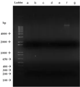
Since the isolates were resistant to many of the antibiotics tested, it is possible for one or more isolates to carry plas-mids bearing antibiotic resistance genes, which may be then acquired by pathogenic organisms. As this outcome is high-ly undesirable, plasmid extraction and gel electrophoresis were performed to determine whether the observed resistan-ces were transmissible. No plasmid presence was detected in the isolates STF4, STF8, STF9, STF10, STF15, and STF26; only STF25 displayed a faint band after gel electrophoresis (Fig.1).
Adhesion and invasion
Abilities of Bacillus isolates to adhere to HT-29 cells were tested. We observed that all isolates tested could adhere to HT-29 cells (Table5). STF9 could not grow in BHI broth; therefore, we could not perform the test for this isolate. STF4 showed the strongest adhesion with a percentage of 2.5 % while STF15 showed the lowest with 0.002 %. Invasion properties of Bacillus isolates were also assayed as mentioned above. Assay results demonstrate that none of
Table 3 Identification and characterization of selected isolates
Isolate 16S rRNA sequence analysis Sporulation efficiency (%) Survival in bile salt Survival in simulated gastric fluid Biofilm formation
(Closest known species) (Vegetative form) (Vegetative form)
STF4 Paenibacillus xylanexedens 79.9 + + − STF8 Bacillus subtilis 127.02 +++ ++++ − STF9 Bacillus subtilis 22.75 +++ +++ + STF10 Bacillus licheniformis 173.91 +++ ++ + STF15 Bacillus pumilus 15.28 + +++++ + STF25 Bacillus licheniformis 35.2 + ++++ − STF26 Bacillus pumilus 36.7 ++ ++++ +
Sporulation efficiency is given as the percentage of survivors
Plus signs under survival columns reflect the degrees of survival in simulated gastric fluid, + denoting the lowest survival and +++++ the highest. Plus signs under the biofilm formation column reflect presence of biofilm formation while minus signs denote absence of biofilm formation
the bacteria were capable of invading HT-29 cells, with the possible exception of STF9, which failed to grow in the assay medium (Table5). Bacillus subtilis ATCC 6633 and B. cereus RSHM709 displayed higher adhesion rates com-pared to our isolates (except STF4), though both reference strains could also invade cells. While B. subtilis ATCC 6633 had minor invasion capacity (0.050 %), the B. cereus strain was highly invasive (0.338 %).
Cell cytotoxicity
The cell cytotoxicities of the supernatants of 18 h grown isolates in BHI were tested by MTT assay. STF9 could not grow in BHI broth and was therefore not a subject for this test. STF8 and STF15 both had heat-stable toxic materials; though their cytotoxicities were different. In addition, STF4, STF25, and STF26 contained low amounts of heat-stable toxic materials in their cell-free supernatants. It is curious
that unmodified STF10 supernatant showed no toxicity to the HT-29 cell line, but the presence of a toxic material was observed after heating (Table5). Bacillus subtilis ATCC 6633 supernatant displayed minor cytotoxicity both prior to and after heating, while B. cereus RSHM709 expressed both heat-labile and heat-stable toxins.
Discussion
Bacillus species are commonly used as probiotics, as these spore-forming bacteria can tolerate the harsh environmental conditions associated with the gastrointestinal tract. Bacillus species have been incorporated in commercial probiotic prod-ucts, either alone or alongside consortia of other bacteria (Cutting 2011). Bactisubtil (B. cereus strain IP5832), Enterogermina (B. clausii), Biosubtyl Nha Trang (B. pumilus), and Bispan (B. polyfermenticus) are examples of commercial-ly available Bacillus probiotics (Duc et al. 2004; Cutting
2011). Further, the production of natto, a fermented soybean product originating in Japan, involves the use of B. subtilis to facilitate the fermentation process, indicating that Bacillus species have been safely used in foodstuffs for a considerable period of time (Hong et al.2008).
Bacillus species potentially suitable for use as probiotics were previously reported from the chicken and fish gastroin-testinal tracts (Barbosa et al. 2005; Vijayabaskar and Somasundaram2008). In a similar vein, we isolated Bacillus strains from bovine chyme and screened the isolates for pro-biotic potential, primarily utilizing antimicrobial activity as a measure of efficiency. Isolation of novel strains with high antimicrobial activity against pathogenic bacteria such as S. aureus, P. aeruginosa, S. enterica, and K. pneumoniae is highly desirable, as prevention of intestinal colonization by pathogenic bacteria is one of the principal ways by which probiotics bolster human and animal health. Other character-istics, such as the ability to survive the passage through the digestive tract and to successfully adhere to the intestinal mucosa, should also be considered in order to evaluate the capacity of a novel isolate as a potential probiotic.
Table 4 Minimum inhibitory concentrations (MIC) of select antibiotics against isolates
A, K, CH, E, V, N, CIP, S, and G refer to ampicillin, kanamycin, chloramphenicol, erytromycin, vancomycin, neomycin, ciprofloxacin, streptomycin, and gentamycin, respectively
Isolate Minimum Inhibitory Concentration (MIC)μg/ml
A K CH E V N CIP S G STF4 <1 <1 <1 <1 <1 4 <1 64 <1 STF8 16 64 8 2 4 64 <1 >128 16 STF9 64 64 64 8 4 64 <1 128 8 STF10 128 <1 8 4 8 64 <1 128 16 STF15 16 64 64 16 4 64 8 >128 8 STF25 128 64 64 128 128 128 16 128 16 STF26 32 16 32 32 32 32 4 32 16
Fig. 1 Agarose gel electrophoresis image of plasmid profile of isolates. a, b, c, d, e, f and g represent STF4 (Paenibacillus xylanexedens), STF8 (Bacillus subtilis), STF9 (Bacillus subtilis), STF10 (Bacillus licheniformis), STF15 (Bacillus pumilus), STF25 (Bacillus licheniformis), and STF26 (Bacillus pumilus), respectively
Spores of certain prospective Bacillus probiotics were found to be sensitive to the gastrointestinal tract (GIT) con-ditions (Duc et al.2004). However, the ability of probiotic strains to survive in the GIT of the animal on which they are used as feed supplement is of great importance, as the pro-biotic bacteria cannot demonstrate their propro-biotic activities if they cannot colonize the gastrointestinal tract. Therefore, re-sistance of isolates to GIT conditions was tested with incuba-tion in simulated gastric and intestinal fluids. Our results revealed that spores of the isolates were resistant to gastric fluid conditions after a 30-min incubation period, exhibiting 100 % survival rates. However, survival rates of STF15 and STF25 spores displayed a marked decrease after 1 h incuba-tion in simulated gastric fluids, while no reducincuba-tion in viability was observed in the other 5 isolates. Similarly, spores of all isolates were found to be tolerant to simulated intestinal fluid conditions, with 100 % survival rates after a 1-h incubation period. However, survival rates decreased in spores of some isolates for longer incubation periods. Vegetative cells, on the other hand, were not as resistant as spores to stress conditions. This result is expected, as spores of the genus Bacillus have protective layers around the nucleoid in the spore core, mak-ing spores extremely resistant to adverse environmental con-ditions (Barbosa et al.2005). Moreover, it has been reported that, once ingested, spores of Bacillus isolates could germinate in the GIT and display activity as vegetative cells in the host animal (Chaiyawan et al.2010; Casula and Cutting2002).
Our results demonstrate that STF9, STF10, STF15, and STF26 are capable of forming biofilm layers. Biofilm pro-duction is desired for prospective probiotics, since it allows the probiotic to remain longer in the GIT (Hong et al.2008). Biofilm formation provides protection for probiotic bacteria against the harsh conditions of the GIT, supports spore formation, and prevents pathogenic bacteria from holding on to the GIT surface (Barbosa et al.2005).
Antibiotic susceptibility is considered as one of the most important characteristics for probiotic bacteria, since the resis-tance genes could be transferred to pathogenic bacteria in the
intestinal tract and thus contribute to the creation of multidrug-resistant pathogens (Patel et al. 2009; Schwarz et al. 2001; Hong et al.2005; Barbosa et al.2005). This transfer mostly occurs by plasmid-borne resistance genes (Schwarz et al.
2001). In this study, seven isolates chosen after the initial screening were tested for antibiotic resistance against nine widely utilized antibiotics. Of those isolates, only STF4 had lower minimal inhibitory concentrations (MICs) than the break-points used by SCAN (von Wright2005) and could be consid-ered susceptible to the antibiotics tested. Other isolates (STF8, STF9, STF10, STF15, STF25, and STF26) were found to be resistant to at least one antibiotic tested. After the observation that most of the isolates displayed resistance to antibiotics, plasmid isolation and visualization was performed to determine whether this resistance is due to plasmid-borne resistance genes. If such genes are not integrated in genomic DNA and are instead carried on plasmids, they may be transferred to other bacteria in the environment and confer on them resistance to the same antibiotic. This constitutes a potential health hazard if the recipient bacteria are pathogenic, and the absence of plasmid DNA should therefore be confirmed for an antibiotic-resistant strain to be recommended as a prospective probiotic. As such, plasmid isolation was performed for all seven bacteria and the presence of plasmid was tested by performing gel electropho-resis. Our results suggest that all the samples tested, with the possible exception of STF25, carry potential antibiotic resis-tance genes in their genomic DNA and bear no risk of transfer-ring them to other pathogenic or non-pathogenic bacteria that may be present in the environment (Hong et al.2008). While it is possible that large, low-copy number plasmids have not been recovered by our isolation protocol, we find this possibility unlikely as cultures were grown to confluence several times in media lacking in antibiotics and, without the environmental stresses selecting for their presence, plasmids encoding antibi-otic resistance genes would likely have been lost.
The ability to adhere to and colonize in the GIT is an important requirement for potential probiotics, as the capacity to adhere increases the mean time of bacteria in the GIT and
Table 5 Adhesion, invasion rates and cytotoxicity of Bacillus isolates against HT-29
All results are given as the percentage of the survivors − sign indicates that no growth on BHI was observed
Isolate Bacterial species Adhesion (%) Invasion (%) Cytotoxicity (%)
Normal Heat treated
STF4 Paenibacillus xylanexedens 2.500 0 2.9 0 STF8 Bacillus subtilis 0.010 0 95.2 68.6 STF9 Bacillus subtilis − − − − STF10 Bacillus licheniformis 0.010 0 0 3.8 STF15 Bacillus pumilus 0.002 0 11.6 7.5 STF25 Bacillus licheniformis 0.100 0 7.8 0 STF26 Bacillus pumilus 0.008 0 8.5 0
ATCC 6633 Bacillus subtilis 0.466 0.050 4.5 2.7
allows greater periods of probiotic activity after oral introduc-tion of the probiotic strain (Jacobsen et al.1999; Saarela et al.
2000). Therefore, probiotic strains displaying stronger adhesion to the GIT are considered to better modify the normal intestinal microbiota, prevent the growth of pathogenic bacteria by com-petitive exclusion, and induce immune system compared to the non-adherent ones (Saarela et al.2000; Patel et al.2009). The HT-29 cell line is used as a model for the testing of adhesion of probiotic bacteria, as these cells differentiate into enterocytes (Saarela et al.2000; Patel et al.2009). Thus, we used the HT-29 colon carcinoma cell line for the assessment of cell adhesion by the prospective probiotic strains. In vitro adhesion assays sug-gest that all isolates except STF9 were able to partially adhere on the colon carcinoma cells, while STF4 had significantly higher adherence ability than the others. The adhesion rates observed were generally similar to the results obtained in other studies related to Bacillus adhesion and our own results on reference strains of B. subtilis and B. cereus (Hong et al.2008; Rowan et al.2001). Though our isolates had relatively low adhesion rates compared to previous records, STF4 again dis-played higher adhesion than many strains in the literature.
Recent research suggests that members of Bacillus species might induce important systemic diseases such as septicemia, peritonitis, endocarditis, liver failure, and meningitis (Rowan et al.2001). Therefore, invasion studies were conducted for our candidate probiotics. The test results suggest that none of the isolates invaded the HT-29 cells, displaying similar results to our B. subtilis reference strain. B. cereus RSHM709, how-ever, was highly invasive. This result shows that our isolates are non-invasive and incapable of causing invasion-related diseases, though live animal tests should be conducted for further confirmation.
MTT assay was used for detection of toxic materials in bacterial supernatants. This assay was chosen as it has been determined to be more reliable and sensitive than other cell viability assays (European Commission2000). In contrast to previous methods, which were based on visual evaluation of toxin-induced cell damage, MTT has the advantage of remov-ing the subjective visual assessment from the assay. MTT is a yellow, water-soluble tetrazolium salt, which is cleaved in the mitochondria of metabolically active cells. In the presence of living cells, MTT solution displays a color change as the insoluble purple formazan is formed due to intramitochondrial metabolization of MTT (Rowan et al.2001; Finlay et al.1999; Beattie and Williams 1999; European Commission 2000). MTT assay is therefore highly sensitive for cellular respiration and suitable for assessing cell viability and cytotoxicity, since only living cells produce formazan reaction products (Beattie and Williams1999; Finlay et al.1999; Rowan et al.2001). In this study, the cytotoxicity of cell-free supernatants was assessed by using MTT salts. STF8 had high levels of heat-stable cytotoxic materials in its cell-free supernatant, while other isolates did not produce a significant level of toxic
materials. STF10 in particular displayed 0 % cytotoxicity. For STF4, STF25, and STF26, heat treatment of the culture supernatants eliminated toxicity to HT-29, suggesting that toxic materials produced by those isolates are protein-based. The relatively low toxicities of our isolates (bar STF8) is expected as our isolates generally belong to the B. subtilis group, which are generally considered safe for human and animal use. Our B. subtilis reference strain likewise displayed low cytotoxicity, while the B. cereus strain was highly toxic prior to heating and possessed a heat-stable toxin that allowed it to retain some toxicity after heat treatment.
In conclusion, Bacillus species were isolated from the GIT of bovine and assessed with regards to their potential capacity as probiotics. Among the isolates, STF4, STF10, STF15, and STF26 appeared to have the highest potential as probiotic candidates compared to other isolates, taking into account their antimicrobial effects, resistance to simulated gastrointes-tinal conditions, antibiotic susceptibility, colonization capaci-ty, and lack of toxicity.
Acknowledgments We would like to thank Selma Bulut for her
assistance in SEM imaging and cell culture studies and Zeynep Ergul Ulger for her help in cell culture maintenance and in carrying out the MTT assay protocol. This work is supported by grants from the State Planning Organization of Turkey (DPT).
References
Barbosa T, Levy S (2000) The impact of antibiotic use on resistance development and persistence. Drug Resist Update 3:303–311 Barbosa T, Serra C, La Ragione R, Woodward M, Henriques A (2005)
Screening for Bacillus isolates in the broiler gastrointestinal tract.
Appl Environ Microb 71:968–978
Beattie SH, Williams AG (1999) Detection of toxigenic strains of Bacillus cereus and other Bacillus spp. with an improved
cyto-toxicity assay. Lett Appl Microbiol 28:221–225
Cartman ST, La Ragione RM, Woodward MJ (2008) Bacillus subtilis spores germinate in the chicken gastrointestinal tract. Appl
Environ Microb 74:5254–5258
Casula G, Cutting S (2002) Bacillus probiotics: spore germination in
the gastrointestinal tract. Appl Environ Microb 68:2344–2352
Chaiyawan N, Tayeeteptaikul P, Wannissorn B, Ruengsomwong S, Klungsupya P, Buaban W, Itsaranuwat P (2010) Characterization and probiotic properties of Bacillus strains isolated from broiler.
Thai J Vet Med 40:207–214
Cutting SM (2011) Bacillus probiotics. Food Microbiol 28:214–220 Duc L, Hong H, Barbosa T, Henriques A, Cutting S (2004)
Characterization of Bacillus probiotics available for human use.
Appl Environ Microb 70:2161–2171
European Commission (2000) Opinion of the scientific committee on animal nutrition on the safety of use of bacillus species in animal nutrition
Finlay WJ, Logan NA, Sutherland AD (1999) Semiautomated metabolic staining assay for Bacillus cereus emetic toxin. Appl Environ
Microb 65:1811–1812
Hamon M, Lazazzera B (2001) The sporulation transcription factor Spo0A is required for biofilm development in Bacillus subtilis.
Henriques A, Beall B, Roland K, Moran C (1995) Characterization of cotj, a sigma(e)-controlled operon affecting the polypeptide com-position of the coat of Bacillus subtilis spores. J Bacteriol
177:3394–3406
Hong H, Duc L, Cutting S (2005) The use of bacterial spore formers as probiotics. FEMS Microbiol Rev 29:813–835
Hong H, Huang J, Khaneja R, Hiep L, Urdaci M, Cutting S (2008) The safety of Bacillus subtilis and Bacillus indicus as food probiotics. J Appl Microbiol 105:510–520
Jacobsen C, Nielsen V, Hayford A, Moller P, Michaelsen K, Paerregaard A, Sandstrom B, Tvede M, Jakobsen M (1999) Screening of probiotic activities of forty-seven strains of Lactobacillus spp. by in vitro techniques and evaluation of the colonization ability of five selected strains in humans. Appl
Environ Microb 65:4949–4956
Kim K, Kim M, Kim D, Park Y, Kang J (2009) Characterization of Bacillus
polyfermenticus KJS-2 as a probiotic. J Microbiol Biotechn 19:1013–
1018
Lalloo R, Ramchuran S, Ramduth D, Gorgens J, Gardiner N (2007) Isolation and selection of Bacillus spp. as potential biological agents for enhancement of water quality in culture of ornamental fish. J Appl Microbiol 103:1471–1479
Lutful Kabir SM (2009) The role of probiotics in the poultry industry. Int J Mol Sci 10:3531–3546
Mazza P (1994) The use of Bacillus subtilis as an antidiarrhoeal microorganism. Boll Chim Farm 133:3–18
Modesto M, D’aimmo M, Stefanini I, Trevisi P, De Filippi S, Casini L,
Mazzoni M, Bosi P, Biavati B (2009) A novel strategy to select Bifidobacterium strains and prebiotics as natural growth
pro-moters in newly weaned pigs. Livest Sci 122:248–258
Nicholson WL, Munakata N, Horneck G, Melosh HJ, Setlow P (2000) Resistance of Bacillus endospores to extreme terrestrial and
ex-traterrestrial environments. Microbiol Mol Biol Res 64:548–572
Patel A, Ahire J, Pawar S, Chaudhari B, Chincholkar S (2009) Comparative accounts of probiotic characteristics of Bacillus
spp. isolated from food wastes. Food Res In 42:505–510
Patterson J, Burkholder K (2003) Application of prebiotics and
pro-biotics in poultry production. Poultry Sci 82:627–631
Rowan N, Candlish A, Bubert A, Anderson J, Kramer K, Mclauchlin J (2000) Virulent rough filaments of Listeria monocytogenes from clinical and food samples secreting wild-type levels of cell-free p60 protein. J Clin Microbiol 38:2643–2648
Rowan NJ, Deans K, Anderson JG, Gemmell CG, Hunter IS, Chaithong T (2001) Putative virulence factor expression by clin-ical and food isolates of Bacillus spp. after growth in reconstituted
infant milk formulae. Appl Environ Microb 67:3873–3881
Saarela M, Mogensen G, Fondén R, Mättö J, Mattila-Sandholm T (2000) Probiotic bacteria: safety, functional and technological
properties. J Biotechnol 84:97–215
Santini C, Baffoni L, Gaggia F, Granata M, Gasbarri R, Di Gioia D, Biavati B (2010) Characterization of probiotic strains: an appli-cation as feed additives in poultry against Campylobacter jejuni.
Int J Food Microbiol 104:S98–S108
Saravanakumari P, Mani K (2010) Structural characterization of a novel xylolipid biosurfactant from Lactococcus lactis and analy-sis of antibacterial activity against multi-drug reanaly-sistant pathogens.
Bioresource Technol 101:8851–8854
Schwarz S, Kehrenberg C, Walsh T (2001) Use of antimicrobial agents in veterinary medicine and food animal production. Int J Antimicrob Agric 17:431–437
Setlow P (2006) Spores of Bacillus subtilis: their resistance to and killing by radiation, heat and chemicals. J Appl Microbiol 101:514–525 The European Parliament and the Council of the European Union (2003)
Regulation (EC) No 1831/2003 of the European Parliament and of the Council of 22 September 2003 on additives for use in animal nutrition
Vijayabaskar P, Somasundaram ST (2008) Isolation of bacteriocin pro-ducing lactic acid bacteria from fish gut and probiotic activity against common fresh water fish pathogen. Aeromonas hydrophilia.
Biotechnol 7:124–128
Von Wright A (2005) Regulating the safety of probiotics—the