Mammalian Telomeric DNA Suppresses Endotoxin-induced
Uveitis
*
□SReceived for publication, March 22, 2010, and in revised form, June 16, 2010Published, JBC Papers in Press, July 14, 2010, DOI 10.1074/jbc.M110.125948
Fuat C. Yagci‡1, Ozlem Aslan§1, Mayda Gursel¶, Gizem Tincer‡, Yasemin O¨ zdamar§, Kutay Karatepe‡, K. Can Akcali‡,
and Ihsan Gursel‡2
From the‡Biotherapeutic Oligonucleotide Laboratory, Department of Molecular Biology and Genetics,¶Merkez Lojmanlari, Bilkent University, 06800 Ankara, Turkey, and the§Ministry of Health, Ulucanlar Eye Research and Education Hospital, 06240 Ankara, Turkey
Telomeric regions of mammalian chromosomes contain sup-pressive TTAGGG motifs that inhibit several proinflammatory and Th1-biased immune responses. Synthetic oligodeoxynucle-otides (ODN) expressing suppressive motifs can reproduce the down-regulatory activity of mammalian telomeric repeats and have proven effective in the prevention and treatment of several autoimmune and autoinflammatory diseases. Endotoxin-in-duced uveitis (EIU) is an established animal model of acute ocu-lar inflammation induced by LPS administration. Augmented expression of proinflammatory cytokines/chemokines such as TNF␣, IL-6, and MCP1 and bactericidal nitric oxide production mediated by LPS contribute to the development of EIU. Sup-pressing these mediators using agents that are devoid of unde-sirable systemic side effects may help prevent the development of EIU. This study demonstrates the selective down-regulatory role of suppressive ODN after (i) local or (ii) systemic treatment in EIU-induced rabbits and mice. Our results indicate that sup-pressive ODN down-regulate at both the transcript and protein levels of several proinflammatory cytokines and chemokines as well as nitric oxide and co-stimulatory surface marker mole-cules when administrated prior to, simultaneously with, or even after LPS challenge, thereby significantly reducing ocular inflammation in both rabbit and mouse eyes. These findings strongly suggest that suppressive ODN is a potent candidate for the prevention of uveitis and could be applied as a novel DNA-based immunoregulatory agent to control other autoimmune or autoinflammatory diseases.
DNA and RNA are the essential components of all living organisms. Accumulated evidence strongly suggests that these nucleic acids have multiple and complex effects on the immune system and are more than a blueprint of life (1, 2). On one hand, due to their high unmethylated CpG motif frequency, bacterial DNAs are recognized as “non-self” via TLR9 (Toll-like receptor 9) and trigger an innate immune response characterized by the proliferation and maturation of B cells, natural killer cells, and plasmacytoid dendritic cells and the secretion of T-helper
1-type cytokines, chemokines, and/or multivalent immuno-globulins (3– 8). On the other hand, telomeric regions of mam-malian chromosomes contain suppressive TTAGGG motifs that can inhibit several TLR-dependent and TLR-independent Th1-mediated immune responses. Of note, these motifs are underrepresented in the prokaryotic genome. Synthetic single-stranded oligodeoxynucleotides (ODN)3containing repetitive
TTAGGG motifs mimic this effect (1, 9 –11). Previous studies revealed that deleterious inflammatory responses to a host can be down-regulated by suppressive ODN. In vitro, suppressive ODN inhibits the production of several proinflammatory cyto-kines and chemocyto-kines induced by bacteria (1, 12–14). Further-more, in vivo suppressive ODN administration reduces the fre-quency and severity of several autoimmune and inflammatory diseases such as arthritis, systemic lupus erythematosus, pul-monary inflammation, toxic shock, silicosis, and experimental autoimmune encephalomyelitis (10, 15–21).
Uveitis is an ophthalmic disorder that causes vision loss in developed countries (22, 23) and is characterized by acute, recurrent, or persistent ocular inflammation, the breakdown of the blood-ocular barrier, and infiltration of leukocytes (24). The underlying causes of uveitis can vary. For example, acute ante-rior uveitis is often associated with (i) Behcet disease, (ii) Reiter syndrome, and (iii) ankylosing spondylitis, as well as other sys-temic inflammatory diseases (25).
Endotoxin-induced uveitis (EIU) is an established animal model of acute ocular inflammation. It is triggered by the administration of LPS, which is a component of the Gram-neg-ative bacterial outer membrane (26). A ligand for TLR4, LPS enhances the expression of various proinflammatory cytokines and chemokines such as IL-6 (27, 28), TNF␣ (29), and MCP1 (monocyte chemoattractant protein 1) (30) and the production of nitric oxide. All of these mediators contribute to the break-down of the blood-ocular barrier and infiltration of leukocytes, resulting in the development of EIU (26). It has been shown that suppressing proinflammatory cytokines, including IL-6, TNF␣, MCP1, and inducible nitric-oxide synthase (iNOS), retards if not prevents the development of EIU (31). Conventional drugs used to control these concerted inflammatory activation are mainly immunosuppressive in character and are associated with undesirable systemic side effects (24). It is of the utmost importance to develop effective, less toxic agents that
selec-*This work was supported by Scientific and Technological Research Council of Turkey (TUBITAK) Grants SBAG106S102, SBAG108S316, EU/FP6/Marie Curie, and EU/FP7 UNAM REGPOT 203953.
□S The on-line version of this article (available at http://www.jbc.org) contains supplemental Figs. 1 and 2.
1Both authors contributed equally to this work.
2To whom correspondence should be addressed. Tel.: 90-312-290-2408; Fax: 90-312-266-5097; E-mail: [email protected].
3The abbreviations used are: ODN, oligodeoxynucleotide(s); EIU, endotoxin-induced uveitis; iNOS, inducible nitric-oxide synthase.
at BILKENT UNIVERSITY on November 9, 2017
http://www.jbc.org/
tively block proinflammatory immune activation while elimi-nating the unwanted systemic side effects.
To date, the inhibitory effect of suppressive ODN on LPS-mediated EIU at both the local and systemic levels has not been studied by others. In this study, a very aggressive form of exper-imental uveitis was initiated via endotoxin administration. We investigated whether the suppressive ODN “A151” can inhibit the induction and development of ocular inflammation (before or at the time of LPS insult or even 2 h after LPS treatment) and help to reduce the symptoms of EIU in rabbits and mice. Our results revealed, for the first time, that A151 is capable of down-regulating the mRNA expression and protein levels of several potentially pathologic chemokines and cytokines at both the local and systemic levels. Consequently, suppressive ODN mimicking telomeric DNA offers a novel nucleic acid-based immunotherapeutic agent to control overexuberant undesir-able immune responses such as seen in autoimmune and auto-inflammatory diseases.
EXPERIMENTAL PROCEDURES
Materials—All cell culture medium components were from HyClone. Cytokine pairs for ELISAs were from Endogen. LPS (isolated from Escherichia coli) was obtained from Sigma. The phosphorothioate-modified suppressive ODN A151 (24-mer, 5⬘-(TTAGGG)4-3⬘) and control ODN
(24-mer, 5⬘-(TTACCC)4-3⬘) were obtained from Alpha DNA
(Montreal, Canada). TRIdity G (AppliChem GmbH, Darms-tadt, Germany) was used for RNA isolation. cDNAs were synthesized using a DyNAmoTM cDNA synthesis kit
(Finnzymes, Espoo, Finland) according to the manufac-turer’s protocol. DyNAzymeTM PCR Master Mix was used
for PCRs.
Maintenance of Animals—Adult female BALB/c mice and adult New Zealand rabbits were used for the experiments. The animals were kept in the animal holding facility of the Depart-ment of Molecular Biology and Genetics at Bilkent University under controlled conditions at 22 °C with 12-h light and 12-h dark cycles. They were provided with unlimited access of food and water.
Induction of Endotoxin-induced Uveitis—Specific pathogen-free 10-week-old female BALB/c mice were injected intraperi-toneally with 25, 50, 100, or 200g of LPS in 200 l of PBS and/or suppressive ODN. Mice were killed at the end of clinical evaluation. Both eyes were enucleated and used for cytokine expression assays. Spleens were removed and split in two; splenocytes were incubated on tissue culture plates for 6, 12, and 24 h; and supernatants were collected for cytokine deter-mination by ELISA. IL-6 was measured as an indicator of EIU response. The other half of the spleen was used to extract total RNA for further cytokine/chemokine gene transcript expres-sion analysis by RT-PCR. In another experiment, rabbits (three to four animal/group,⬃1500 g each; housed in the Ankara Hos-pital animal facility) were separated into different treatment groups, and EIU was initiated via intraocular LPS injection (100 g) with or without suppressive ODN treatment. Eyes were removed, and further analyses as described for mice were conducted.
Clinical Evaluation and Histopathological Investigation —Ani-mals were subjected to blind investigation by an ophthalmolo-gist under a dissection microscope 18 –24 h after injection, cor-responding to the time of maximal severity of EIU. Clinical ocular inflammation was graded on a scale from 0 to 4 for each animal described previously (32): no sign of inflammation⫽ 0; discrete inflammation in iris and conjunctiva⫽ 1; dilatation of iris and conjunctiva vessels⫽ 2; hyperemia in iris associated with Tyndall effect in anterior chamber⫽ 3; in addition to the signs in scale 3, synechia or fibrin is formed⫽ 4 (32). For his-topathological investigations, enucleated eyes were fixed in 10% formalin for 24 h, washed with running tap water for 1 h, and placed in 60% ethyl alcohol for an extra 3 h. Eyes were embed-ded in paraffin, which was sectioned and stained with hematox-ylin and eosin. Sections were examined blindly by a his-topathologist, using score systems of severity ranging from 0 to 4. Focal non-granulomatous monocytic infiltration in the cho-roid, the ciliary body and retina were scored as 0.5. Retinal perivascular infiltration and monocytic infiltration in the vitre-ous were scored as 1. Granuloma formation in the uvea and retina and the presence of occluded retinal vasculitis along with photoreceptor folds, serous detachment, and loss of photore-ceptor were scored as 2. In addition, the formation of Dalen-Fuchs nodules (granuloma at the level of the retinal pigmented epithelium) and the development of subretinal neovasculariza-tion were scored as 3 and 4 according to the number and size of the lesions (33).
Cytokine and IgM ELISAs and NO Assays—Immulon 2 HB microtiter plates (Thermo Scientific) were coated with anti-cytokine or anti-IgM antibodies (BD Pharmingen) and then blocked with PBS and 1% BSA (1, 34). Serially diluted stand-ards and culture supernatants or mouse sera were added to these plates for 2 h. Cytokine was detected using biotinylated anti-cytokine antibody followed by phosphatase-streptavi-din (Perbio), whereas bound IgM was detected using phos-phatase-conjugated anti-IgM antibodies (Southern Biotech-nology Associates, Birmingham, AL) as described (1). Nitric oxide detection by the Griess method was conducted on murine peritoneal exudate cells (106/ml) after 12–36 h of ex vivo
incu-bation as described by the supplier (Promega).
Analysis of Cell-surface Molecule Expression by FACS—2⫻ 106spleen cells/ml were isolated from 24-h post-treated mice.
Cells were washed, fixed, and co-stained with one of the phy-coerythrin-labeled anti-CD40, anti-CD86, and anti-ICAM-1 and FITC-labeled cell-specific antibodies (i.e. CD11c for den-dritic cells, CD11b for macrophages, and B220 for B cells (BD Pharmingen)) for 30 min at room temperature. Following washing, they were studied using a FACSCalibur (BD Bio-sciences) and analyzed with CellQuest Pro software.
Cytokine and Chemokine RT-PCR—Animals were injected with LPS and/or suppressive ODN. Total RNA was extracted from the eyes or spleens of the mice 4 – 6 h later (or from the irises or corneas of the rabbits), reverse-transcribed, and ampli-fied to obtain cDNA in a standard PCR for 30 cycles using primers for mouse- or rabbit-specific target genes (Table 1) as described previously (1, 34). PCR-amplified material was sepa-rated on 1.5% agarose gels and visualized under UV light after ethidium bromide staining.
at BILKENT UNIVERSITY on November 9, 2017
http://www.jbc.org/
Statistical Analysis—Assays were performed in triplicate on at least three to five different cell preparations. Statistical sig-nificance between untreated (or control) and treated groups was evaluated using Student’s t test.
RESULTS
EIU is an established animal model of acute ocular inflam-mation. It is induced by either systemic or intravitreal administration of LPS, the major component of Gram-neg-ative bacteria. LPS acts through the TLR4-triggering proin-flammatory signaling cascade. The expression of Th1 cyto-kines and chemocyto-kines, including IL-6, IL-1, and MIP3␣ (macrophage inflammatory protein 3␣), contributes to the development of EIU.
This study was performed with 82 mice and 26 rabbits. Initial experiments were conducted to optimize the induction of EIU (supplement Fig. 1). For the mouse experiments, systemic admin-istration of LPS doses between 25 and 100g/mouse were suffi-cient to induce uveitic eyes within 24 h as judged by clinical and histopathological investigations (supplement Figs. 1 and 2). For the rabbit experiments, intraocular 100-g LPS injection was found to be optimal to induce EIU. Following local or intraperito-neal LPS and or suppressive ODN administration, rabbit and mouse eyes were removed, and RNAs from the irises, vitreous, and corneas of the rabbit eyes were obtained. PCR was run with the cDNA from each sample, and the mRNA levels of IL-6, IL-15, IP10 (interferon-␥-inducible protein 10), iNOS, MIP1␣, IL-18, MIP3␣, CXCL16(CXC chemokine ligand 16), MIP1, and IL-1 were monitored. In addition, 24 h post-LPS and/or A151 treatment, splenocyte suspensions were incubated ex vivo for 6 –24 h, and IgM, IL-6, IL-10, IL-12, and IFN␥ levels from the super-natants were determined by ELISA. FACS analyses were conducted on spleen cells to monitor co-stimulatory/surface marker molecule expressions.
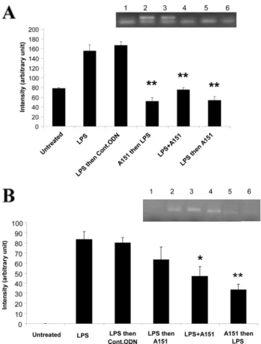
The results indicated that in rabbits, suppressive ODN administered before or after 100-g LPS treatment or co-in-jected with LPS significantly down-regulated the expression of IL-1 message from the iris (Fig. 1A). In cornea, IL-6 was down-regulated when suppressive ODN was given before or simulta-neously with LPS administration (Fig. 1B). There was no
sig-nificant inhibitory effect when A151 was given post-LPS treatment. In all these experiments, the mRNA message reduc-tion was suppressive ODN-dependent because control ODN administration did not show any benefit for alleviation of LPS reactogenicity.
TABLE 1
Oligonucleotide PCR primers used in mouse or rabbit experiments m, mouse; rb, rabbit.
Primer Forward Reverse Product
bp
m-actina GTATGCCTCGGTCGTACCA CTTCTGCATCCTGTCAGCAA
450
mIP10a GCCGTCATTTTCTGCCTCAT GCTTCCCTATGGCCCTCATT
127
miNOSa CAGCTGGGCTGTACAAACCTT CATTGGAAGTGAAGCGTTTCG
95
mMIP1␣b ACCATGACACTCTGCAACCA AGGCATTCAGTTCCAGGTCA
238
mIL-5a AGCACAGTGGTGAAAGAGACCTT TCCAATGCATAGCTGGTGATTT
117
mIL-15a CATCCATCTCGTGCTACTTGTGTT CATCTATCCAGTTGGCCTCTGT
126
mIL-18b GATCAAAGTGCCAGTGAACC ACAAACCCTCCCCACCTAAC
384 mMCP1b AGGTCCCTGTCATGCTTCTG TCTGGACCCATTCCTTCTTG 249 mMIP3␣b CGTCTGCTCTTCCTTGCTTT CCTTTTCACCCAGTTCTGCT 250 mCXCL16b CCTTGTCTCTTGCGTTCTTC GGTTGGGTGTGCTCTTTGTT 384
mMIP1b CCAGCTCTGTGCAAACCTAA CTGTCTGCCTCTTTTGGTCA
250
rbGAPDHc TCACCATCTTCCAGGAGCGA CACAATGCCGAAGTGGTCGT
319
rbIL-6c GCTCCTGGTGGTGGCTAC GGGTGGCTTCTTCATTCAAA
450
rbIL-1c GCCGATGGTCCAATTACAT ACAAGACCTGCCGGAAGCT
121
a
Taken from Ref. 43.
b
In house-designed primers.
c
Taken from Ref. 44.
FIGURE 1. Suppressive A151 ODN administration after LPS challenge
sig-nificantly down-regulates IL-1 and IL-6 expression in the iris and cor-nea, respectively. Rabbits were injected intraocularly with 100g of LPS and
250g of suppressive ODN. The average of densitometric measurements of four animals for IL-1 mRNA from iris (A) and IL-6 mRNA from cornea (B) is shown. Insets are the representative gel image of each group labeled from untreated to A151 and then LPS as 1 to 6. *, p⬍ 0.05; **, p ⬍ 0.01 between LPS-treated and A151 ODN-co-administered groups.
at BILKENT UNIVERSITY on November 9, 2017
http://www.jbc.org/
In the murine EIU model, mice tolerated up to 100-g LPS intraperitoneal treatment. Doses⬎150g caused animals to succumb to endotoxin treatment. The mouse experiments were conducted with three doses of LPS: 25, 50, and 100g. Suppressive A151 ODN and control ODN (2 h before and at the time of LPS injection and 2 h after LPS treatment) were used in the range of 100 –250g. Although in rabbits, the injection of ODN and endotoxin was intraocular, in mice, injections were given intraperitoneally in 200l of PBS.
The results showed that when 250g of suppressive ODN was administered before LPS injection (2 h), it significantly down-regulated the expression of IP10, iNOS, MIP3␣, CXCL16, and MIP1 in the 100-g LPS-injected mouse EIU model (Figs. 2, A and B). Other cytokines such as MIP1␣ and IL-18 also showed substantial but insignificant down-regula-tion at these doses (data not shown). To understand the sys-temic effect of suppressive A151, IL-6 secreted from murine splenocytes after ex vivo incubation for up to 24 h in culture was monitored by ELISA (Fig. 3). Our results revealed that suppres-sive ODN was able to reduce⬎65% of the secreted IL-6 (430 ⫾ 70 and 135⫾ 55 ng/ml for LPS and A151 ⫹ LPS groups, respec-tively). Co-administration of suppressive ODN with LPS signif-icantly decreased cytokine mRNA levels in vivo or cytokine pro-duction in ex vivo spleen cells (p⬍ 0.01) (Figs. 2 and 3). These effects were attributable to the activity of suppressive motifs because control ODN did not reduce the cytokine production elicited by co-administered LPS (Figs. 1–3).
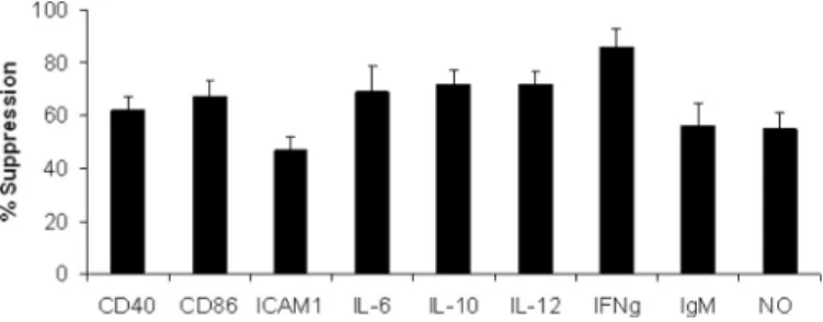
The (TTAGGG)4 multimers inhibited LPS-dependent
up-regulation of co-stimulatory and surface marker molecules on
antigen-presenting cells (CD40, CD86, and ICAM-1), IgM pro-duction by B cells, and NO release from peritoneal macro-phages (p⬍ 0.01) (Fig. 4). Furthermore, co-administration of LPS (50g) with A151 ODN (250 g) inhibited ⬎65% of several immunoregulatory and inflammatory cytokines (i.e. IL-6, IL-10, and IL-12; p⬍ 0.001) (Fig. 4). This reduction reached ⬎85% for IFN␥ (176 ⫾ 29 and 26 ⫾ 15 ng/ml for LPS and A151 ⫹ LPS groups, respectively).
DISCUSSION
In this study, we examined the effect of synthetic telomeric repeat units (suppressive A151 ODN) localized at the end of mammalian chromosomes on EIU, which is an established ani-mal model of acute ocular inflammation in both mouse and rabbit models. The results indicated that suppressive ODN was able to down-regulate the expression and protein levels of sev-eral proinflammatory and immunoregulatory cytokines/che-mokines at local and systemic levels when administrated (i) prior to, (ii) simultaneously with, or (iii) even after LPS chal-lenge (Figs. 2– 4).
FIGURE 2. A, suppressive A151 ODN treatment after 100-g LPS challenge significantly down-regulates IP10, MIP3␣, iNOS, MIP1, and CXCL16 expres-sion levels in the eyes of mice. Mice were injected intraperitoneally with 100 g of LPS and 250 g of suppressive ODN and killed 18 h after injection. *, p ⬍ 0.05; **, p⬍ 0.01 between LPS-treated and LPS ⫹ A151 ODN-co-administered groups. B, representative gel image.
FIGURE 3. Suppressive A151 ODN administration significantly
sup-presses IL-6 release from murine splenocytes. Mice were injected
intrap-eritoneally with 100g of LPS and 250 g of suppressive or control ODN as further indicated. Spleen cells were removed (24 h post injection) and incu-bated 6 –24 h, and supernatants were collected for cytokine ELISA. IL-6 was measured as an indicator of EIU response. *, p⬍ 0.05, between LPS and LPS ⫹ A151 groups.
FIGURE 4. Inhibitory effect of suppressive ODN on LPS-mediated immune
activation. The levels of CD40, CD86, and ICAM-1 expression (mean
fluores-cence intensity) were determined by FACS 24 h after in vivo injection of LPS or LPS plus A151 (50g of LPS and 250 g of ODN). IL-6, IL-10, IL-12, IFN␥, and IgM levels in culture supernatants were determined by ELISA following 36 h of
ex vivo incubation. The Griess method was used to detect nitric oxide in
isolated peritoneal exudate cells supernatants 24 h post-treatment. %
Sup-pression was calculated by the following formula: (1⫺ ((activation by LPS ⫹
suppressive ODN)⫺ (background)/(activation by LPS ⫹ control ODN) ⫺ (background)))⫻ 100.
at BILKENT UNIVERSITY on November 9, 2017
http://www.jbc.org/
Compared with local injection of (intraocular) LPS and/or LPS- and control ODN-treated rabbits, suppressive ODN-ad-ministrated animals exhibited reduced levels of IL-1 and IL-6 expression in the iris and cornea, respectively. In the mouse model, the results revealed that pretreatment with 250g of suppressive ODN reduced the expression of IP10, iNOS, MIP1␣, IL-18, MIP3␣, CXCL16, and MIP1 in 100-g LPS-injected mice. In another experiment, with different doses of suppressive ODN and LPS, suppressive ODN also down-regu-lated the expression of MCP1, which is an important chemo-kine for monocyte chemoattraction (data not shown). The sup-pressive action of this class of ODN was not only on the mRNA levels of several Th1-type cytokines and chemokines but also on the secreted protein level. ELISA experiments showed that sup-pressive ODN pre- and post-treatments significantly dimin-ished IL-6 secretion at 6 and 24 h; simultaneous administration of suppressive ODN also reduced IL-6 production.
Several studies indicated that suppressing proinflammatory cytokines, including IL-6, TNF␣, MCP1, and iNOS, prevents the development of EIU (26, 29, 30, 31). Here, we have shown that either local (intraocular) or systemic (intraperitoneal) administration of suppressive A151 ODN can significantly reduce several proinflammatory cytokines and chemokines even 2 h after in vivo LPS challenge.
Corticosteroids and chemotherapeutic agents are currently in use in uveitis therapy (35). However, long-term treatment with these drugs may have grave side effects such as increased intraocular pressure (36) and cytotoxicity (37) and thus limit their use (25, 33, 35). Therefore, a new therapeutic strategy is urgently needed (38, 39). The mechanism of action of this novel ODN-based immunosuppressive drug candidate is currently unknown. Previous studies revealed that suppressive ODN can inhibit immune response by blocking the stimulatory effects of CpG motifs (1, 15). It also has been shown by Shirota et al. (10) that suppressive A151 ODN can also protect mice from lethal endotoxic shock that is induced by LPS. It has been shown that suppressive ODN can also inhibit several signal transduction cascades related to the production of Th1 cytokines such as IFN␥ and IL-12 by binding and inhibiting the phosphorylation of STAT1 (signal transducer and activator of transcription 1) and STAT4 proteins (10, 13). Our study has demonstrated that suppressive ODN can block immune responses mediated by endotoxin in the eye (an immune privileged site), an established animal model of acute ocular inflammation. Recently, Fujimoto
et al.(40) reported that suppressive A151 ODN can inhibit ocular inflammation in two murine models, IRBP (interpho-toreceptor retinoid-binding protein)-induced experimental autoimmune uveitis and adoptively transferred ocular in-flammation. These forms are antigen-driven and, compared with LPS, are significantly less aggressive forms of experimental uveitis models. The control of LPS-mediated EIU at both the local and systemic levels has not been studied by others and increases the breadth of the suppressive ODN-mediated ther-apy for the eye. Collectively, these observations support the provocative possibility that the evolutionary expansion of TTAGGG repeats in telomeres, in addition to known proper-ties such as, protecting genomic DNA from degradation, and chromosome capping (41, 42) may also be linked to their ability
to down-regulate sustained/pathologic microbe-associated molecular pattern-induced immunity. In conclusion, we have provided evidence that suppressive A151 ODN is able to signif-icantly reduce the ocular inflammatory responses in both rabbit and murine EIU models.
Acknowledgments—We thank Tamer Kahraman and Erdem Erikci for excellent technical support and Burcu C. Insal for assistance dur-ing animal procedures. We greatly appreciate Dr. Onder Bozdogan for guidance during histopathological investigations.
REFERENCES
1. Gursel, I., Gursel, M., Yamada, H., Ishii, K. J., Takeshita, F., and Klinman, D. M. (2003) J. Immunol. 171, 1393–1400
2. Ishii, K. J., and Akira, S. (2005) Int. J. Cancer 117, 517–523
3. Yamamoto, S., Yamamoto, T., Kataoka, T., Kuramoto, E., Yano, O., and Tokunaga, T. (1992) J. Immunol. 148, 4072– 4076
4. Klinman, D. M., Yi, A. K., Beaucage, S. L., Conover, J., and Krieg, A. M. (1996) Proc. Natl. Acad. Sci. U.S.A. 93, 2879 –2883
5. Sparwasser, T., Koch, E. S., Vabulas, R. M., Heeg, K., Lipford, G. B., Ellwart, J. W., and Wagner, H. (1998) Eur. J. Immunol. 28, 2045–2054
6. Krieg, A. M. (2000) Curr. Opin. Immunol. 12, 35– 43
7. Yamada, H., Gursel, I., Takeshita, F., Conover, J., Ishii, K. J., Gursel, M., Takeshita, S., and Klinman, D. M. (2002) J. Immunol. 169, 5590 –5594 8. Klinman, D. M., Klaschik, S., Sato, T., and Tross, D. (2009) Adv. Drug
Deliv. Rev. 61,248 –255
9. Klinman, D. M., Zeuner, R., Yamada, H., Gursel, M., Currie, D., and Gur-sel, I. (2003) Ann. N.Y. Acad. Sci. 1002, 112–123
10. Shirota, H., Gursel, I., Gursel, M., and Klinman, D. M. (2005) J. Immunol.
174,4579 – 4583
11. Hu, D., Su, X., Sun, R., Yang, G., Wang, H., Ren, J., Sun, L., Wu, X., Hu, X., Yu, Y., and Wang, L. (2009) Mol. Immunol 46, 1387–1396
12. Zhu, F. G., Reich, C. F., and Pisetsky, D. S. (2002) J. Leukocyte Biol. 71, 686 – 694
13. Shirota, H., Gursel, M., and Klinman, D. M. (2004) J. Immunol. 173, 5002–5007
14. Klinman, D. M., Tross, D., Klaschik, S., Shirota, H., and Sato, T. (2009)
Ann. N.Y. Acad. Sci. 1175,80 – 88
15. Zeuner, R. A., Ishii, K. J., Lizak, M. J., Gursel, I., Yamada, H., Klinman, D. M., and Verthelyi, D. (2002) Arthritis Rheum. 46, 2219 –2224 16. Ho, P. P., Fontoura, P., Ruiz, P. J., Steinman, L., and Garren, H. (2003)
J. Immunol. 171,4920 – 4926
17. Zeuner, R. A., Verthelyi, D., Gursel, M., Ishii, K. J., and Klinman, D. M. (2003) Arthritis Rheum. 48, 1701–1707
18. Dong, L., Ito, S., Ishii, K. J., and Klinman, D. M. (2004) Arthritis Rheum. 50, 1686 –1689
19. Dong, L., Ito, S., Ishii, K. J., and Klinman, D. M. (2005) Arthritis Rheum. 52, 651– 658
20. Ho, P. P., Fontoura, P., Platten, M., Sobel, R. A., DeVoss, J. J., Lee, L. Y., Kidd, B. A., Tomooka, B. H., Capers, J., Agrawal, A., Gupta, R., Zernik, J., Yee, M. K., Lee, B. J., Garren, H., Robinson, W. H., and Steinman, L. (2005)
J. Immunol. 175,6226 – 6234
21. Sato, T., Shimosato, T., Alvord, W. G., and Klinman, D. M. (2008) J.
Im-munol. 180,7648 –7654
22. Rothova, A., Suttorp-van Schulten, M. S., Frits Treffers, W., and Kijlstra, A. (1996) Br. J. Ophthalmol. 80, 332–336
23. Durrani, O. M., Tehrani, N. N., Marr, J. E., Moradi, P., Stavrou, P., and Murray, P. I. (2004) Br. J. Ophthalmol. 88, 1159 –1162
24. Sande, P. H., Fernandez, D. C., Aldana Marcos, H. J., Chianelli, M. S., Aisemberg, J., Silberman, D. M., Sa´enz, D. A., and Rosenstein, R. E. (2008)
Am J. Pathol. 173,1702–1713
25. Hafezi-Moghadam, A., Noda, K., Almulki, L., Iliaki, E. F., Poulaki, V., Thomas, K. L., Nakazawa, T., Hisatomi, T., Miller, J. W., and Gragoudas, E. S. (2007) FASEB J. 21, 464 – 474
26. Nagai, N., Oike, Y., Noda, K., Urano, T., Kubota, Y., Ozawa, Y., Shinoda,
at BILKENT UNIVERSITY on November 9, 2017
http://www.jbc.org/
H., Koto, T., Shinoda, K., Inoue, M., Tsubota, K., Yamashiro, K., Suda, T., and Ishida, S. (2005) Invest. Ophthalmol. Vis. Sci. 46, 2925–2931 27. Hoekzema, R., Verhagen, C., van Haren, M., and Kijlstra, A. (1992) Invest.
Ophthalmol. Vis. Sci. 33,532–539
28. Ohta, K., Kikuchi, T., Miyahara, T., and Yoshimura, N. (2005) Exp. Eye Res.
80,401– 412
29. Koizumi, K., Poulaki, V., Doehmen, S., Welsandt, G., Radetzky, S., Lappas, A., Kociok, N., Kirchhof, B., and Joussen, A. M. (2003) Invest. Ophthalmol.
Vis. Sci. 44,2184 –2191
30. Mo, J. S., Matsukawa, A., Ohkawara, S., and Yoshinaga, M. (1999) Exp. Eye
Res. 68,333–340
31. Mandai, M., Yoshimura, N., Yoshida, M., Iwaki, M., and Honda, Y. (1994)
Invest. Ophthalmol. Vis. Sci. 35,3673–3680
32. Lajavardi, L., Bochot, A., Camelo, S., Goldenberg, B., Naud, M. C., Behar-Cohen, F., Fattal, E., and de Kozak, Y. (2007) Invest. Ophthalmol. Vis. Sci.
48,3230 –3238
33. Chan, C. C., Caspi, R. R., Ni, M., Leake, W. C., Wiggert, B., Chader, G. J., and Nussenblatt, R. B. (1990) J. Autoimmun. 3, 247–255
34. Gursel, I., Gursel, M., Ishii, K. J., and Klinman, D. M. (2001) J. Immunol.
167,3324 –3328
35. Dunn, J. P. (2004) Curr. Opin. Ophthalmol. 15, 293–298
36. Moorthy, R. S., Mermoud, A., Baerveldt, G., Minckler, D. S., Lee, P. P., and Rao, N. A. (1997) Surv. Ophthalmol. 41, 361–394
37. Lightman, S. (1997) Eye 11, 222–226
38. Avunduk, M. C., Avunduk, A. M., Oztekin, E., Baltaci, A. K., Ozyazgan, Y., and Mogolkoc, R. (2004) Exp. Eye Res. 79, 357–365
39. Adamus, G., Burrows, G. G., Vandenbark, A. A., and Offner, H. (2006)
Invest. Ophthalmol. Vis. Sci. 47,2555–2561
40. Fujimoto, C., Klinman, D. M., Shi, G., Yin, H., Vistica, B. P., Lovaas, J. D., Wawrousek, E. F., Igarashi, T., Chan, C. C., and Gery, I. (2009) Clin. Exp.
Immunol. 156,528 –534
41. Rudolph, K. L., Chang, S., Lee, H. W., Blasco, M., Gottlieb, G. J., Greider, C., and DePinho, R. A. (1999) Cell 96, 701–712
42. Hackett, J. A., Feldser, D. M., and Greider, C. W. (2001) Cell 106, 275–286 43. Giulietti, A., Overbergh, L., Valckx, D., Decallonne, B., Bouillon, R., and
Mathieu, C. (2001) Methods 25, 386 – 401
44. Sobajima, S., Shimer, A. L., Chadderdon, R. C., Kompel, J. F., Kim, J. S., Gilbertson, L. G., and Kang, J. D. (2005) Spine J. 5, 14 –23
at BILKENT UNIVERSITY on November 9, 2017
http://www.jbc.org/
Karatepe, K. Can Akcali and Ihsan Gursel
Fuat C. Yagci, Ozlem Aslan, Mayda Gursel, Gizem Tincer, Yasemin Özdamar, Kutay
doi: 10.1074/jbc.M110.125948 originally published online July 14, 2010 2010, 285:28806-28811.
J. Biol. Chem.
10.1074/jbc.M110.125948
Access the most updated version of this article at doi: Alerts:
When a correction for this article is posted
•
When this article is cited
•
to choose from all of JBC's e-mail alerts
Click here
Supplemental material:
http://www.jbc.org/content/suppl/2010/07/14/M110.125948.DC1 http://www.jbc.org/content/285/37/28806.full.html#ref-list-1This article cites 44 references, 20 of which can be accessed free at
at BILKENT UNIVERSITY on November 9, 2017
http://www.jbc.org/