ROLE OF ENVIRONMENTAL FACTORS IN MESENCHYMAL STEM CELL BIOLOGY
A THESIS SUBMITTED TO
THE DEPARTMENT OF MOLECULAR BIOLOGY AND GENETICS AND THE INSTITUTE OF ENGINEERING AND SCIENCE OF
BILKENT UNIVERSITY
IN PARTIAL FULFILLMENT OF THE REQUIREMENTS FOR THE DEGREE OF MASTER OF SCIENCE
BY
SINAN GÜLTEKIN AUGUST 2009
SIGNATURE PAGE
I certify that I have read this thesis and that in my opinion it is fully adequate, in scope and in quality, as a thesis for the degree of Master of Science.
Assist. Prof. Dr. K. Can Akçalı
I certify that I have read this thesis and that in my opinion it is fully adequate, in scope and in quality, as a thesis for the degree of Master of Science.
Assoc. Prof. Dr. İhsan Gürsel
I certify that I have read this thesis and that in my opinion it is fully adequate, in scope and in quality, as a thesis for the degree of Master of Science.
Prof. Dr. Yakup Arıca
Approved for the Institute of Engineering and Science
Director of Institute of Engineering and Science Prof. Dr. Mehmet Baray
ABSTRACT
ROLE OF ENVIRONMENTAL FACTORS IN MESENCHYMAL STEM CELL BIOLOGY
Sinan Gültekin
M.S. in Molecular Biology and Genetics Supervisor: Associate. Prof. Dr. K. Can Akçalı
August, 104 Pages
Mesenchymal Stem Cells (MSCs) have the abilities of self-renewal and differentiation into fat, bone, cartilage, and muscle tissues. Besides intrinsic mechanisms that control the fate of the MSCs, extrinsic physiological factors also play role in this decision. Therefore, our aim is to explore the effects of possible environmental factors, involved in MSC maintenance by using rat MSCs as a model. We studied the effects of hypoxia and estrogen on growth regulation and cellular proliferation in MSCs. MSCs cells exhibited high colony number in hypoxic conditions and the expansion of MSCs was increased addition of the estrogen. In addition, estrogen prevents apoptosis, under hypoxic condition. The effects of estrogen on the expression levels of Notch genes (Notch1, Notch2, Notch3 and Notch4) were also investigated. In order to understand the possible mechanism of estrogen response, an experimental and in silico approach are used. The expression levels of Notch1 and Notch 3 were decreased treatment and the expression level of Notch 4 was increased upon estrogen treatment. In addition, bioinformatics analysis showed that, estrogen upregulates ERG family transcription factors, ELF family transcription factors, HOXL4 family transcription factors, KLF family transcription factors and transcription factor SOX3, which bind to Notch 1 transcriptional regulatory region, implying indirect effects of estrogen on Notch 1 expression. Twenty biomaterials were also investigated in order to assess whether they provide an appropriate environment for MSCs expansion. It was found that eight of the biomaterials out of twenty designated as, CA-1, CA-2, CA-3, CI-K, CI-A, CIII-1, CIII-2 and CIII-3, were appropriate candidates to expand MSCs. The combination polymers designated as HPMA/PEG provided appropriate conditions when prepared in the proportion of 1:0 (CA-1), 1:1 (CA-2) and 2:1 (CA-3). The appropriate proportion of polymers designated as HEMA/PEG/HPC was 2:1:1(CIII-1), 3:0:1 (CIII-2) and 1:1:0 (CIII-5).
ÖZET
ÇEVRESEL FAKTÖRLERİN MEZENKİMAL KÖK HÜCRE BİYOLOJİSİ ÜZERİNDEKİ ROLÜ
Sinan Gültekin
Moleküler Biyoloji ve Genetik Yüksek Lisans Tez Yöneticisi: Doç. Dr. K. Can Akçalı
Ağustos 2009, 104 Sayfa
Mezenkimal Kök Hücreler (MKH) hem kendilerini yenileyebilmekte hem de yağ, kemik, kıkırdak ve kas hücrelerine farklılaşabilmektedirler. MKH’ların yenilenebilme ve farklılaşabilme özellikleri, içsel mekanizmaların yanısıra, fizyolojik dış faktörler tarafından da kontrol edilir. Buradan hareketle sıçan MKH modelinde, çevre faktörlerinin, MKH
devamlılığındaki olası etkilerini belirlemek amaçlanmıştır.Farklılaşma, büyüme ve çoğalma kapasitesi üzerindeki etkisi göz önüne alınarak, hipoksi ve östrojenin, MKH’ler üzerindeki roller araştırıldı. MKH’lerin koloni oluşturma özelliklerinin hipoksik ortamda artığı ve bu artışın östrojenin eklenmesiyle daha da fazlalaştığı gözlemlendi. Buna ek olarak, östrojenin hipoksik ortamda, MKH’lerin apoptoza girmelerini engellediği gösterildi. Östrojenin Notch reseptör ifadesindeki etkisi hem deneysel, hem de bioinformatik yöntemle araştırıldı.
Deneysel olarak Notch1 ve Notch3 ekspresyonunun östrojenle birlikte azaldığı, ancak Notch4 ifadesinin arttığı gözlendi. Bununla birlikte, bioinformatik yöntemle östrojenin, ERG, ELF, HOXL4 transkripsiyon faktör aile gruplarının ve SOX3 transkripsiyon faktörü ifadesini arttırdığı, bu transkripsiyon faktörlerinin Notch1’in transkripsiyonu regüle eden bölgelerine bağlandığı gösterildi. MKH’lerin büyümelerine uygun ortam oluşturmak için önceden
hazırlanmış 20 tane biyomateryal kullanıldı, bunlardan sadece sekiz tanesi (CA-1,CA-2,CA-3, CI-A,CI-K,CIII-I,CIII-II ve CIII-2) MKH’lerin büyümesi ve gelişmesi için iyi bir ortam oluşturduğu saptanmıştır.
Anahtar Kelimeler: Mezenkimal kök hücreler, hipoksia, östrojen, notch reseptör ailesi ve biomalzeme.
ACKNOWLEDGEMENTS
I would like to express my deepest gratitude to my supervisor Assoc. Prof. K. Can Akçalı. Without his endless support, encouragement and patience I would not be able to learn so much about life and my studies. His personal and academic advices besides his guidance and patience, was very valuable for me.
I would also like to thank Zeynep Tokcaer Keskin, Fatma Ayaloğlu, Sumru Bayın and Hande Koçak for their incredible help in my crisis moments and their support in the lab.
I would like to thank Burcu for helping me in all my animal work. I could not have completed my thesis without her valuable assistance.
I would like to thank Onur, Atıl, Derya, Gurbet, Nilüfer, Tamer, Rümeysa, Ahmet Raşit, Fırat, Duygu and Ender for their support and friendship.
I would like to thank Gonca, Zeynep, Hande and Murat for their moral support.
I would like to thank The Scientific and Technological Research Council of Turkey (TÜBİTAK) for their financial support throughout my studies.
In addition, I would like to thank MBG family members for their support, friendship, and help.
Finally, I would like to thank my family for being there whenever I needed them and supporting me in every decision I gave. Without them and their unconditional love, nothing would be possible.
TABLE OF CONTENTS SIGNATURE PAGE...ii ABSTRACT ...iii ÖZET... iv ACKNOWLEDGEMENTS ... v TABLE OF CONTENTS ... vi LIST OF TABLES ... ix LIST OF FIGURES... xi ABBREVIATIONS...xiii 1. INTRODUCTION ... 1 1.1 Stem Cells... 2
1.1.1 Embryonic Stem Cells... 2
1.1.2 Embryonic Germ Cells... 4
1.1.3 Induced Pluripotent Stem Cells... 4
1.1.4 Adult Stem Cells ... 6
1.1.5 Mesenchymal Stem Cells ... 8
1.1.6 Stem Cell Niches... 11
1.2 Hypoxia ... 12
1.2.1 Hypoxia and Stem Cells... 16
1.2.2 Bone Marrow Microenvironment... 17
1.2.2.1. Effects of Hypoxia on Haematopoietic and Embryonic Stem Cells... 18
1.2.2.2. Effects of Hypoxia on Bone Marrow-Derived Mesenchymal Stem Cells.. 18
1.2.3 The role of HIFs in regulating stem cells... 18
1.3 Estrogen ... 19
1.3.1 Estrogen Signals... 20
1.3.1.1. Ligand-Dependent Genomic Pathways... 20
1.3.1.2. Nongenomic Pathways... 20
1.3.2 Estrogen and Hypoxia ... 21
1.3.3 Estrogen and Notch ... 22
1.3.4 Estrogen and Stem Cells ... 22
1.4 Notch Signaling Pathway ... 23
1.4.2 Epigenetic regulators... 26
1.4.3 Notch Signaling Pathway and Stem Cells... 26
1.4.4 Notch Pathway and Hypoxia in Stem Cells ... 28
1.5 Biomaterials... 29
2. AIM OF STUDY ... 31
3. MATERIALS AND METHODS... 32
3.1 Animals... 32
3.2 Isolation of the Bone Marrow from the Animals ... 32
3.3 Cell Culture... 33
3.3.1 Cell Number Detection with Cell Count ... 33
3.3.2 MSC Culture ... 33
3.3.3 Estrogen Treatment ... 33
3.4 Hypoxic Condition ... 33
3.5 Biomaterials... 34
3.5.1 Preparation of Biomaterials... 34
3.5.2 Growing Rat Bone Barrow Cells on Biomaterials ... 35
3.6 Standard Solutions and Buffers can be found in Appendix A... 35
3.7 Colony Forming Assay... 35
3.8 Determination of the Gene Expression... 36
3.8.1 Total RNA Isolation from Rat MSCs... 36
3.8.2 Concentration and Integrity of RNA... 36
3.8.3 Denaturing Gel Electrophpresis ... 36
3.8.4 The cDNA Synthesis... 37
3.9 RT- PCR ... 37
3.9.1 Primer Design... 37
3.10 Agarose Gel Electrophoresis ... 41
3.11 Protein Expression... 41
3.11.1 Total Protein Isolation from the MSCs ... 41
3.11.2 Protein Quantification ... 41
3.12 Western Blotting... 42
3.12.1 SDS Polyacrylamide Gel Electrophoresis (SDS-PAGE) ... 42
3.12.2 Transfer of Proteins to the Membrane... 42
3.12.4 Coomassie Blue Staining of the Gel and Membrane ... 43
3.13 Immunohistochemistry Staining... 43
3.14 Immunofluorescent Staining... 44
3.15 In Situ Cell Death Detection (TUNEL ASSAY)... 45
3.16 BrdU incorporation assay ... 45
3.17 In Silico Analysis... 46
3.17.1 Finding Transcription Factors ... 46
3.17.2 Finding Estrogen induced Transcription Factors ... 47
3.17.3 Finding Common Genes... 49
3.18 Statistical Analysis ... 50
4. RESULTS ... 51
4.1 Characterization of Rat MSCs... 51
4.2 Role of Hypoxia and Estrogen in MSCs Biology... 52
4.3 Characterization of MSCs after CoCl2treatment ... 52
4.4 Expression of HIF-1upon CoCl2 treatment. ... 53
4.5 Expression of ERand ERin Mesenchymal Stem Cells. ... 54
4.6 Colony Forming Assay... 54
4.7 TUNEL Assay ... 55
4.8 BrdU Incorporation Assay... 56
4.9 Expression of Notch Receptors in hypoxic condition and upon estrogen treatment in MSCs. ... 57
4.9.1 Expression of Notch Receptors ... 58
4.10 Bone Marrow Isolated Cells Grown on Biomaterials ... 62
4.11 Bioinformatics Analysis ... 67
4.11.1 Transcription Factors on Notch 1 10000bp upstream region... 68
4.11.2 Estrogen Induced Genes... 69
4.11.3 Common Genes between TFs found by MatInspector and Estrogen Up-Regulated Genes. ... 75
5. DISCUSSION ... 78
6. FUTURE PERSPECTIVES... 86
7. REFERENCES ... 87
LIST OF TABLES
Table 3. 1: Different Composition With Varying Monomer Ratio... 35
Table 3. 2: The Sequences and The Sizes of The Primers ... 38
Table 3. 3: PCR mixture With Taq Polymerase... 39
Table 3. 4: PCR mixture With With Phire Hot Start Dna Polymerase ... 39
Table 3. 5: The Standard Curve for Protein Concentration Detection ... 42
Table 3. 6: Publicly Available Gene Expression Data From Estrogen Studies on Cell Lines. 47 Table 4. 1: Different Composition of Biomaterials with Varying Monomer Ratio... 62
Table 4. 2: Summary of Observed Cell Morphology and Observed Colony Formation on Each Biomaterials. ... 67
Table 4. 3: Transcription Factors Binding To 10000bp Upstream Region of Notch 1... 69
Table 4. 4: Number of Estrogen Up-Regulated Genes in Gse 1153 in Different Experimental Setups... 70
Table 4. 5: Number of Estrogen Up-Regulated Genes in Gse 1153 in Different Experimental Setups... 71
Table 4. 6: Number of Estrogen Up-Regulated Genes in Gse9757, Gse9758 And Gse9759 in Different Experimental Setups... 71
Table 4. 7: Number of Estrogen Up-Regulated Genes in Gse 11352 in Different Experimental Setups... 72
Table 4. 8: Number of Estrogen Up-Regulated Genes in Gse 11506 in Different Experimental Setups... 72
Table 4. 9 Number of Estrogen Up-Regulated Genes in Gse 2251 in Different Experimental Setups... 73
Table 4. 10: Number of Estrogen Up-Regulated Genes in Gse4025 in Different Experimental Setups... 73
Table 4. 11 Number of Estrogen Up-Regulated Genes in Gse4668 in Different Experimental Setups... 74
Table 4. 12: Number of Estrogen Up-Regulated Genes in Gse4006 in Different Experimental Setups... 74
Table 4. 13: Number of Estrogen Up-Regulated Genes in Gse11324 in Different Experimental Setups. ... 75
Table 4. 14: Common Genes Between Reanalyzed Gse1153 Datasets And MatInspector ... 75
Table 4. 15: Common Genes Between Reanalyzed Gse2292 Datasets And MatInspector ... 76
Table 4. 16: Common Genes Between Reanalyzed Gse9757,Gse9758 And Gse9859 Datasets And MatInspector. ... 76
Table 4. 17: Common Genes Between Reanalyzed Gse11352 Datasets And MatInspector ... 76
Table 4. 18: Common Genes Between Reanalyzed Gse11506 Datasets And MatInspector ... 76
Table 4. 19: Common Genes Between Reanalyzed Gse2251 Datasets And MatInspector ... 76
Table 4. 20: Common Genes Between Reanalyzed Gse4025 Datasets And MatInspector ... 77
Table 4. 21: Common Genes Between Reanalyzed Gse4668 Datasets And MatInspector ... 77
Table 4. 22: Common Genes Between Reanalyzed Gse4006 Datasets And MatInspector ... 77
LIST OF FIGURES
Figure 1. 1: Adult Stem Cells... 7
Figure 1. 2 Pluripotent Capacity Of Msc To Differentiate İnto Mesodermal And Non-Mesodermal Cell Lineages, ... 10
Figure 1. 3 Physiological And Pathophysiological Oxygen Partial Pressures (Po2)... 13
Figure 1. 4 Domain Structures Of Human Hıf- And Hıf-1.. ... 15
Figure 1. 5. Oxygen Tension In Bone Marrow ... 17
Figure 1. 6: Outline Of Estrogen Signaling Pathways. ... 21
Figure 1. 7: A Proposed Model of The Effects of E2And Hypoxia on Transactivation of ERs. ... 22
Figure 1. 8: Effects Of Estrogen On Various Stem Cells And Progenitor Cells... 23
Figure 1. 9 Dsl Ligands And Notch Receptors... ... 25
Figure 1. 10: Schematic İllustration Of Notch And The Pathway ... 25
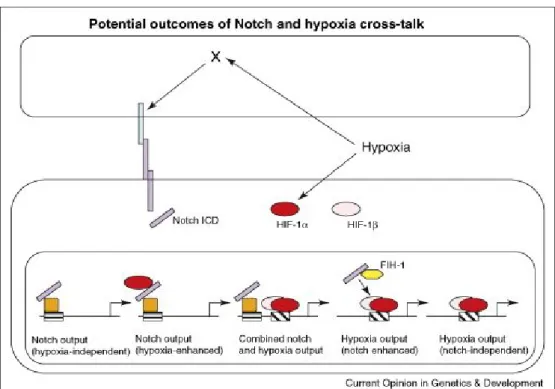
Figure 1. 11: Schematic Depiction Of Different Levels Of İnteraction Between Notch And The Cellular Hypoxic Response And The Potential Transcriptional Outcome Of This Cross-Talk... 29
Figure 3. 1: The Isolation of The Bone Marrow of The Rats... 32
Figure 3. 2 PCR Conditions and Cycles... 41
Figure 4. 1: The Expression of CD Markers in Rat Bone Marrow isolated Cells After 14 Days. ... 51
Figure 4. 2: The Expression of CD90 and CD34 in Bone Marrow Isolated Cells After 14 Days. ... 52
Figure 4. 3: Expression Levels of CD90, CD29, CD71, CD34, CD45... 53
Figure 4. 4: Expression Level of Hydroxylated Hif-1alpha... 54
Figure 4. 5: The Expression Level of ErAnd Er... 54
Figure 4. 6: The Number of Colonies. ... 55
Figure 4. 7: The Apoptotic Cell Ratios of Cells... 56
Figure 4. 8: The Proliferation Rate of Msc. ... 57
Figure 4. 9: The Expression Profile of Notch Receptors. ... 58
Figure 4. 10 :The Expression of Notch 1 ... 59
Figure 4. 11: The Expression of Notch 3. ... 60
Figure 4. 13: On The 14thDay Pictures of Rat Female Bone Marrow isolated Cells Grown on Biomaterials “CIII”... 63 Figure 4. 14: Colonies Formed on Biomaterials Designated As CIII. ... 64 Figure 4. 15: On The 14thDay Pictures of Rat Female Bone Marrow Isolated Cells Grown on
Biomaterials “CI”... 65 Figure 4. 16: Colonies Formed on CI-A And CI-K Biomaterials After 14 Days Culture.. ... 65 Figure 4. 17: On The 14thDay Pictures of Rat Female Bone Marrow isolated Cells Grown on
Biomaterials Designated as “CA”... 66 Figure 4. 18: Colonies Formed on CA-2 And CA-3 Biomaterials After 14 Days Culture... 67
ABBREVIATIONS
AIBN A-A-Azoisobisbutyronitrile
ASC Adult Stem Cells
bFGF Basic Fibroblast Growth Factor
bHLH Basic Helix Loop Helix
BM Bone Marrow
bp Base Pair
BSA Bovine Serum Albumin
cDNA Complementary Deoxyribonucleic Acid
CoCl2 Cobalt Chloride
CSL [Named After CBF1, Su(H) and LAG-1]
ddH2O Double distilled water
DEPC Diethylpyrocarbonate
dH2O Distilled Water
DMEM Dulbecco’s Modified Eagle Medium
DNA Deoxyribonucleic Acid
DNase Deoxyribonuclease
EB Embryoid Bodies
ECM Extra Cellular Matrix
EGF The Epidermal Growth Factor
ERE Estrogen Response Element
FBS Fatal Bovine Serum
FIH-1 The Protein Factor Inhibiting HIF-1α
GSC Germ-line Stem Cells
HEMA Hydroxyethylmethacrylate
HIF Hypoxia-Inducible Factor
HPC Hydroxypropylchitosan
HPMA Hydroxypropylmethacrylate
HSCs Hematopoietic Stem Cells
ICM Inner Cell Mass
IDO Indoleamine 2,3- Dioxygenase
IGF The Insulin-Like Growth Factors
iPS Induced Pluripotent Stem Cells
LIF Leukemia Inhibitory Factor
M Molar
MEF Mouse Embryonic Fibroblast
MeOH Methyl Alcohol
mM milliMolar
mmHg milli meter mercury
MOPS 3-(N-morpholino)propanesulfonic acid
MSC Mesenchymal Stem Cells
Nicd Notch intracellular domain
OD Optical Density
PBS Phosphate Buffered Saline
PCR Polymerase Chain Reaction
PDGF Platelet-Derived Growth Factor
PEG Polyethylene Glycol
PFA Paraformaldehyde
PGC Primordial Germ Cell
PHD Prolyl Hydroxylase Domain
pO2 Oxygen Partial Pressure
RNA Ribonucleic acid
RT Room Temperature
RT-PCR Reverse-Transcriptase Polymerase Chain Reaction
SDS Sodium Dodecyl Sulfate
TA Transit Amplifying
TGF-b Transforming Growth Factor-beta
VEGF Vascular Endothelial Growth Factor
VHL Von Hippel-Lindau
μg Microgram
1. INTRODUCTION
In the last century, the lifespan of humans has increased due to better life conditions. Advanced technology led to progressive research results for improved clinical applications and treatments. This longer life phenomenon has brought together the concept of a better life quality, since the body functions and health conditions decline with time. The recent research areas mainly focus on different intrinsic and extrinsic factors, which might help in the
treatments of diseases and disorders which are results of a longer life time; such as cancer, osteoporosis, stroke and heart attack, in addition to neurodegenerative diseases like
Parkinson’s disease.
Stem cells, which are an evolving research topic,have the ability for prolonged self-renewal and differentiation into mature cells of various lineages, which make them important cell sources for therapeutic applications. Their remarkable ability to replenish and
differentiate in vivo is regulated not only by intrinsic mechanisms but also by extrinsic physiological factors. These environmental factors are important for adult stem cell maintenance. We plan to examine the effects of hypoxia and estrogen during this period.
Hypoxia is one of the important environmental factors that plays role in the stem cell behavior. The niche structures where stem cells reside are quite hypoxic, and differentiation of stem cells is inhibited in hypoxic condition, while their “stemness” feature is maintained (Poellinger and Lendahl 2008). It has been shown in other cells that one of the major
pathways that participate in hypoxic response is notch pathway. Notch pathway also leads to differentiation of one type of stem or progenitor cells into their specific lineages, while
prevent the differentiation of other stem/progenitor cells and keep them in a self renewal state. Estrogen is another important physiological factor and plays key role in development and maintenance of normal sexual and reproductive functions. It has also effect on the
proliferation and migration of different stem cells. Use of biomaterials during the maintenance of cells is drawing interest in recent years since because they mimic the extra cellular matrix (ECM) component (Tabata Y. 2009). Identification of new biomaterials has critical
importance especially in stem cells since this recapitulates in vivo niche structures and allows manipulations in vitro.
To elucidate the effects of hypoxia and estrogen, mesenchymal stem cells (MSCs) were examined at hypoxic condition in the presence of estrogen. Then, we investigated the effect of estrogen on notch expression in order to understand a possible mechanism. We used
both experimental and in silico approaches to shed light to its mechanism. Finally, use of appropriate biomaterials for MSCs’ expansion to mimic their niche structure was discussed.
1.1 Stem Cells
Stem cells are unspecialized cells which are capable of self-renewal and give rise to differentiated cells (Till and McCulloch. 1961). They are found in various sources such as the embryo, bone marrow, blood, cornea and retina of the eye, brain, skeletal muscle, dental pulp, liver, skin, lining of the gastrointestinal tract, and pancreas. Although self-renewal and
differentiation to other cells are their common features; stem cells vary in their potential to differentiate, duration and pathways of self-renewal, places they are mostly found in, and division properties (Morrison et al. 1997).
Stem cells can be classified due to their source of origin and their potential to
differentiate to different cell types. There are three main types of stem cells: Embryonic Stem Cells (ESCs), Germ Stem Cells (GSCs), and Adult Stem Cells (ASCs). In addition, a new type of stem cell- Induced Pluripotent Stem Cells (iPSs) were identified in recent years generated by reprogramming of mouse embryonic fibroblast cells by inducing four defined factors ( Takashashi andYamanka. 2006).
1.1.1 Embryonic Stem Cells
ESCs were first isolated from the inner cell mass of the mouse in 1981 by Kaufman and Martin, and from humans in 1998 by Thomson et al. ESCs are not embryos themselves but they can form cells from all three germ-layers and undergo an unlimited number of symmetrical divisions without differentiating in vitro (Burdon et al. 2002). In order to generate cultures of mouse and human ESCs, the inner cell mass is removed from the
trophectoderm and is transferred to the top of mouse embryonic fibroblast (MEFs) cells on the culture medium. These MEFs which are called feeder layer are inactivated so that they cannot divide, but they provide a sticky surface for the ESCs to attach besides releasing essential factors to the culture medium. Both mouse ESCs and human ESCs can be grown both with and without a feeder layer (Xu et al. 2001). A single ESC can give rise to a colony of genetically identical cells, or clones under appropriate conditions (Sell. 2003)
ESCs can stay undifferentiated for a long period of time in vitro, and there are several examples to prove the pluripotentiality of ESCs. When mouse ESCs derived from one
blastocyst are injected to another blastocyst and is transferred to the uterus of a
pseudopregnant mouse in vivo, chimeras form which are a mixture of tissues and organs derived both from the host and the donor blastocyst (Prelle et al. 1999). Also when ESCs are
injected into adult immune-deficient mice, they develop into tumors called teratomas which contain cells from all three germ layers (Martin. 1981). In vitro when the culture conditions are changed, ESCs form embryoid bodies (EB) which have a similar structure to teratomas. They form large structures which contain partially differentiated cells from all three germ layers in a disorganized manner (Evans and Kaufman. 1981). Both mouse and human ESCs express oct-4 which is an important gene in the maintenance of pluripotency.
ESCs have high telomerase activity, which adds telomere repeats to the ends of chromosomes resulting in long telomeres (Armstrong et a., 2000) They have stable
karyotypes, and X inactivation does not occur in the undifferentiated ESCs (Xu et al. 2001). Unlike differentiated cells, ESCs do not require any external stimulus in order to initiate DNA replication. ESCs lack the G1 checkpoint in the cell cycle and spend most of their time in the S phase of the cell cycle synthesizing DNA. When they start to differentiate, the G1 phase of the cell cycle becomes longer with the increase in cyclin D expression and the rate of cell division slows (Burdon et al. 2002).
ESCs can be used in several ways in basic and clinical research. Studies on ESCs are very informative on the complex events that occur during human development. Understanding the genetic and molecular control mechanisms behind the stem cell regulation may provide an understanding on how genetic and growth abnormalities arise and suggest new strategies and methods for their therapy.
In addition to all the positive aspects of ESCs, there are several handicaps in their research and applications. Since there are many different cellular pathways regulating them, it is hard to figure out the interactions between them. ESCs cannot give rise to a full embryo but the fact that they are isolated from embryos creates problems, therefore there are ethical problems arising. In addition, when injected to immunodeficient mice, they form tumors. This indicates that due to their high differentiation potential, they can form any type of cell so they have to be controlled very strictly and all the factors affecting the differentiation of ESCs should be figured out before using them in any clinical application. Also ESCs have a
potential to cause immune rejection, which is another obstacle. Until the big gaps are filled in the area of stem cell regulation and differentiation besides the ethical issues being solved, ESCs cannot be used in therapeutic applications.
1.1.2 Embryonic Germ Cells
EGCs were first identified in 1998 by John Gearheart and they were cultured from primordial germ cells (PGCs) obtained from the gonadal ridge and mesentery of the 5thto 9th week of fetal tissue. They are pluripotent cells and express oct-4 besides other pluripotency markers similar to ESCs, and they have the capacity for long-term self-renewal. In addition they have a normal stable karyotype. The location and maintenance of germ-line stem cells are among the most studied areas and are clearly identified. Especially Drosophila is the best studied model in this area both in male and in female (Gilboa and Lehmann. 2004).
PGCs are diploid germ cell precursors which transiently exist in the embryo before they are committed as germ cells. In order to obtain EGCs, PGC cultures are grown in fetal bovine serum supplemented media. The PGCs are plated on a feeder layer consisting of STO
fibroblasts which are non-dividing. They are cultured in a growth medium which includes the cytokine, leukemia inhibitory factor (LIF), basic fibroblast growth factor (bFGF), and
forskolin. At the end of three weeks, the PGCs form dense, multilayered colonies of cells resembling EGCs. Although both are pluripotent, cultures derived from embryoid bodies generated from human EGCs have less capacity for proliferation compared to ESCs. EGCs will proliferate for 40 population doublings while human ESCs can proliferate for two years through 300 population doublings or even 450 population doublings (Pedersen. 1999).
Another major difference between the pluripotency of ESC and EGC is that EGC do not form teratomas when injected to immunodeficient mouse (Pedersen. 1999).
Although both are pluripotent, EGCs do not have the same proliferation and differentiation capacity as ESCs do, which makes EGCs less appealing for therapeutic applications. Also they do not form teratomas when injected to immunosuppressed mouse, and they do not participate in the formation of chimeras in vivo; which suggest that EGC’s differentiation potential is not as wide as ESC’s. In addition there is not much known about their telomerase activity either, which might be a reason why their doublings are shorter than ESCs (http://stemcells.nih.gov/).
1.1.3 Induced Pluripotent Stem Cells
iPS cells are pluripotent cells that are derived from adult stem cells using reprogramming. In 2006, a group of Japanese scientists made pluripotent stem cells by introducing of Oct4, Sox2, Klf4, and c-Myc transcription factors into murine somatic cells (Takahashi and Yamanaka 2006). The first generation iPS cells were similar to ES cells in morphology, proliferation, the expression of some ES cell marker genes, and the formation of teratomas. However, these iPS cells had a different global gene expression pattern from ES
cells and failed to produce adult chimeric mice. In 2007, germline transmission was achieved with mouse iPS cells (Yamanaka 2007), and iPS cells were generated from human fibroblasts (Yamanaka 2009). Then four groups generated iPS cells from patients with various
neurodegenerative diseases—amyotrophic lateral sclerosis (ALS), spinal muscular atrophy (SMA) and Parkinson’s disease and a variety of genetic diseases with either Mendelian or complex inheritance (Feng et al. 2009). In addition, iPS cells have been generated from both monkey and rat (Yamanaka 2009).
In addition, Thomson and colleagues were also able to reprogram human fibroblasts with a distinct set of transcription factors comprising OCT4, SOX2, NANOG, and LIN28 (OSNL), indicating that KLF4 and c-MYC could be substituted with NANOG and LIN28 (Feng et al. 2009). It was found that the orphan nuclear receptor, Esrrb, can replace Klf4 in the reprogramming of mouse embryonic fibroblasts (MEFs) when cotransduced with either OSM or OS (Feng et al. 2009). Besides transcription factors and epigenetic modifiers, micro-RNAs may play an equally important role in reprogramming, commensurate with their
emerging role in the maintenance of ESCs. Only one particular microRNA, mir-302, which is expressed abundantly in human ESCs, has been implicated in reprogramming (Feng et al. 2009). In addition, several chemicals have recently been reported to either enhance reprogramming efficiencies or substitute for specific reprogramming factors. Among the reported chemicals, some are known to affect chromatin modifications, such as DNA methyltransferase inhibitor and histone deactylase inhibitors (Feng et al. 2009)
iPS cell research does not raise any ethical problems since blastocysts or embryos are not destroyed. Therefore, it has opened the possibility of autologous regenerative medicine whereby patient-specific pluripotent cells could be derived from adult somatic cells
(Yamanaka 2009) However, several limitations of most existing iPSCs prohibit their usage in the clinical setting. First, virus-mediated delivery of reprogramming factors introduces
unacceptable risks of permanent transgene integration into the genome. The resulting genomic alteration and possible reactivation of viral transgenes pose serious clinical concerns. Second, reprogramming factors Klf4 and c-Myc are oncogenic. Third, iPSC reprogramming is an inefficient and slow process. During the course of reprogramming, a substantial reduction in efficiency may also result from incomplete reprogramming (Feng et al. 2009).
1.1.4 Adult Stem Cells
In the pioneering experiments with ASCs in 1940-1950’s, the researchers were able to repopulate the blood cells of a mouse by the infusion of bone marrow cells from a different mouse. In 1960; two different kinds of stem cell population have been identified in the bone marrow; hematopoietic stem cells (HSCs) which form all types of blood cells and bone marrow stromal cells which generate bone, cartilage, fat, and fibrous connective tissue. In the 1990’s neural stem cells were discovered which are able to generate the brain's three major cell types: astrocytes, oligodendrocytes, and neurons.
ASCs are found in tissues and organs, and their primary role is to maintain
homeostasis. They are important for the organ or tissue to fulfill their required task besides supplying them with the sufficient amount of stem cells for a life-time (Serakinci and Keith, 2006). ASCs are mostly found in the bone marrow, blood, cornea and retina of the eye, brain, skeletal muscle, dental pulp, liver, skin, the lining of the gastrointestinal tract, and pancreas (Figure1.1). They usually divide to generate progenitor or precursor cells, which then give rise to specialized cells with specific shapes and functions (Weissman 2000).
The number of ASCs is rare compared to other types of cells and they do not divide very often unless there is a stimulus such as tissue injuries or diseases. ASCs are scattered throughout the tissues of the mature organism and have very different roles depending on their local environment. Currently, cell surface markers and observations on stem cell differentiation patterns in test tubes and culture dishes are being used for the characterization of adult stem cells. It is also hard to expand ASCs in culture in an undifferentiated state. Many factors including growth factors, extracellular matrix contacts, cell-cell interactions, and intrinsic cell kinetics (Sherley 2002) effect their maintenance and differentiation.
Most of the ASCs are stored in specific cell compartments called niches, which are the microenvironments where ASCs are located with the same type of stem cells and
differentiated cell types. These niches provide an extracellular matrix, adherens junctions, and integrins besides allowing physical interactions between cells. This enables stem cells to stay in an undifferentiated state and give rise to differentiated cell types when necessary. Niches are crucial by means of affecting the properties of stem cells but are not irreversible. They are also effective in the determination of symmetric division (Fuchs et al. 2004). In addition to differentiating into cells which they are programmed to, ASCs may also form the specialized cell types of other germlayers, which is known as plasticity (Horwitz 2003). If the
cells from different sources might be used in the repair of diseased tissues and cells of other origins.
Figure 1. 1: Adult Stem Cells (Raghunath J. et.al.2005)
The most commonly studied ASCs are HSCs, MSCs, neural stem cells, epithelial stem cells, and skin stem cells. HSCs give rise to red blood cells, B lymphocytes, T lymphocytes, natural killer cells, neutrophils, basophils, eosinophils, monocytes, macrophages, and platelets. MSCs give rise to osteocytes, chondrocytes, adipocytes, and other kinds of connective tissue cells such as those in tendons. Neural stem cells in the brain give rise to three major cell types: nerve cells (neurons) and two categories of non-neuronal cells: astrocytes and
oligodendrocytes. Epithelial stem cells in the lining of the digestive tract give rise to absorptive cells, goblet cells, paneth cells, and enteroendocrine cells. Skin stem cells are found in the basal layer of the epidermis and at the base of hair follicles. They give rise to keratinocytes, hair follicle, and epidermis (http://stemcells.nih.gov/).
ASCs can be used in the generation of cells and tissues in cell-based therapies. The organs and tissues donated are often used to replace malfunctioning or destroyed tissues, but the need for transplantable tissues and organs is a lot more compared to the available supply. ASCs offer the possibility of a renewable source for replacement of cells and tissues to treat several diseases. ASCs are potential future treatments for various diseases such as cardiac disorders, diabetes, Alzheimer disease and many others (http://stemcells.nih.gov/).
There are still many unknowns and questions to be answered such as how they stay undifferentiated in a differentiated environment, the signals regulating their differentiation and proliferation, the stimuli leading them to sites of injury, and how they can be increased in number in order to reach an adequate number to heal injuries. In addition, due to the large set of unknowns in this area, it is hard to identify, isolate, and maintain ASCs (Badge 2001). Also there are big gaps in the detection of the differentiation factors affecting their specialization, which prevent the in vitro tissue and organ synthesis. Mainly, the signals affecting ASC self-renewal and differentiation should be revealed in order to consider ASCs for possible treatments.
1.1.5 Mesenchymal Stem Cells
Adult MSCs were first discovered by Friedenstein et al., which were described as bone marrow-derived clonogenic, plastic-adherent cells capable of differentiating into osteoblasts, adipocytes, and chondrocytes. ( Friedenstein et. al. 1966). After that, different populations or subsets of bone marrow stromal cells were obtained. These other cells are referred to as bone marrow stromal stem cells (BMSSC) (Gronthos et. al. 2003), mesenchymal stem
cells/marrow stromal cells (MSC) (Caplan 1994) marrow-isolated adult multipotent inducible cells (MIAMI) (D'Ippolito et. al. 2004), multipotent adult progenitor cells (MAPC), and mesenchymal adult stem cells (MASCS) (Belema-Bedada et. al. 2008).
MSCs, in general, display significant heterogeneity. So far, there are no accepted specific surface markers for the isolation of MSCs. Instead, MSCs are defined retrospectively by a constellation of characteristics in vitro, including a combination of phenotypic markers and multipotent differentiation and functional properties (Liu et. al. 2009).
The minimal requirement for a population of cells to qualify as MSCs is to meet the three criteria, including the plastic adherence of the isolated cells in culture, the expression of CD105, CD73, and CD90 in greater than 95% of the culture, and their lack of expression of markers including CD34, CD45, CD14 or CD11b, CD79 or CD19 and HLA-DR in greater than 95% of the culture, and the differentiation of the MSCs into bone, fat and cartilage (Dominici et. al. 2006).
Although MSCs and MSCs-like cells are isolated from the bone marrow, they are also located in other sites including adipose tissue, periosteum, synovial membrane, synovial fluid (SF), skeletal muscle, dermis, deciduous teeth, pericytes, trabecular bone, infrapatellar fat pad, articular cartilage, umbilical cord blood, placenta, liver, spleen, and thymus (Bianco et. al. 2008). Among the MSCs obtained from different sources, there is not much difference by
means of yield, growth kinetics, cell senescence, multi-lineage differentiation capacity, and gene transduction capacity (Dominici et. al. 2006).
MSCs are studied in various model organisms such as human, canine, rabbit, rat, and mouse and it is shown that MSCs isolated mostly from the bone marrow differentiate to different types of cells both in vitro and in vivo (Barry and Murphy 2004). The MSC isolation from every animal is from different sites of the bone marrow using different methods. In humans it is mostly from the pelvis (Digirolamo et al. 1999), tibia and femur. In larger
animals it is from the same site but in rodents it is usually harvested from the mid-diaphysis of the tibia and femur (Barry and Murphy 2004).
MSCs are a very small fraction of the nucleated cells in the bone marrow; only %0.01-%0.1 of the total population (Pittenger et al. 1999). They can be isolated and expanded in tissue culture conditions with high efficiency and they can be induced to differentiate under specific conditions. They have fibroblastic morphology and they are adherent spindle-shaped cells. Under appropriate media conditions, MSCs adhere to the culture plate leading to the formation of colonies. Usually basal mediums with serum support are used to expend MSCs in tissue culture and growth factors are added in order for MSCs to differentiate (Barry and Murphy 2004)
MSCs have a highly variable expansive potential in subculturing. Some MSC cultures can expand more than fifteen cell doublings, while others can replicate only about four cell doublings. There are several reasons for this difference such as the procedure to harvest the marrow, the low frequency of MSCs in marrow harvests, and the age or condition of the donor. Although MSCs have a high ex vivo expansion potential, they do not lose their normal karyotype and telomerase activity. However, in extensive subcultivation, signs of senescence and apoptosis are observed (Minguell et al. 2001).
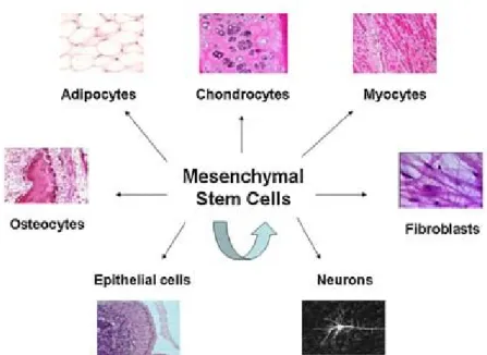
MSCs are characterized by their intrinsic self-renewal capacity, which is reflected in its clonogenic property and multi-lineage differentiation potential (Fig.1.2). Although not immortal, they have the ability to expand numerous times in culture while retaining their growth and pluripotent potential. In addition to their capacity to differentiate into
chondrocytes, osteoblasts, and adipocytes, MSCs may serve as hematopoiesis-supporting stromal cells (Pittenger et. al. 1999). MSCs can differentiate into myocytes and
cardiomyocytes and even into cells of non-mesodermal origin, including hepatocytes, insulin-producing cells, and neurons (Jiang et. al. 2002).
Figure 1. 2 Pluripotent capacity of MSC to differentiate into mesodermal and non-mesodermal cell lineages, including osteocytes, adipocytes, chondrocytes, myocytes, fibroblasts, epithelial cells, and neurons In addition, MSCs are capable to self-renewal. (Chen F et.al.2008)
The specific lineage commitment of MSCs is largely influenced by culture condition, especially growth factors. Growth factors that have regulatory effects on MSCs include members of the transforming growth factor-beta (TGF-b) superfamily, the insulin-like growth factors (IGF) , the fibroblast growth factors (FGF), the epidermal growth factor (EGF), the platelet-derived growth factor (PDGF), the vascular endothelial growth factor (VEGF), and the family of growth factors known as Wnt (Liu et al. 2009).
MSCs actively inhibit T-cell proliferation and due to this reason they are considered as nonimmunogenic or hypoimmunogenic which is important in host response to allogeneic MSC therapy (Uccelli et al. 2008). There are few mechanisms in immunospuression by MSCs. Contact-dependent mechanisms and soluble factors are thought to collaborate for the induction of MSC-mediated immunosupression. The interaction between MSCs and their target cells involves cell-cell contact mediated by adhesion molecules, such as PD1 and its ligands. Several soluble immunosuppressive factors have reported which either produced by MSCs or released following cross-talk with target cells, such as nitric oxide and indoleamine 2,3- dioxygenase (IDO).
Another important feature of MSCs is long-term homing and engraftment of implanted cells to various tissues even after the development of immunocompetence. The implanted MSCs can migrate to the specific site of injury by the help of factors that are released from the wound. This has been observed in cases like bone fractures, myocardial infarction, meniscus, and ischemic cerebral injury. Also at specific sites it has been observed
that the stem cells injected differentiate according to the local signals at the local site of injury (Miguell et al. 2001). MSCs have a high potential for regenerative medicine and tissue
engineering besides being used as a gene delivery method, which makes their therapeutic value very high. Researchers are looking for different ways to use specialized cells derived from MSCs in targeting specific cancerous cells and delivering treatments that will destroy them or make them benign.
Besides these advantages of MSC in therapeutic applications, there are some
unknowns like long-term effect and safety which require more toxicology studies. In addition, the efficiency is not clear. There are some evidences on homing and differentiation, but they are insufficient. Also large-scale culture, storage and distribution are important for their applications of MSCs (Miguell et al. 2001).
1.1.6 Stem Cell Niches
Stem cells unique ability to replenish themselves during the development, maintenance of tissue homeostasis, and repair of many tissues through self-renewal and differentiation are regulated by both intrinsic programming and input from their local
environment, often referred to as the stem cell niche or microenvironment (Watt et. al.2000).
Germ stem cells in the Drosophila ovariole and testis require physical interaction with supporting cap or hub cells, respectively, to retain stem cell identity. In the C. elegans gonad, the niche consists primarily of a single distal tip cell whose long cytoplasmic processes make extensive physical contact with germ stem cells (Keith et. al. 2007).
In mammals, spatially defined stem cell niches have also been identified in multiple tissues, including the gonad, skin, intestine, and brain. A number of molecular factors that control stem cell identity have identified. These factors typically supplied by the supporting cells of the niche include components of the BMP, Notch, Wnt, JAK-STAT, and Sonic hedgehog (Shh) signaling pathways, which provide intercellular cues that regulate stem cell identity and differentiation (Keith et. al. 2007).
In addition, niche structure provides mechanical stimuli and low oxygen tension. These features are shared by many types of adult stem cells. Indeed, increasing evidence demonstrates that oxygen tension is not only a metabolic substrate but also a powerful signaling molecule that regulates stem cell proliferation and differentiation (Ma et. al. 2009). Further information about effects of hypoxia on stem cell is discussed later.
1.2 Hypoxia
Oxygen is one of the essential elements that influence the evolution of life. Primitive life is considered to have started less than 4 billion years ago, and the first organisms that had the ability to convert and store the energy of light in the form of biochemical molecules with higher energetic potential appeared less than 3 billion years ago. The concentration of the oxygen started to increase in the atmosphere through the photosynthesis done by these
organisms, during the next billion years. About 2 billion years ago, the concentration of O2in
atmosphere reached a level that influence life. Several adaptation mechanisms have been evolved by living organisms in order to detoxify the toxic effects of oxygen in parallel with the increase of O2 concentrations in atmosphere which continued until 500 million years ago
(Ivanovic 2009).
Therefore, the eukaryotes, whose physiology based on the aerobic metabolism, are built during the evolution and became dependent on oxygen. Since inadequate oxygen availability can lead to cellular dysfunction and, since it causes cell death if its level is sufficiently profound in aerobic organisms, sophisticated systems are required to adjust the oxygen level, such as the cardiovascular system and the respiratory system (Kaelin and Ratcliffe 2008).
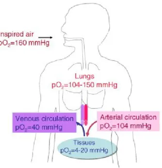
The blood circulation carries oxygen to tissues and a physiological tissue distribution of oxygen occurs as a result of progressive consumption of the oxygen in the blood circulation as it passes through different organs. The oxygen partial pressure (pO2) of inspired air is
around 160 mmHg at sea level (Fig. 1.3). This drops progressively first in the lungs; in part due to water vapor and diffusion, then in the blood flowing from the alveolar capillaries that carry the oxygen, at a pO2of around 104 mmHg, towards organs and tissues for their
oxygenation. A further drop in the pO2is observed in the venous system. The pO2of a given
tissue depends on the type of organ; rat spleen has a measured pO2of around 16 mmHg, while
the thymus was measured at 10 mmHg (Brahimi-Horn and Pouyssegur 2007). This “normal” tissue pO2can be considered as “hypoxic” from a molecular standpoint. The low
vascularization of the rat retina also makes it relatively hypoxic (2–25 mmHg) (Brahimi-Horn and Pouyssegur 2007), while tissues of the rat brain is even more hypoxic, 0.4–8.0 mmHg (Brahimi-Horn and Pouyssegur 2007). The diffusion distance of oxygen in a tissue is
approximately 100–200 μm and an oxygen partial pressure of almost zero has been reported at only 100 μm from blood vessels (Brahimi-Horn and Pouyssegur 2007)
Figure 1. 3 Physiological and pathophysiological oxygen partial pressures (pO2). The pO2of inspired air drops progressively as oxygen passes from the lungs into the circulation, which irrigates the tissues of major organs, and then in the venous system before re-oxygenation in the lungs. (Brahmi et al. (2009)).
In mammals an increase in altitude results in an initial physiological response that increases the rate and depth of breathing. Low levels of oxygen in tissues (hypoxia) also arise in normal development and as a consequence of a number of pathophysiological conditions where there is a diminished oxygen supply due to a defective vasculature. Such conditions include ischemic disorders (cerebral or cardiovascular), diabetes, atherosclerosis,
inflammatory diseases, psoriasis, pre-eclampsia, chronic obstructive pulmonary disease and cancer (Brahimi-Horn and Pouyssegur 2007).
In these responses, at the molecular level, cells have ability to sense the oxygen availability whether it is insufficient, and thus undergo adaptive changes in gene expression that either enhance oxygen delivery or promote survival in a low oxygen (hypoxic)
environment. An evolutionarily conserved pathway mediated by oxygen-dependent
posttranslational hydroxylation of a transcription factor called hypoxia-inducible factor (HIF) plays a pivotal role in this process (Kaelin and Ratcliffe 2008).
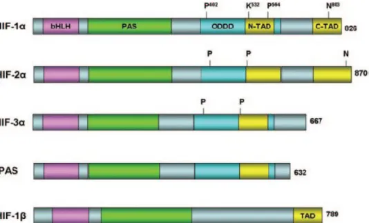
There are three isoforms of hypoxia inducible factors. All three factors bind to a hypoxia response element (HRE; 5’-RCGTG-3’) found in the target genes. (Semenza et. al., 1991). HIF-1 belongs to the basic helix-loop-helix Per-ARNT-Sim (bHLH–PAS) protein family (Fig. 1.4) (Wang et. al. 1995), where it composes, in hypoxic condition, a
heterodimeric complex with HIF-1which is also known as the aryl hydrocarbon nuclear translocator (ARNT).
HIF-2is a closely related protein to HIF1-and is also termedendothelial PAS protein, HIF-like factor (HLF), HIF-related factor (HRF), and member of the PAS
superfamily 2 (MOP2) (Tian et. al. 1997). 48% amino acid sequence identity of HIF-2is similar to HIF-1It also share structural and biochemical similarities with HIF-1, such as heterodimerization with HIF-1 and binding HREs. Although HIF- 1is ubiquitously expressed, expression of HIF-2is tissue specific such as in the lung, endothelium, and carotid body (Ke and Costa 2006).
HIF-3 is the latest discovered isoform between hypoxia inducible factors. This transcription factor is also expressed in a different tissues and can dimerize with HIF-1 and binds to HREs. HIF-3has splice variant, inhibitory PAS (IPAS), which is predominantly expressed in the Purkinje cells of the cerebellum and corneal epithelium. IPAS possesses no endogenous transactivation activity. Instead, it interacts with the amino-terminal region of HIF-1 and prevents its DNA binding, acting as a dominant negative regulator of HIF-1. (Ke and Costa 2006)
The bHLH and PAS motifs are required for heterodimer formation between the HIF-1 and HIF-1 subunits, and the downstream basic regions required for specific binding to the HRE DNA sequence (Crews 1998). HIF-1has two transactivation domains, called N-terminal (N-TAD) and C-N-terminal (C-TAD), in the C-N-terminal half of the HIF-1 protein (Ruas et. al. 2002). The C-TAD interacts with coactivators such as CBP/p300 to activate gene transcription (Lando et. al. 2002). HIF-1_ also contains an oxygen-dependent degradation domain (ODDD) that mediates oxygen-regulated stability (Pugh et. al. 1997).
Figure 1. 4 Domain structures of human HIF- and 1. ( 1, 2, 3 IPAS) and HIF-belong to the bHLH and PAS protein family. HIF- contains an ODDD that mediates oxygen-regulated stability through the hydroxylation of two proline (P) residues and the acetylation of a lysine (K). HIF-1 and HIF-2 also contain two transaction domains (C-TAD and N-TAD), whereas HIF-1 has only one TAD (KE et.al. (2006).
Despite of the fact that HIF-1 beta protein is constitutively expressed and its mRNA and protein level are constant regardless of oxygen availability, HIF-1 alpha protein is highly controlled by oxygen (Salceda and Caro 1997). In normoxia, HIFα becomes hydroxylated at one (or both) of two highly conserved prolyl residues located near the NTAD by prolyl hydroxylase domain (PHD) family (Kaelin 2005). In normoxia, hydroxylation of two proline residues and acetylation of a lysine residue in its ODDD promote interaction of HIF-1 with the von Hippel-Lindau (pVHL) ubiquitin E3 ligase complex, which is a component of a ubiquitin ligase complex (Masson et. al. 2001). As a result, HIFα is polyubiquitylated and subjected to proteasomal degradation by 26S proteasome when oxygen is available.
The PHD proteins belong to the Fe(II) and 2-oxoglutarate-dependent oxygenase superfamily, whose activity is dependent on oxygen. Under low oxygen conditions, HIFα accumulates then dimerizes with an HIFβ family member, translocates to the nucleus, and transcriptionally activates 100–200 genes, including genes involved in erythropoiesis, angiogenesis, autophagy, and energy metabolism (Kaelin and Ratcliffe 2008)
FIH1, like the PHD family members, is an Fe(II)- and 2-oxoglutarate-dependent dioxygenase. When oxygen is available, FIH1 hydroxylates a conserved asparaginyl residue within the HIF1α and HIF2α CTADs, and thus prevents the recruitment of the coactivators p300 and CBP (Kaelin and Ratcliffe 2008).FIH1 remains active at lower oxygen
concentrations than the PHDs and so might suppress the activity of HIFα proteins that escape destruction in moderate hypoxia (Dayan et. al. 2006).
Moreover, HIF-1 is acetylated by an acetyltransferase named arrest-defective-1 (ARD1) at Lysine residue located in the ODDD domain, which favors the interaction of HIF-1 with pVHL, and thus destabilizes HIF-HIF-1 (Jeong et. al. 2002).
Despite the central importance of hydroxylases in sensing oxygen tension and
regulating HIF-1 activity, there are other mechanisms that contribute to the control of HIF-1. Phosphorylation is well known to be crucial in controlling protein activities. Direct
phosphorylation of HIF-1 has been reported, and the mitogen-activated protein kinase (MAPK) pathway seems to play a role (Minet et. al. 2001). Phosphorylation does not affect stability or DNA binding of HIF-1; instead, it increases the transcriptional activity of HIF-1. In addition to the post-translational modification of HIF-1 , SUMOylation of HIF-1
contributes to repressing transactivation (Brahimi-Horn et. al. 2005).
HIF-1 is also regulated in an oxygen-independent manner. Cytokines, growth factors, environmental stimuli, and other signaling molecules have been implicated in controlling HIF-1 under non-hypoxic condition. Although complex and cell-type dependent, some stimulate HIF-1 transactivation or synthesis by activation of the MAPK or the phosphatidylinositol 3-kinase (PI3K) signaling pathways (Ke and Costa 2006).
The PHDs and FIH1, like other 2-oxoglutarate-dependent oxygenases, employ a two-histidine-one-carboxylate facial triad to coordinate the catalytic Fe(II) center, leaving two positions for binding 2-oxoglutarate, and one for molecular oxygen. During catalysis, the splitting of molecular oxygen is coupled to the hydroxylation of HIFα and to the oxidative decarboxylation of 2-oxoglutarate to succinate and CO2. The reaction proceeds via the
formation of a highly reactive ferryl (FeIV= O) intermediate that oxidizes the target amino acid residue. Ascorbate is required for full catalytic activity and likely functions to reduce the catalytic iron in the event of uncoupled turnover, in which failure to oxidize the HIFα
substrate leaves the iron center in an oxidized and inactive form (Schofield and Ratcliffe, 2004). Therefore, The HIF hydroxylases are also inhibited in vivo by iron chelators (Page et. al. 2007).
1.2.1 Hypoxia and Stem Cells
The effect of oxygen tension on stem cell physiology has been studied for over 30 years (Toya et. al.,1976). For hematopoietic stem cells (HSCs), it has been found that
colony initiating cells (LTC-ICs) relative to cultures under ambient (20%, v/v) oxygen concentrations.(Cipolleschi et. al.2000).Recently, it has also been observed, in several other stem and progenitor cell populations, that cultivation under hypoxic conditions resulted in enhanced proliferation and maintenance of their naïve states (Csete et. al.2006).In vivo studies have shown that mesenchymal stem and progenitor cells home specifically to hypoxic niches (Ma et. al. 2009). Although the identity of cellular oxygen sensor is being debated, emerging evidence indicate that some of the effects of hypoxia on stem cell function are directly regulated by hypoxia-inducible factor (HIF) proteins (Keith et. al.2007).
1.2.2 Bone Marrow Microenvironment
The oxygen distribution within the bone marrow has been studied almost exclusively in the context of hematopoietic stem cells (HSCs) and to a much lesser extent in the context of MSCs. However, bone marrow has a hierarchical structure, in which the haematopoietic compartments are bound by stromal elements (Weiss et. al. 1976) - mainly mesenchymal stem cells (MSCs) - and such that the two cell types form an integral part of each other's niche (Figure 1.5). It has long been proposed that hematopoietic progenitors exist at high concentration at the endosteal surface and release via the central venous sinus as they differentiate and mature (Lord et. al. 1990). Early direct measurement revealed that bone marrow in general is hypoxic, where some regions are as low as 1-2% O2(Ma et. al. 2009).
Figure 1. 5. Oxygen tension in bone marrow is well below ambient oxygen environment in the range of 3–7%
1.2.2.1. Effects of Hypoxia on Haematopoietic and Embryonic Stem Cells
Embryonic stem (ES) cells grow more efficiently under low O2conditions, as opposed to in ambient air that is supplemented with 5% carbon dioxide. It was previously noted that bovine blastocysts produced under reduced O2tensions exhibited significantly more inner cell
mass (ICM) cells than those that were maintained at higher O2levels(Simon M. C. and Keith
B. 2008). The ICM and its ES cell counterparts are pluripotent. Roberts et al.demonstrated that human ES cells proliferate at a similar rate when cultured at 3–5% O2as they do under
21% O2. The authors concluded that hypoxic conditions are required to maintain the full
pluripotency of mammalian ES cells (Simon M. C. and Keith B. 2008). HSCs and their proliferating progenitors are naturally occupying the most hypoxic niches. Furthermore, Danet et al. demonstrated that culturing bone marrow HSCs at 1.5% O2promoted their ability
to engraft and repopulate the haematopoietic organ of immunocompromised recipient mice (Simon M. C. and Keith B. 2008)
1.2.2.2. Effects of Hypoxia on Bone Marrow-Derived Mesenchymal Stem Cells
In general, MSCs exhibited greater colony-forming potential, proliferated faster, and longer, and maintained their undifferentiated characteristics better under low oxygen
conditions (Ma et. al. 2009). It has been shown that hMSCs grown in 3D scaffolds under extended hypoxic conditions (2% O2) increased their expression of pluripotent genes Oct-4
and Rex-1 and had elevated CFU-F ability while maintaining their ability to differentiate along osteogenic or adipogenic lineages (Grayson et. al. 2006). In two-dimensional (2D) culture studies, hMSCs under hypoxic conditions exhibited 30-fold greater expansion potential over a 6-week period than normoxic cells, homogenously maintained their spindle morphology, and formed multiple cell-layers with high expression of connexin-43 (Grayson et. al. 2007). Hypoxia affects the differentiation characteristics of MSCs in ways that may be correlated with the physiological oxygen requirements of the differentiated cells (Fink et. al. 2004).
1.2.3 The role of HIFs in regulating stem cells
Some of the effects of hypoxia on stem cell function are directly mediated by the HIF proteins. Targeted mutation of the ARNT subunit eliminates both HIF-1α and HIF-2α
function and results in a decreased number of progenitor cells of all hematopoietic lineages in the embryonic yolk sac of Arnt−/−mouse embryos (Keith et. al. 2007).
Recently, new molecular mechanisms by which HIFs directly modify cellular
differentiation and stem cell function have been defined. Lendahl, Poellinger, and colleagues reported that hypoxia blocked the differentiation of myogenic satellite cells, a myogenic cell line (C2C12), and primary neural stem cells in a Notch-dependent manner (Gustafsson et. al. 2005). It was shown that HIF-1α was physically recruited to a DNA-binding complex
containing the Notch intracellular domain in hypoxic cells. Hypoxic induction of Notch target genes was dependent on the Notch intracellular domain and also required the functional C-terminal transactivation domain of HIF-1α, which interacts directly with p300/CBP.
The links between the HIFs, Notch, and Oct4 reveal specific molecular mechanisms whereby oxygen responses can inhibit differentiation and, possibly, promote stem cell identity. They also raise the possibility of crosstalk between hypoxia and other stem cell signaling pathways. TGF-β has been reported to induce HIF-α stabilization by inhibiting PHD2 (McMahon et. al. 2006). In addition, a recent paper describing physical interaction between β-catenin and HIF-1α suggests at least one mechanism by which Wnt signaling might affect HIF activity in stem cells (Kaidi et. al. 2007).
1.3 Estrogen
Estrogen is a hormone which is produced by the ovaries and testes. It stimulates the development of secondary sexual characteristics, induces menstruation, and regulates growth, differentiation, cell proliferation, metabolic activities, reproduction, homeostasis,
cardiovascular health, bone integrity, cognition, and behavior. It targets many organs like the brain, heart, bone, breast, uterus, and prostate. This wide range of organs, which estrogen is involved in the functioning of, makes estrogen a very important hormone for therapy and therefore it is crucial to understand the imbalances of estrogen, gene networks controlled by estrogen, and regulation of its targets (Deroo and Korach 2006).
Estrogen controls the expression of a wide variety of genes through distinct genomic and nongenomic pathways.In the classical genomic pathway, estrogen signals are mediated through the estrogen receptor (ER), which functions as a transcription factor for target genes. In the nongenomic pathway, estrogen regulates the functions of factors in cells through various mechanisms, including protein phosphorylation (Norman et. al. 2004).
1.3.1 Estrogen Signals
1.3.1.1. Ligand-Dependent Genomic Pathways
Estrogen exerts its biological effects by binding to ER, which mainly exists in the nucleus as a member of the nuclear receptor superfamily of transcription factors (Figure 1.6). ER acts through the formation of homo- or heterodimers of ERα and ERβ. In the classical pathway, estrogen-bound ERs dimerize and function as a transcription factor which binds to a specific DNA sequence named the estrogen response element (ERE) present in the promoter or enhancer regions of target genes. ER binds to ERE through its DNA-binding domain (DBD) and recruits coactivators such as SRC-1, AIB1 and p300/CBP to form a functional ER complex. It is now known that ER target genes which have full or half ERE sites (Smith et. al. 2004).
In the genomic pathway, ER can also regulate transcription without binding directly to DNA. ER acts as a coactivator, and interacts with other transcription factors such as AP-1, SP-1 and NF-κB via protein–protein interactions, and it could regulate the transcription of genes that lack ERE but has a binding element for its partner’s protein (Hayashi et. al. 2009).
1.3.1.2. Nongenomic Pathways
Estrogen also exerts its effects that are not accounted for by transcriptional
mechanisms (Figure 1.6). There is accumulating evidence that estrogen receptors are also located at the plasma membrane and are responsible for nongenomic actions (Hayashi et. al. 2008)Membrane ER associates with many growth factor receptors, such as IGF-1R, EGFR, HER2. In the activation of IGF-1R, E2 induces the formation of a ternary complex among ERα, IGF1-R and Shc, the adaptor protein, in the plasma membrane, which induces phosphorylation of IGF-1R (Song et. al. 2004). The estrogen-bounded membrane ER activates several signals in a cell type-specific manner, including calcium currents, cAMP, inositol phosphate, G proteins, Src, and Shc, which leads to the activation of downstream kinases, such as mitogen-activated protein kianse (MAPK) and phosphatidylinositol 3-kinase (PI3K)/Akt (Norman et. al. 2004).
Figure 1. 6: Outline of estrogen signaling pathways. Estrogen evokes genomic and nongenomic actions via nuclear ER and membrane-associated ER. Moreover, these signals are also stimulated or modulated by crosstalk with the intracellular protein kinase-mediated phosphorylation signaling cascade (Hayashi S. et.al 2008). 1.3.2 Estrogen and Hypoxia
The relationship between hypoxia and estrogen-mediated pathways comes from breast cancer. It has been shown that, in T47 human breast cancer cells, the expression level of 31 genes out of 8000 changes in hypoxic condition increase in the presence of estrogen. These genes are involved in cell growth, differentiation, angiogenesis, protein transport, metabolism, and apoptosis (Yi. et. al. 2009).
So far, there have been two possible mechanistic explanations for the relationship between estrogen and hypoxia. One mechanism is that HIF-1α is a coactivator for ERα itself. Other is that HIF-1α activates intermediary cofactors for ERα that further facilitate ERα transactivation (Fig. 1.7), such cofactors as SRC, CBP, ASC (Seifeddine et. al., 2007). In addition, HIF-1α directly controls ERs activation and degradation, where the level of ERα decreases in hypoxia. The mechanisms of ERa downregulation during hypoxia are probably dependent on both increased proteasomal degradation of ERa and decreased transcriptional ERa-activation. The transcriptional inhibition of ERa during hypoxia seems to be mediated by the ERK pathway (Kronblad et. al. 2005).
However, there is no interplay between ERβ and HIF-1α in contrast to ERα. It was shown that estrogen-occupied ERα activates HIF-1α by the MAPK and PI3K pathways in the uterus (Kazi and Koos 2007). Even if mechanism is not known HIF-1α is a coactivator only for ERα, but not ERβ. HIF-1 α mediated ERα activation involves the C-terminal domain of estrogen in which C-terminal AF-2 is found, and is important for the transcription activation. Some coregulators, such as SRC-1, TIF-2, and AIB-1, interact with the AF-2 of ERα, whereas
other cofactors, such as p68 RNA helicase protein, interact with AF-1 in potentiating ERα function (Yi.et. al. 2009)
Figure 1. 7: A proposed model of the effects of E2and hypoxia on transactivation of ERs. Unknown factor(s) recruited by HIF1α specifically interact(s) with ERα and synergistically activate(s) transactivation of ERα (A). Such synergism is not observed with ERβ (B) (Yi et. al. 2009).
1.3.3 Estrogen and Notch
There are no direct results showing the relationship between Notch pathway and estrogen. The direct link between Notch signaling pathway and estrogen comes from breast cancer. It has been shown that the amount of Notch-1 was increased by 17β-estradiol
treatment in breast cancer cell lines such as T47D or MCF-7 (Rizzo et. al. 2008). Rizzo et. al. (2009) showed that loss of estrogen signaling caused Notch re-activation because Notch-1 is found primarily on cell surface and nuclear levels NICare reduced when cells are treated with estrogen, suggesting that cross-talk between estrogen and Notch signaling leads to Notch re-activation.
1.3.4 Estrogen and Stem Cells
Gender differences, by the differential effects of sex-specific hormones, exist in a variety of cardiovascular, cardiopulmonary, neurodegenerative, endocrine and metabolic bone diseases such as osteoporosis (Figure 1.8). Recent studies reported the presence of estrogen receptor on stem cells, suggesting that estrogen may modify the function of those cells (Ray R. et. al. 2008). 17β-estradiol enhances the proliferation and migrations of some stem cells, such as endothelial progenitor cells (EPCs), to the injured vessels, or ischemic myocardial tissues through the process of homing and help in repair and regeneration to compensate for the lost tissue. Thus, modification of the function of stem cells through estrogenic stimulus may increase the function of stem cells (Ray R et. al. 2008).
Figure 1. 8: Effects of estrogen on various stem cells and progenitor cells. ESC (embryonic stem cell); EPC (endothelial progenitor cell); MSC (mesenchymal stem cell); HSC (hematopoietic stem cell); CF (cardiac fibroblast); BMP (bone matrix protein); RUNX2/ CBFA1 (runt-related transcription factor 2/core-binding factor alpha) (Ray R. et.al.2008)
Estrogens may enhance the protective function of MSCs by increasing or decreasing cytokine and growth factor production (Crisostomo et. al.2008). Female MSCs express less amount of proinflammatory cytokines, TNF-α and IL-6, when compared with male MSCs in inflammatory reactions (Crisostomo et. al. 2006).
Furthermore, estrogen plays a role in osteogenic differentiation of MSCs through the ERα receptor, and support growth and differentiation (Wang et. al. 2006). When bone marrow MSCs are subjected to osteogenic differentiation medium supplemented with 17β-estradiol, the expression levels of bone morphogenetic protein (BMP) and osteocalsin increase together with increase calcium deposition (Hong et. al. 2006). 17β-estradiol also stimulates the
expression of osteogenic genes for ALP, collagen I, and TGF-β1 in MSCs.
1.4 Notch Signaling Pathway
Notch signaling has been shown to regulate a broad range of events during embryonic and post-natal development, including proliferation, apoptosis, border formation, and cell fate decisions. In self-renewing organs in vertebrates, inhibition of differentiation, lineage