Address for correspondence: Dr. Muhammed Ulvi Yalçın, Selçuk Üniversitesi Tıp Fakültesi, Kardiyoloji Anabilim Dalı, Konya-Türkiye
Phone: +90 332 221 00 00 Fax: +90 332 323 67 23 E-mail: [email protected] Accepted Date: 10.06.2019 Available Online Date: 26.07.2019
©Copyright 2019 by Turkish Society of Cardiology - Available online at www.anatoljcardiol.com DOI:10.14744/AnatolJCardiol.2019.88555
Kadri Murat Gürses, Muhammed Ulvi Yalçın
1, Duygu Koçyiğit
2, Muhammed Said Beşler*,
Hande Canpınar**, Banu Evranos*, Hikmet Yorgun*, Mehmet Levent Şahiner*,
Ergün Barış Kaya*, Necla Özer*, Dicle Güç**, Kudret Aytemir*, Lale Tokgözoğlu*
Department of Basic Medical Sciences, Faculty of Medicine, Adnan Menderes University; Aydın-Turkey
1Department of Cardiology, Faculty of Medicine, Selçuk University; Konya-Turkey 2Cardiology Clinics, Afyonkarahisar Dinar State Hospital; Afyonkarahisar-Turkey
Departments of *Cardiology, and **Basic Oncology, Cancer Institute, Faculty of Medicine, Hacettepe University; Ankara-Turkey
The association between serum angiogenin and osteopontin levels
and coronary collateral circulation in patients
with chronic total occlusion
Introduction
Ischemic heart disease (IHD) is one of the leading causes of death worldwide. A well-functioning coronary collateral circu-lation has been shown to have a favorable impact on mortality, myocardial infarction recurrence, and adverse cardiovascular events in patients with chronic IHD (1-3). Angiogenesis and arte-riogenesis are the main mechanisms for coronary collateral de-velopment. Previous studies have shown a relationship between
biomarkers related to angiogenesis and arteriogenesis and coro-nary collateral development (3-7).
Angiogenin (AGN) is a ribonuclease and has been shown to induce blood vessel formation (8). Transplantation of autologous mesenchymal stem cells overexpressing the AGN gene to the heart in a chronic ischemia model was associated with the im-provement of heart perfusion and function (9). Previous studies have also demostrated that AGN is a marker of three-vessel cor-onary artery disease (CAD) (10). In addition, AGN level increases
Objective: A well-developed coronary collateral circulation lowers both in-hospital and long-term morbidity and mortality limiting the infarct. Angiogenin (AGN) and osteopontin (OPN) are known to be potent inducers of angiogenesis. The aim of the present study was to investigate the relationship between serum ANG and OPN levels and collateral filling grade in subjects with stable coronary artery disease (SCAD).
Methods: A total of 122 age- and gender-matched consecutive patients who were found to have total occlusion (n=70) and no significant stenosis in epicardial coronary arteries (n=52) who underwent coronary angiography due to SCAD between January 2015 and July 2017 were included in the study. AGN and OPN levels were measured using enzyme-linked immunosorbent assay. Coronary collateral circulation was graded using Rentrop’s classification of collateral filling.
Results: A total of 52 patients (61.60±11.78 years, 61.5% male) without significant epicardial coronary artery stenosis and 70 patients (62.87±8.24 years, 65.7% male) with totally occluded coronary arteries were included in the study. Subjects with total occlusion had significantly higher levels of AGN [122.00 (79.00–623.00) pg/mL vs. 98.00 (18.00–160.00) pg/mL, p<0.001] and OPN [1863.50 (125.00–6500.00) pg/mL vs. 451.00 (112.00– 1850.00) pg/mL, p<0.001] than those without significant stenosis. In addition, AGN [127.00 (87.00–623.00) pg/mL vs. 110.00 (79.00–188.00) pg/mL, p=0.011] and OPN [2681.00 (126.00–6500.00) pg/mL vs. 649.00 (125.00–4255.00) pg/mL, p=0.001] levels were significantly higher in patients with better developed collaterals. Serum AGN and OPN levels were found to be significantly associated with coronary collateral development. Conclusion: AGN and OPN are associated with better developed coronary collateral circulation and may have therapeutic implications for the promotion of coronary collateral development. (Anatol J Cardiol 2019; 22: 77-84)
Keywords: coronary collateral circulation, osteopontin, angiogenin
A
BSTRACTin acute coronary syndrome (ACS) and has a prognostic value in patients with ACS (11).
Osteopontin (OPN) is a 34 kDa, phosphorylated sialic acid-rich non-collagenous matricellular protein that functions in a va-riety of biological processes, including inflammation, immunity, wound repair, tumorigenesis, cell adhesion, cell migration, bone mineralization, and remodeling (12). It is a unique component of the extracellular matrix that may play an important role in the control of vascular growth. The role of OPN in ischemic limb re-vascularization has been demostrated in a previous study (13). In addition, decreased OPN expression has been related with impaired neovascularization, whereas overexpression of OPN has been found to be increased during angiogenesis (14).
The relationship between serum AGN and OPN levels with coronary collateral circulation in patients with stable CAD (SCAD) has not been evaluated yet in the literature. The aim of the present study was to compare AGN and OPN levels in pa-tients who were found to have either chronic total occlusion (CTO) or no significant epicardial coronary artery stenosis dur-ing coronary angiography due to SCAD. In addition, the present study aimed to investigate the relationship between AGN and OPN levels and collateral filling grade in subjects with CTO.
Methods
This was an observational study. A total of 122 patients who were admitted to our outpatient clinics with stable angina pec-toris and scheduled for elective coronary angiography between January 2015 and July 2017 were enrolled in the study. Of the 122 patients, 70 had total occlusion in at least one major coronary artery, and 52 age- and gender-matched subjects with no signifi-cant epicardial coronary artery stenosis were classified as the control group. Stable angina pectoris was diagnosed according to the American College of Cardiology/American Heart Asso-ciation criteria (15). The decision of coronary angiography was made based on a positive non-invasive stress test or presence of high clinical suspicion for severe coronary artery stenosis. The study was approved by the Institutional Ethics Committee in compliance with the principles outlined in the Declaration of Helsinki. Informed consent was obtained from all participants.
Baseline demographic and clinical characteristics including age, gender, body mass index (BMI), history of smoking, hyperten-sion, diabetes mellitus, and family history of CAD were recorded for all patients. Diabetes mellitus was defined as plasma glucose level ≥126 mg/dL at fasting or ≥200 mg/dL at any measurement or the current use of a glucose-lowering agent. Hypertension was defined as either the current use of anti-hypertensive medica-tion or the documentamedica-tion of a systolic blood pressure ≥140 mm Hg and/or a diastolic blood pressure ≥90 mm Hg. Family history of CAD was defined as the presence of CAD or sudden cardiac death in a first-degree relative before the age of 55 years for men and 65 years for women.
Patients with recent ACS (within the last 6 months), previous revascularization, heart failure with reduced ejection fraction (left ventricular ejection fraction <40%), sign and/or symptoms of decompensated heart failure, symptomatic peripheral vascular disease (transient ischemic attack, stroke, intermittent claudica-tion, peripheral revascularizaclaudica-tion, or amputation), evidence of ongoing infection or inflammation, chronic kidney disease (se-rum creatinine >1.4 mg/dL), chronic obstructive pulmonary dis-ease, previous diagnosis of malignancy, BMI <20 kg/m2 or >30 kg/
m2, and patients with diabetes receiving insulin treatment were
excluded from the study. Coronary angiography
Standard Judkins technique was used for coronary artery vi-sualization. At least two orthogonal plane images were obtained for each coronary artery. The coronary angiograms of the study population were examined again for collateral vessels by two experienced interventional cardiologists from our institute who were totally blinded to the study. CTO was defined as a complete interruption of coronary artery flow.
Coronary collateral grading was determined according to Rentrop’s method (16), with grade 0, no filling of any collateral vessels; grade 1, filling of side branches of the artery to be per-fused by collateral vessels without visualization of epicardial segment; grade 2, partial filling of the epicardial artery via col-lateral vessels; and grade 3, complete filling of the epicardial ar-tery via collateral vessels (17). The CTO group was divided into two groups according to the degree of collateral development. Patients who were graded as 0 or 1 were classified as the poor collateral group, whereas patients graded as 2 or 3 were classi-fied as the better developed collateral group.
Laboratory analysis
Venous peripheral blood samples for complete blood count and biochemistry panel were withdrawn following 12 h of fasting before coronary angiography. Samples were centrifuged at 1600 rpm for 15 min and stored at –80 °C. After the completion of patient recruitment, frozen serum samples were rapidly thawed, brought to room temperature of 24 °C, and assayed for the presence of OPN (Human OPN PicoKine™ ELISA Kit; Boster, CA, USA) and AGN (Human ANG PicoKine™ ELISA Kit; Boster) by using ELISA kits according to the manufacturer’s instructions. Serial dilutions of known concentrations of human OPN and AGN were used to construct a standard curve of the analytes. The intensity of the col-or in each well was measured on a microplate reader (Molecular Devices, SpectraMax Plus, UK). Serum OPN and AGN levels were then estimated by extrapolation from a log:log linear regression curve determined from the serially diluted OPN ranging from 5000 pg/mL to 156 pg/mL and AGN ranging from 5000 pg/mL to 78 pg/mL.
Statistical analysis
Statistical analyses were performed using Statistical Pack-age for Social Sciences software (IBM SPSS Statistics for
Windows, version 20.0; IBM Corp., Armonk, NY, USA). Normally distributed parameters were presented as mean ± standard deviation, and skewed parameters were expressed as median (interquartile range: minimum–maximum). Categorical variables were presented as percentage (%). Kolmogorov–Smirnov test was used to test the normality of distribution. Mann–Whitney U test or Student’s t-test was used to compare continuous vari-ables, where appropriate. Chi-square test was used to compare categorical variables. Binary logistic regression analysis was used to determine the independent associates of better devel-oped coronary collaterals. The validity of the multiple regression model was tested by calculating the Variance Inflation Factors for the variables included in the models. A p value <0.05 was con-sidered statistically significant.
Results
In the patient group, there were 70 patients with coronary total occlusion in at least one major coronary artery. The age of the patient group was 62.87±8.24 years. The patient group was composed of 65.7% male. In the control group, there were 52
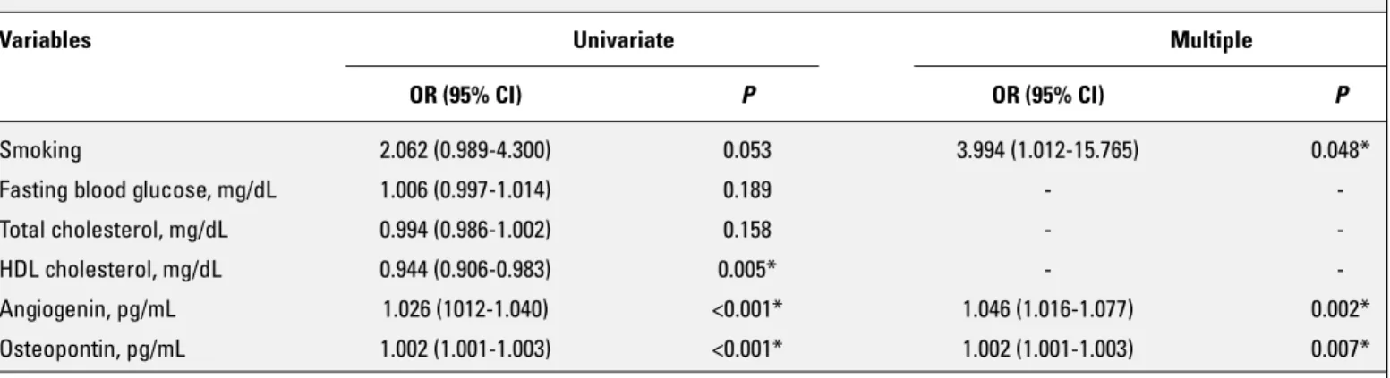
patients with no significant epicardial coronary artery stenosis. The age of the control group was 61.60±11.78 years. The con-trol group was composed of 61.5% male. Baseline demographic, clinical, laboratory, and echocardiographic parameters of the study population are shown in Table 1. Baseline characteristics did not differ between the two groups except high-density lipo-protein cholesterol, AGN, and OPN levels. Subjects with CTO had significantly higher levels of AGN [122.00 (79.00–623.00) vs. 98.00 (18.00–160.00) pg/mL, p<0.001] and OPN [1863.50 (125.00–6500.00) vs. 451.00 (112.00–1850.00) pg/mL, p<0.001] than the control group. Smoking and AGN and OPN levels were found to be in-dependently associated with the presence of CTO in the multiple binary logistic regression analysis (Table 2).
When patients with CTO were grouped according to their Rentrop grade, 23 patients were classified in the poor collateral group (grades 0 and 1), and 47 patients were classified in the bet-ter developed collabet-teral group (grades 2 and 3). Baseline charac-teristics were similar between these two groups except for the duration of ischemic symptoms and the number of affected coro-nary arteries (Table 3). Among laboratory parameters, only AGN and OPN levels differed significantly between the two groups (Table 3). AGN [127.00 (87.00–623.00) vs. 110.00 (79.00–188.00)
Table 1. Baseline demographic, clinical, laboratory, and echocardiographic parameters of the study population
Control group (n=52) Chronic total occlusion group (n=70) P
Clinical parameters
Gender, male, n (%) 32 (61.5) 46 (65.7) 0.635
Age, years 61.60±11.78 62.87±8.24 0.483
BMI, kg/m2 26.07±3.16 26.65±3.53 0.357
Smoking, n (%) 19 (36.5) 38 (54.3) 0.052
Family history of CAD, n (%) 19 (36.5) 28 (40.0) 0.698
Diabetes mellitus, n (%) 13 (25.0) 25 (35.7) 0.206
Hypertension, n (%) 32 (61.5) 47 (67.1) 0.522
Laboratory parameters
WBC count, ×109/L 8.30±1.96 7.92±2.33 0.964
Fasting blood glucose, mg/dL 109.56±51.30 121±43.19 0.184
Total cholesterol, mg/dL 201.96±45.42 190.05±46.05 0.158 LDL cholesterol, mg/dL 125.88±33.58 119.06±38.53 0.309 HDL cholesterol, mg/dL 44.50±12.16 39.16±7.22 0.003 Triglyceride, mg/dL 157.86±70.95 159.17±80.00 0.926 Angiogenin, pg/mL 98.00 (18.00-160.00) 122.00 (79.00-623.00) <0.001* Osteopontin, pg/mL 451.00 (112.00-1850.00) 1863.50 (125.00-6500.00) <0.001* Echocardiographic parameters LV end-diastolic diameter, mm 4.84±0.63 4.94±0.50 0.337 LVEF, % 60.57±4.52 59.25±8.04 0.290 *P<0.0, statistically significant
BMI - body mass index; CAD - coronary artery disease; HDL - high-density lipoprotein; LDL - low-density lipoprotein; LV - left ventricular; LVEF - left ventricular ejection fraction; WBC - white blood cell
pg/mL, p=0.011] and OPN [2681.00 (126.00–6500.00) vs. 649.00 (125.00–4255.00) pg/mL, p=0.001] levels were significantly higher in patients with better developed coronary collaterals than in those with poor collateral circulation. Box plot graphs depicting OPN and AGN levels in the control, poor, and good coronary col-lateral groups, including p values, are shown in Figures 1 and 2. The multiple binary logistic regression analysis revealed that AGN [odds ratio (OR) 1.032; 95% confidence interval (CI) 1.008– 1.057, p=0.010] and OPN (OR 1.001; 95% CI 1.000–1.001, p<0.001) levels were independently associated with better developed coronary collateral circulation in patients with CTO (Table 4).
Discussion
The present study demonstrated an association between serum AGN and OPN levels and the presence of coronary CTO.
In addition, serum AGN and OPN levels were found to be signifi-cantly associated with better coronary collateral circulation. To the best of our knowledge, this is the first study demonstrating the relationship between AGN and OPN levels and the presence of coronary CTO and degree of coronary collateral development. AGN is a potent angiogenic growth factor related to endothe-lial cell proliferation. It was first isolated as a tumor angiogenic factor based on its angiogenic activity; therefore, subsequent studies have focused mainly on its angiogenic capacity (18). It has been shown to interact with all steps of angiogenesis, in-cluding migration, proliferation, and tube formation (19). Previous studies have suggested that AGN also plays a role in regulating rRNA transcription in various conditions, such as cancer, chron-ic heart failure, ACS, wound healing, and asthma (11, 20-23).
Tello-Montoliu et al. (11) have demonstrated that plasma AGN levels are significantly increased in ACS, and that high AGN levels are predictive of adverse events during follow-up. AGN-modified mesenchymal stem cells (MSCs) have been shown to
Table 2. Binary logistic regression analyses to determine the independent associates of chronic total occlusion
Variables Univariate Multiple
OR (95% CI) P OR (95% CI) P
Smoking 2.062 (0.989-4.300) 0.053 3.994 (1.012-15.765) 0.048*
Fasting blood glucose, mg/dL 1.006 (0.997-1.014) 0.189 -
-Total cholesterol, mg/dL 0.994 (0.986-1.002) 0.158 -
-HDL cholesterol, mg/dL 0.944 (0.906-0.983) 0.005* -
-Angiogenin, pg/mL 1.026 (1012-1.040) <0.001* 1.046 (1.016-1.077) 0.002*
Osteopontin, pg/mL 1.002 (1.001-1.003) <0.001* 1.002 (1.001-1.003) 0.007*
*P<0.05, statistically significant
CI - confidence interval; HDL - high-density lipoprotein; OR - odds ratio
Figure 1. Box plot graph depicting osteopontin levels in the control, poor, and good coronary collateral groups
8000.00
6000.00
4000.00
2000.00
Control Group
Serum Osteopontin Level (pg/mL)
Poor Collateral Group Groups
Good Collateral Group P=0.001 P=0.001 P<0.001 *13 0.00 56 63 62 61
Figure 2. Box plot graph depicting angiogenin levels in the control, poor, and good coronary collateral groups
800.00 600.00 400.00 200.00 Control Group Serum Ang iogenin Level (pg/mL)
Poor Collateral Group Groups
Good Collateral Group P=0.143 P=0.011 P<0.001 *79 *101 *111 0.00 57 74 114 108 107
enhance the tolerance of engrafted MSCs to hypoxia injury in vitro and improve their viability in infarcted hearts, thus helping to preserve the left ventricular (LV) contractile function and at-tenuate LV remodeling through vasculogenesis (19). Krecki et al. (10) have reported that AGN is a novel marker of three-vessel CAD, showing a relationship with the angiographic severity of the
disease. In our study, AGN levels were higher in patients with coronary CTO and were independently associated with better developed coronary collateral circulation.
OPN is a transformation-related phosphorylated acidic gly-coprotein (12). It is a key player in essential biological phenom-ena, such as inflammation, autoimmune disease progression,
Table 3. Baseline demographic and clinical parameters of the study population regarding Rentrop’s classification of coronary collateral filling
Poor collateral (n=23) Better developed collateral (n=47) P
Clinical parameters
Gender, male, n (%) 15 (65.2) 31 (66.0) 0.951
Age, years 63.30±7.85 62.66±8.5 0.761
BMI, kg/m2 26.66±2.91 26.65±3.82 0.996
Smoking, n (%) 14 (60.9) 24 (51.1) 0.439
Family history of CAD, n (%) 10 (43.5) 18 (38.3) 0.678
Diabetes mellitus, n (%) 6 (26.1) 19 (40.4) 0.240
Hypertension, n (%) 16 (69.6) 31 (66.0) 0.763
Gensini score 32 (2-32) 32 (4-32) 0.193
Duration of angina pectoris, months 3 (3-72) 25 (3-350) 0.001*
No. of coronary arteries with severe stenosis, n (%)
1 13 (56.5) 40 (85.1) 0.022*
2 9 (39.1) 7 (14.9)
3 1 (4.3) 0 (0.0)
Medications
Renin–angiotensin system blockers, n (%) 14 (60.9) 28 (59.6) 0.917
Beta blockers, n (%) 16 (69.6) 28 (59.6) 0.416
Calcium channel blockers, n (%) 2 (8.7) 7 (14.9) 0.467
Statins, n (%) 16 (69.6) 26 (55.3) 0.253
ASA, n (%) 19 (82.6) 34 (72.3) 0.347
Glucose-lowering drugs, n (%) 3 (13) 13 (27.7) 0.171
Laboratory parameters
WBC count, ×109/L 8.21±2.69 7.78±2.15 0.473
Fasting blood glucose, mg/dL 113.13±43.30 124.85±43.08 0.290
Total cholesterol, mg/dL 185.35±48.28 192.35±45.27 0.554 LDL cholesterol, mg/dL 114.65±39.24 121.21±38.41 0.507 HDL cholesterol, mg/dL 38.48±8.97 39.49±6.28 0.586 Triglyceride, mg/dL 161.09±105.89 158.23±65.08 0.890 Angiogenin, pg/mL 110.00 (79.00-188.00) 127.00 (87.00-623.00) 0.011* Osteopontin, pg/mL 649.00 (125.00-4255.00) 2681 (126.00-6500.00) 0.001* Echocardiographic parameters LV end-diastolic diameter, mm 5.0±0.64 4.9±0.42 0.458 LVEF, % 59.87±4.83 58.95±9.25 0.656 *P<0.05, statistically significant
ASA - acetylsalicylic acid; BMI - body mass index; CAD - coronary artery disease; HDL - high-density lipoprotein; LDL - low-density lipoprotein; LV - left ventricular; LVEF - left ventricular ejection fraction; WBC - white blood cell
bone remodeling, angiogenesis, aortic stenosis, and tumor cell metastasis (24-28). Although first isolated from mineralized bone matrix, OPN can also be synthesized by several other cells, such as cardiomyocytes, vascular endothelial cells, and fibroblasts. OPN has been detected in human atherosclerotic plaques in the aorta, carotid, and coronary arteries, and it has been shown to be implicated in the development and progression of atheroscle-rosis (29). Increased levels of OPN have been found to be related to the presence and extent of CAD and to restenosis following percutaneous coronary revascularization (29, 30). Leaw et al. (31) have shown that rat carotid neointimal lesions induced by bal-loon catheter denudation can be reduced by in vivo neutraliza-tion of OPN.
Some studies have implicated the role of OPN in postnatal neovascularization, such as increased mRNA expression at the sites of ischemia-induced retinal neovascularization in mice and impaired neovascularization in blunted OPN expression in a mu-rine model of hind limb ischemia (14, 32). In addition, OPN was shown to be associated with tumor-related angiogenesis, me-tastasis, and healing after bone fractures (33). In our study, OPN levels were higher in the coronary CTO group and were indepen-dently associated with better developed coronary collaterals.
A significant number of patients with coronary CTO are ei-ther ineligible or demonstrate suboptimal responses to surgical and percutaneous revascularization approaches. A non-invasive approach aimed at promoting the growth of coronary collateral blood vessels has been proposed as a new treatment strategy in such patients (34). Therefore, identifying new molecules that promote the growth of coronary collateral blood vessels is very important. OPN and AGN were widely studied for their angio-genic potential in different disease conditions (23, 26, 32, 33). In the current study, AGN and OPN levels were revealed to be independently associated with the coronary collateral develop-ment in patients with coronary CTO. In addition, OPN itself and molecules affecting OPN expression were shown to improve the angiogenic properties of stem cells in recent studies (35, 36). All these findings suggest that these molecules can constitute
fu-ture therapeutic targets for medical revascularization, and their exact role in coronary collateral development should be clarified in further studies.
Our study has several limitations. First, this is a single-center study, and the small number of the study population has limit-ed some statistical analyses to be performlimit-ed. Second, this is a hypothesis-generating clinical study, and a mechanistic link be-tween levels of AGN and OPN and the presence of coronary total occlusion and degree of collateral circulation development can-not be proposed with these data.
Conclusion
In conclusion, the present study highlights the association between AGN and OPN levels and coronary collateral circula-tion in patients with coronary total occlusion. However, the un-derlying mechanisms remain largely unknown. The results of our study merit further studies to reveal if AGN and OPN are biomark-ers or moderators in coronary collateral development.
Acknowledgments: This study was presented at the EAS 2018 Con-gress as a poster presentation.
Funding: This research was supported by Hacettepe University Sci-entific Research Projects Coordination Unit (project no.: 845).
Conflict of interest: None declared. Peer-review: Externally peer-reviewed.
Authorship contributions: Concept – K.M.G., M.U.Y., D.K., B.E., H.Y., M.L.Ş., E.B.K., N.Ö., D.G., K.A., L.T.; Design – K.M.G., M.U.Y., D.K., H.C., N.Ö., D.G., K.A., L.T.; Supervision – H.C., B.E., H.Y., M.L.Ş., E.B.K., N.Ö., D.G., K.A., L.T.; Fundings – E.B.K., N.Ö., D.G., K.A., L.T.; Materials – K.M.G., M.U.Y., D.K., M.S.B., H.C., B.E., H.Y., M.L.Ş., E.B.K.; Data collection &/or process-ing – K.M.G., M.U.Y., D.K., M.S.B., H.C., B.E., H.Y., M.L.Ş., E.B.K., N.Ö., D.G., K.A., L.T.; Analysis &/or interpretation – K.M.G., M.U.Y., D.K., M.S.B., H.C.,
Table 4. Binary logistic regression analyses to determine the independent associates of better developed coronary collateral circulation
Variables Univariate Multiple
OR (95% CI) P OR (95% CI) P
Gensini score 1.054 (0.969-1.147) 0.222 -
-Duration of angina pectoris, months 1.018 (1.00-1.036) 0.051 1.023 (1.00-1.047) 0.052
No. of coronary arteries with severe stenosis, n 0.232 (0.077-0.700) 0.009* -
-Glucose-lowering drugs, n 0.392 (0.100-1.546) 0.181 -
-Angiogenin, pg/mL 1.018 (1.00-1.037) 0.046* 1.032 (1.008-1.057) 0.010*
Osteopontin, pg/mL 1.001 (1.00-1.001) 0.002* 1.001 (1.000-1.001) <0.001*
*P<0.05, statistically significant CI - confidence interval; OR - odds ratio
B.E., H.Y., M.L.Ş., E.B.K., N.Ö., D.G., K.A., L.T.; Literature search – K.M.G., M.U.Y., D.K., N.Ö., D.G., K.A., L.T.; Writing – K.M.G., M.U.Y., D.K., M.S.B., H.C., B.E., H.Y., M.L.Ş., E.B.K., N.Ö., D.G., K.A., L.T.; Critical review – B.E., H.Y., M.L.Ş., E.B.K., N.Ö., D.G., K.A., L.T.
References
1. Seiler C, Engler R, Berner L, Stoller M, Meier P, Steck H, et al. Prog-nostic relevance of coronary collateral function: confounded or causal relationship? Heart 2013; 99: 1408-14. [CrossRef]
2. Meier P, Hemingway H, Lansky AJ, Knapp G, Pitt B, Seiler C. The impact of the coronary collateral circulation on mortality: a meta-analysis. Eur Heart J 2012; 33: 614-21. [CrossRef]
3. Seiler C, Stoller M, Pitt B, Meier P. The human coronary collateral circulation: development and clinical importance. Eur Heart J. 2013; 34: 2674-82. [CrossRef]
4. Helisch A, Schaper W. Angiogenesis and arteriogenesis--not yet for prescription. Z Kardiol 2000; 89: 239-44. [CrossRef]
5. Meier P, Gloekler S, de Marchi SF, Indermuehle A, Rutz T, Traupe T, et al. Myocardial salvage through coronary collateral growth by granu-locyte colony-stimulating factor in chronic coronary artery disease: a controlled randomized trial. Circulation 2009; 120: 1355-63. [CrossRef]
6. Zorkun C, Akkaya E, Zorlu A, Tandogan I. Determinants of coronary collateral circulation in patients with coronary artery disease. An-adolu Kardiyol Derg 2013; 13: 146-51. [CrossRef]
7. Oğuz D, Atmaca Y, Ozdöl C, Ozdemir AO, Kaya CT, Erol C. The rela-tionship between coronary collateral artery development and in-flammatory markers. Anadolu Kardiyol Derg 2014; 14: 336-41. 8. Shestenko OP, Nikonov SD, Mertvetsov NP. Angiogenin and its role
in angiogenesis. Mol Biol (Mosk) 2001; 35: 349-71. [CrossRef]
9. Huang SD, Lu FL, Xu XY, Liu XH, Zhao XX, Zhao BZ, et al. Trans-plantation of angiogenin-overexpressing mesenchymal stem cells synergistically augments cardiac function in a porcine model of chronic ischemia. J Thorac Cardiovasc Surg 2006; 132: 1329-38. 10. Kręcki R, Krzemińska-Pakuła M, Drożdż J, Szcześniak P, Peruga JZ,
Lipiec P, et al. Relationship of serum angiogenin, adiponectin and resistin levels with biochemical risk factors and the angiographic severity of three-vessel coronary disease. Cardiol J 2010;17: 599-606.
11. Tello-Montoliu A, Marin F, Patel J, Roldan V, Mainar L, Vicente V, et al. Plasma angiogenin levels in acute coronary syndromes: implica-tions for prognosis. Eur Heart J 2007; 28: 3006-11. [CrossRef]
12. Icer MA, Gezmen-Karadag M. The multiple functions and mecha-nisms of osteopontin. Clin Biochem 2018; 59: 17-24. [CrossRef]
13. Duvall CL, Weiss D, Robinson ST, Alameddine FM, Guldberg RE, Tay-lor WR. The role of osteopontin in recovery from hind limb ischemia. Arterioscler Thromb Vasc Biol 2008; 28: 290-5. [CrossRef]
14. Lyle AN, Joseph G, Fan AE, Weiss D, Landazuri N, Taylor WR. Reac-tive oxygen species regulate osteopontin expression in a murine model of postischemic neovascularization. Arterioscler Thromb Vasc Biol 2012; 32: 1383-91. [CrossRef]
15. Fihn SD, Blankenship JC, Alexander KP, Bittl JA, Byrne JG, Fletcher BJ, et al.; American College of Cardiology/Americal Heart Associa-tion Task Force on Practice Guidelines; American AssociaAssocia-tion for Thoracic Surgery; Preventive Cardiovascular Nurses Association; Society for Cardiovascular Angiography and Interventions; Society of Thoracic Surgeons. 2014 ACC/AHA/AATS/PCNA/SCAI/STS fo-cused update of the guideline for the diagnosis and management of patients with stable ischemic heart disease: a report of the
Ameri-can College of Cardiology/AmeriAmeri-can Heart Association Task Force on Practice Guidelines, and the American Association for Thoracic Surgery, Preventive Cardiovascular Nurses Association, Society for Cardiovascular Angiography and Interventions, and Society of Tho-racic Surgeons. J Thorac Cardiovasc Surg 2015; 149: e5-23. 16. Rentrop KP, Cohen M, Blanke H, Phillips RA. Changes in collateral
channel filling immediately after controlled coronary artery occlu-sion by an angioplasty balloon in human subjects. J Am Coll Cardiol 1985; 5: 587-92. [CrossRef]
17. Cohen M, Rentrop KP. Limitation of myocardial ischemia by col-lateral circulation during sudden controlled coronary artery oc-clusion in human subjects: a prospective study. Circulation 1986; 74: 469-76. [CrossRef]
18. Fett JW, Strydom DJ, Lobb RR, Alderman EM, Bethune JL, Riordan JF, et al. Isolation and characterization of angiogenin, an angio-genic protein from human carcinoma cells. Biochemistry 1985; 24: 5480-6. [CrossRef]
19. Liu XH, Bai CG, Xu ZY, Huang SD, Yuan Y, Gong DJ, et al. Therapeutic potential of angiogenin modified mesenchymal stem cells: angio-genin improves mesenchymal stem cells survival under hypoxia and enhances vasculogenesis in myocardial infarction. Microvasc Res 2008; 76: 23-30. [CrossRef]
20. Tsuji T, Sun Y, Kishimoto K, Olson KA, Liu S, Hirukawa S, et al. An-giogenin is translocated to the nucleus of HeLa cells and is involved in ribosomal RNA transcription and cell proliferation. Cancer Res 2005; 65: 1352-60. [CrossRef]
21. Patel JV, Sosin M, Gunarathne A, Hussain I, Davis RC, Hughes EA, et al. Elevated angiogenin levels in chronic heart failure. Ann Med 2008; 40: 474-9. [CrossRef]
22. Steed DL, Trumpower C, Duffy D, Smith C, Marshall V, Rupp R, et al. Amnion-derived cellular cytokine solution: a physiological combi-nation of cytokines for wound healing. Eplasty 2008; 8: e18. 23. Hoshino M, Takahashi M, Aoike N. Expression of vascular
endo-thelial growth factor, basic fibroblast growth factor, and angiogenin immunoreactivity in asthmatic airways and its relationship to angio-genesis. J Allergy Clin Immunol 2001; 107: 295-301. [CrossRef]
24. Senger DR, Wirth DF, Hynes RO. Transformed mammalian cells se-crete specific proteins and phosphoproteins. Cell 1979; 16: 885-93. 25. Gimba ER, Tilli TM. Human osteopontin splicing isoforms: known
roles, potential clinical applications and activated signaling path-ways. Cancer Lett 2013; 331: 11-7. [CrossRef]
26. Blasberg JD, Goparaju CM, Pass HI, Donington JS. Lung cancer osteopontin isoforms exhibit angiogenic functional heterogeneity. J Thorac Cardiovasc Surg 2010; 139: 1587-93. [CrossRef]
27. Grau JB, Poggio P, Sainger R, Vernick WJ, Seefried WF, Branchetti E, et al. Analysis of osteopontin levels for the identification of asymp-tomatic patients with calcific aortic valve disease. Ann Thorac Surg 2012; 93: 79-86. [CrossRef]
28. Scatena M, Liaw L, Giachelli CM. Osteopontin: a multifunctional molecule regulating chronic inflammation and vascular disease. Arterioscler Thromb Vasc Biol 2007; 27: 2302-9. [CrossRef]
29. O'Brien ER, Garvin MR, Stewart DK, Hinohara T, Simpson JB, Schwartz SM, et al. Osteopontin is synthesized by macrophage, smooth muscle, and endothelial cells in primary and restenotic hu-man coronary atherosclerotic plaques. Arterioscler Thromb 1994; 14: 1648-56. [CrossRef]
30. Ohmori R, Momiyama Y, Taniguchi H, Takahashi R, Kusuhara M, Na-kamura H, et al. Plasma osteopontin levels are associated with the presence and extent of coronary artery disease. Atherosclerosis 2003; 170: 333-7. [CrossRef]
31. Liaw L, Lombardi DM, Almeida MM, Schwartz SM, deBlois D, Giachelli CM. Neutralizing antibodies directed against osteopontin inhibit rat carotid neointimal thickening after endothelial denuda-tion. Arterioscler Thromb Vasc Biol 1997; 17: 188-93. [CrossRef]
32. Takagi H, Suzuma K, Otani A, Oh H, Koyama S, Ohashi H, et al. Role of vitronectin receptor-type integrins and osteopontin in ischemia-induced retinal neovascularization. Jpn J Ophthalmol 2002; 46: 270-8. 33. Duvall CL, Taylor WR, Weiss D, Wojtowicz AM, Guldberg RE.
Im-paired angiogenesis, early callus formation, and late stage remodel-ing in fracture healremodel-ing of osteopontin-deficient mice. J Bone Miner Res 2007; 22: 286-97. [CrossRef]
34. Lavine KJ, Ornitz DM. Rebuilding the coronary vasculature: hedge-hog as a new candidate for pharmacologic revascularization. Trends Cardiovasc Med 2007; 17: 77-83. [CrossRef]
35. Carvalho MS, Cabral JM, da Silva CL, Vashishth D. Synergistic effect of extracellularly supplemented osteopontin and osteocalcin on stem cell proliferation, osteogenic differentiation, and angiogenic properties. J Cell Biochem 2019; 120: 6555-69. [CrossRef]
36. Tang Z, Xie H, Jiang S, Cao S, Pu Y, Zhou B, et al. Safflower yel-low promotes angiogenesis through p-VHL/ HIF-1α/VEGF signaling pathway in the process of osteogenic differentiation. Biomed Phar-macother 2018; 107: 1736-43. [CrossRef]