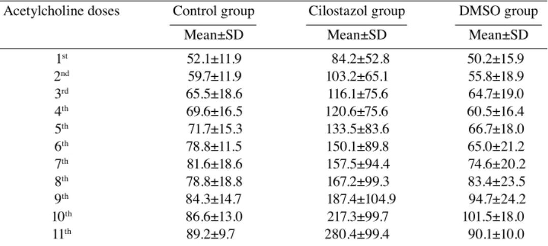
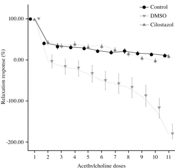
Cilostazol enhances endothelium-dependent vasodilatationof intact endothelium in isolated rat aortic rings
Tam metin
Şekil
Benzer Belgeler
Beliefs about being a donor includedreasons for being a donor (performing a good deed, being healed, not committing a sin), barriers to being a donor (beingcriticized by others,
It was observed at the 60 th day that microvilli-like structures formed on the apical surface of endothelium cells of the rats exposed to cigarette smoke.. At this stage, cells
Our data imply that -NF, at lower concentrations, induces endothelium-dependent vasorelaxation by promoting extracellular Ca2+ influx in endothelium and the activation of the
It was retrospectively evaluated whether there was a difference in the severity and course of stroke in acute ischemic stroke patients diagnosed with type-2 DM and taking
Additionally, partially lower clamping pressures lead to increased vascular endothelial growth factor expression with cellular damage, which can cause late stenosis
In this review, specific documentation on the various ambiences of the physiological environment, i.e., hypobaria, chronic ionizing radiation, and hypergravity pull, would be
The effects of two different antioxidants, sodium selenate and omega-3E treatments of diabetic rats on endothelium-mediated responses of aortas were evaluated by measuring
In this study, our aim was to evaluate anterior segment biometry using OLCR and corneal endothelial changes with confocal microscopy in PEX syndrome/glaucoma eyes before
