http://journals.tubitak.gov.tr/biology/ © TÜBİTAK
doi:10.3906/biy-1401-28
Phylogenetic analysis of peste des petits ruminants virus from outbreaks in
Turkey during 2008–2012
Leyla GÜLER1,*, Murat ŞEVİK2, Mustafa HASÖKSÜZ3
1Department of Microbiology, Faculty of Veterinary Medicine, Mehmet Akif Ersoy University, Burdur, Turkey 2Konya Veterinary Control Institute, Konya, Turkey
3Department of Virology, Faculty of Veterinary Medicine, İstanbul University, İstanbul, Turkey
1. Introduction
Peste des petits ruminants (PPR) is a contagious disease of small ruminants caused by a single-stranded negative-sense RNA virus classified within the genus Morbillivirus in the family Paramyxoviridae. PPR occurs in most African countries, except Southern Africa, and in Central and Southeast Asia, the Middle East, the Near East, and the Arabian peninsula. It is one of the main transboundary animal diseases that affect small ruminant production in many developing countries (Banyard, 2010; OIE, 2013). The PPR virus is closely related to rinderpest virus (Bailey et al., 2005). After global eradication of the cattle disease rinderpest, announced by the OIE and FAO in June 2011, international bodies have started to consider PPR as the next target for eradication (Baron et al., 2011; Albina et al., 2013).
PPR was first detected serologically in 1992 in Southeast Anatolia and has rapidly spread in all regions of Turkey to become an endemic disease. It has been a compulsorily notifiable disease since 1997. The initial occurrence of PPR in Turkey also coincided with eradication of rinderpest from the country. The last reported cases of rinderpest were in 1991, and Turkey was declared rinderpest free by
the OIE in 2003. In the following years serological virus detection and immunohistochemical studies carried out in different regions of Turkey indicated the presence of PPR virus infection in all regions of Turkey (Alçığır et al., 1996; Tatar and Alkan, 1999; Özkul et al., 2002; Yener et al., 2004; Yeşilbağ et al., 2005; Albayrak and Alkan, 2009; Sağlam and Temur, 2009). After the first detection of PPR in Turkey, rinderpest vaccine was used for control of the disease. A live, attenuated PPR vaccine from the Nigeria 75/1 strain produced in Turkey has been used for control of the disease since 2002. A mass vaccination program was implemented throughout the country by an EU-supported project during 2010–2012. Because PPR is endemic and extensive vaccination campaigns have been implemented in Turkey, it has not always been possible to observe typical clinical signs of the disease or high mortality rates in recent years. In addition, atypical clinical and pathological findings such as abortions/stillbirths (Toplu, 2004; Kul et al., 2007, 2008) and brain localization have been reported (Toplu et al., 2012). Mixed infections with pestiviruses (Kul et al., 2008; Toplu et al., 2012), sheep and goat pox virus, bluetongue virus (Ozmen et al., 2009), and secondary bacterial agents of pneumonia, probably as a
Abstract: Peste des petits ruminants (PPR) is an important viral disease of sheep and goats and is endemic in all regions of Turkey. In this study, PPR virus infection was investigated by RT-PCR assay based on the fusion (F) gene in PPR-suspected sheep and goat samples. PPR virus RNA was detected in 65 small ruminants (51 sheep, 14 goats) from independent outbreaks during 2008–2012 in provinces in the central and Mediterranean regions and the central-west part of the Aegean region in Turkey. The virus was detected in an aborted sheep fetus sample by RT-PCR, and diagnosis was also confirmed by virus isolation. Vaccine strain Nigeria 75/1 was differentiated from field isolates by restriction fragment length polymorphism analysis of RT-PCR products using EcoRI. Phylogenetic analysis of 16 viruses indicated that all viruses, including the one from the aborted sheep fetus, belonged to lineage IV, as had the PPR viruses previously isolated in Turkey. Nucleotide sequence identity among 16 viruses was 99.1%–100%. Results showed that PPR virus lineage IV has been in circulation in Turkey since the first detection of the disease.
Key words: Peste des petits ruminants virus, RT-PCR, sheep, goat, phylogenetic analysis, abortion, Turkey
Received: 08.01.2014 Accepted: 25.06.2014 Published Online: 05.09.2014 Printed: 30.09.2014 Research Article
result of the immunosuppressive effects of the virus (Diallo et al., 2007), have been frequently observed. Therefore, laboratory diagnosis is essential for confirmation of the disease. Differentiation of the vaccine strain is also a necessity. Molecular diagnostic techniques such as reverse-transcription polymerase chain reaction (RT-PCR) based on the fusion (F) gene (Forsyth and Barrett, 1995) and the nucleoprotein (N) gene (Couacy-Hymann et al., 2002), and real-time RT-PCR targeting the N gene (Kwiatek et al., 2010; Batten et al., 2011), have provided reliable, easy, and fast diagnosis of the disease. Identification of virus lineage is also essential for understanding epidemiology and for control of the disease. Phylogenetically, the PPR virus is classified into 4 lineages based on partial sequencing of F (Özkul et al., 2002) and N genes (Kwiatek et al., 2007; Munir et al., 2012a, 2012b). The lineages are generally correlated with geographic distribution of the virus (Shaila et al., 1996). PPR viruses belonging to lineages I and II have been isolated exclusively from west and central African countries including the Ivory Coast, where the PPR virus was first detected in the 1940s (Chard et al., 2008; Luka et al., 2011; Munir et al., 2012b). Lineage III has been isolated from eastern Africa and the Arabian peninsula (Shaila et al., 1996; Kwiatek et al., 2011); lineage IV has been isolated in Asia (Kwiatek et al., 2007; Kerur et al., 2008; Wang et al., 2009; Balamurugan et al., 2010; Munir et al., 2012a; Anees et al., 2013), in the Middle East (Esmaelizad et al., 2011), and more recently in northern Africa (Sanz-Alvarez et al., 2008; Kwiatek et al., 2011; De Nardi et al., 2012).
Due to its unique geographic situation as a gateway between Asia and Europe, continuous monitoring of PPR viruses circulating in Turkey is crucial for initiation of proper disease control measures in Turkey and risk monitoring and disease preparedness in Europe. In this
study, sequencing and phylogenetic analysis of the PPR virus based on the F gene were carried out in order to investigate the genetic relationship of viruses isolated from different regions of Turkey with those from other countries.
2. Materials and methods 2.1. Samples
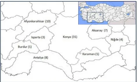
Samples from 190 animals (147 sheep, 43 goats) were tested for PPR virus by RT-PCR. The samples were submitted to the Konya Veterinary Control Institute from several provinces (Konya, Aksaray, Antalya, Afyonkarahisar, Burdur, Isparta, Karaman, and Niğde) located in the central and Mediterranean regions and the central-west part of the Aegean region of Turkey (Figure 1) during 2008–2012. Each animal was from an epidemiologically independent flock and had no vaccination history. The animal breeds were Akkaraman sheep and Turkish hair goat. Animals were brought to the laboratory generally with signs of nasal discharge and/or mouth lesions and sometimes with diarrhea, death, or abortion in the flock. Macroscopically, pneumonia was present in the lungs of most of the animals. Tissue samples included spleen, lymph node, lung, liver, and internal organ specimens of aborted fetuses belong to 22 sheep and 4 goats.
2.2. Detection of PPR virus by RT-PCR
Viral RNA was extracted from tissue samples by RNeasy Mini Kit (QIAGEN) according to the manufacturer’s instructions. One-step RT-PCR was performed with primers PPRVF1b (AGTACAAGAGATTGCTGATCACAGT) and PPRVF2d (GGGTCTCGAAGGCTAGGCCCGAATA) chosen from the F gene sequence of the Turkey 2000 PPR virus genome (GenBank accession no.: AJ849636) at
Figure 1. Map of provinces where samples were collected for PPR virus investigation in the study. Numbers in parentheses indicate the numbers of outbreaks.
locations 5737-5761 and 6184-6160, respectively (Özkul et al., 2002). The RT-PCR reaction was performed with a 1-step RT-PCR kit (QIAGEN) in a final volume of 20 µL, which contained 4 µL of 5X 1-step RT-PCR buffer, 0.4 µM of each primer, 0.8 µL of 10 mM of each dNTP, 0.8 µL enzyme mix, 4 µL of 5X Q solution, 7 µL of PCR-grade water, and 2.6 µL of sample RNA. Amplification was carried out in a MJ Research thermal cycler with the following conditions: 50 °C for 30 min (reverse transcription), 95 °C for 15 min (RT inactivation), and 40 cycles of 94 °C for 1 min, 50 °C for 1 min, and 72 °C for 2 min with a final extension of 10 min at 72 °C. RT-PCR products were electrophoresed in 1.5% agarose gel and visualized by ethidium bromide staining. A 448-bp fragment was amplified in positive reactions. Reactions with weak bands were always repeated.
2.3. Virus isolation
Virus isolation was carried out only in tissues from aborted fetuses, 1 with strong positive and 5 with weak bands in RT-PCR assay. Tissue samples were suspended in PBS at 1/10, and the suspension was centrifuged at 3000 × g for 10 min at 4 °C. Supernatants were filtered through a 0.2-μm membrane and then inoculated on Vero cells grown in tissue culture flasks. The cell growth medium was Dulbecco’s modified Eagle medium supplemented with 10% fetal calf serum (FCS) and 1% mixed antibiotic– antimycotic solution. Cell culture medium was refreshed every 2 days using maintenance medium with 2% FCS. Vero cells were examined daily until cytopathic changes suggestive of PPR virus were observed. After 6 days, 2 blind passages were carried out. Cytopathic-effect–positive cell culture was examined for nucleic acid of PPR virus using RT-PCR (OIE, 2013).
2.4. Restriction fragment length polymorphism (RFLP) analysis of RT-PCR products
For differentiation of vaccine strains from field strains, RT-PCR products were subjected to restriction endonuclease digestion using EcoRI (Özkul et al., 2002). The analysis was performed in 25 µL of reaction volume containing 2.5 µL of 10X RE buffer, 1 µL (10 U) of EcoRI enzyme (Fermentas), 14.5 µL of ultrapure water, and 7 µL of RT-PCR product. The reaction mix was incubated for 2 h at 37 °C, then digested products were analyzed by electrophoresis in a 1.5% agarose gel and visualized by ethidium bromide staining.
2.5. Sequence analysis
For determination of lineage of viruses, RNA from 16 positive samples was subjected to the RT-PCR amplification of a 448-bp segment of the F gene. Amplified PCR products were sequenced by Refgen Biotechnology (Ankara, Turkey). The obtained nucleic acid sequences were aligned with reference sequences of PPR viruses present in the NCBI GenBank. Alignment of nucleotide sequences was performed using Clustal W. The neighbor-joining method in MEGA 4 version 4.1 was used with 1000 bootstrap replications to determine phylogenetic relationships.
3. Results
PPR virus RNA was detected by one-step RT-PCR in samples belonging to 65 out of 190 sheep and goats (51 sheep, 14 goats). Distribution of outbreaks according to year and province is shown in the Table. In 2008, 5 outbreaks were detected (1 goat, 4 sheep); in 2009, 8 (2 goats, 6 sheep); in 2010, 12 (2 goats, 10 sheep); in 2011, 28
Table. Distribution of peste des petits ruminants (PPR) outbreaks in provinces of Turkey from which samples were collected. Province 2008 2009 2010 2011 2012 Total Konya 1 6 3 18 3 31 Afyonkarahisar 2 1 5 2 10 Antalya 2 4 2 8 Aksaray 2 3 2 7 Niğde 1 3 4 Isparta 2 1 3 Burdur 1 1 Karaman 1 1 Total 5 8 13 28 11 65
(5 goats, 23 sheep); and in 2012, 12 (4 goats 8 sheep). The majority of PPR-virus–positive animals, 41 (80.3%) of 51 sheep and 8 (57.1%) of 14 goats, were <1 year old. In 15 animals, lung, spleen, liver, and lymph samples were tested separately in RT-PCR. In 10 PPR-infected animals each of these organ/tissue samples was found positive, whereas in 5 PPR-negative animals, these samples were negative. Therefore, a sample from a mixture of all these tissues, or from those tissues available, was tested by RT-PCR assay in the other samples.
PPR virus was isolated from tissue samples of an aborted sheep fetus in cell culture. The CPE was observed on day 5 after inoculation, and presence of the virus was confirmed in culture by RT-PCR. In 5 fetus samples with weak bands in RT-PCR the virus was not isolated. These samples had given negative results in repeated RT-PCR experiments and were accepted as negative.
No seasonal pattern was observed in the study. During the 5 years (2008–2012) of the study, distribution of the total number of PPR-positive animals in 3-month periods were as follows: December-January-February: 17, March-April-May: 15, June-July-August: 20, and September-October-November: 13.
In the restriction fragment length polymorphism analysis of the RT-PCR products by EcoRI, while PPR virus vaccine strain Nigeria 75/1 produced 2 fragments of 203 bp and 245 bp, field isolates were not digested by this restriction enzyme, indicating nucleotide substitutions in the EcoRI recognition sequence site in the amplified genome region of field isolates. Sequence analysis showed the presence of an EcoRI restriction site (GAATTC) in the amplified genome region of vaccine strain Nigeria 75/1 (accession no.: X74443). A nucleotide substitution (GAACTC) was present in the Turkey 2000 strain (accession no.: AJ849636) and in all of our sequenced virus strains.
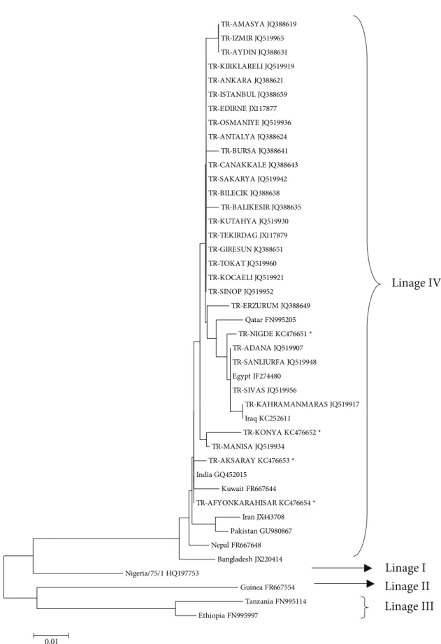
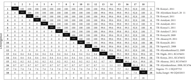
After sequencing a 448-bp fragment of the F gene from 16 animals, each from different outbreaks, the 4 sequences having a difference of at least 1 nucleotide were sent to GenBank and deposited under accession numbers KC476651 (Niğde, 2011; from a kid), KC476652 (Konya, 2009; from an aborted sheep fetus), KC476653 (Aksaray, 2012; from a lamb), and KC476654 (Afyonkarahisar, 2008; from a sheep). When compared with the sequences of 26 PPR viruses in GenBank that were isolated from different provinces in different regions of Turkey, all isolates were defined in the same lineage group: IV. The phylogenetic analysis of the Turkish isolates together with isolates from 13 countries (Qatar, Egypt, Iraq, India, Kuwait, Iran, Pakistan, Nepal, Bangladesh, Nigeria, Guinea, Tanzania, and Ethiopia) is shown in Figure 2. Nucleotide sequence identity among 16 viruses was 99.1%–100% (Figure 3).
4. Discussion
First detected in the 1990s, PPR became one of the major viral diseases of small ruminants during the 2000s in Turkey. Molecular detection and characterization of the virus was done by Özkul et al. (2002), and 2 virus isolates were reported in lineage IV. The full genome sequence of 1 of these PPR viruses isolated in Turkey was determined by Bailey et al. (2005).
For routine diagnosis of PPR, virus isolation is time-consuming and is not possible in many diagnostic laboratories. Submission of suitable material for isolation is not always possible under field conditions. Molecular diagnostic methods, such as conventional and real-time RT-PCR assays, have facilitated diagnosis of PPR and have been commonly used in many diagnostic laboratories in Turkey. Accordingly, with the increase in diagnostic facilities using molecular methods, more cases have been reported.
Control of PPR in Turkey is based mainly on vaccination in all regions of Turkey. Presently, newborn lambs, kids over 3 months of age, and previously unvaccinated adult animals are vaccinated. In the case of outbreaks, susceptible animals in the area are vaccinated. All sheep and goats have to be vaccinated against PPR once in their lifetime for animal movement permissions. Although these vaccination programs exist, vaccination coverage has been insufficient in some areas, and in this study the animals in which the PPR virus was detected were from unvaccinated flocks. Although there was a mass vaccination campaign during 2010–2012, the highest number of outbreaks was detected in 2011. The number of PPR outbreaks reported to the OIE (http://www.oie. int/wahis_2/public/wahid.php/Diseaseinformation/ statusdetail) from Turkey also showed that the number of outbreaks was high countrywide that year. In addition to the vaccination campaign, the conscientiousness of farmers, interest of veterinarians, and availability of molecular techniques made the disease more recognizable and caused previously undiagnosed cases to be diagnosed. Therefore, at the beginning of the campaign there was some increase in the number of outbreaks together with the vaccination of more animals. However, in the following years, and particularly during 2013, a clear decline was observed in the number of outbreaks.
For the control of PPR, live attenuated vaccines with Nigeria 75/1 (lineage I) and Indian strains (lineage IV) are commercially available (Saravanan et al., 2010). Vaccinated animals develop an antibody response that cannot be distinguished serologically from that produced after natural infection. Therefore, vaccination causes difficulties in the serological diagnosis of disease and surveillance studies as well as in molecular diagnosis. In this study, RT-PCR products were subjected to EcoRI digestion,
TR-AMASYA JQ388619 TR-IZMIR JQ519965 TR-AYDIN JQ388631 TR-KIRKLARELI JQ519919 TR-ANKARA JQ388621 TR-ISTANBUL JQ388659 TR-EDIRNE JX117877 TR-OSMANIYE JQ519936 TR-ANTALYA JQ388624 TR-BURSA JQ388641 TR-CANAKKALE JQ388643 TR-SAKARYA JQ519942 TR-BILECIK JQ388638 TR-BALIKESIR JQ388635 TR-KUTAHYA JQ519930 TR-TEKIRDAG JX117879 TR-GIRESUN JQ388651 TR-TOKAT JQ519960 TR-KOCAELI JQ519921 TR-SINOP JQ519952 TR-ERZURUM JQ388649 Qatar FN995205 TR-NIGDE KC476651 * TR-ADANA JQ519907 TR-SANLIURFA JQ519948 Egypt JF274480 TR-SIVAS JQ519956 TR-KAHRAMANMARAS JQ519917 Iraq KC252611 TR-KONYA KC476652 * TR-MANISA JQ519934 TR-AKSARAY KC476653 * India GQ452015 Kuwait FR667644 TR-AFYONKARAHISAR KC476654 * Iran JX443708 Pakistan GU980867 Nepal FR667648 Bangladesh JX220414 Nigeria/75/1 HQ197753 Guinea FR667554 Tanzania FN995114 Ethiopia FN995997 0.01
Linage IV
Linage I
Linage II
Linage III
Figure 2. Phylogenetic relationships between PPR viruses detected in Turkey and reference strains from GenBank. The bootstrap test neighbor-joining method in MEGA 4 software (1000 replicates) was used to draw the tree. *: Samples were sequenced in this study.
as stated by Özkul et al. (2002). This allowed for the differentiation of the vaccine strain of Nigeria 75/1 from field strains. Sequence analysis of 16 viruses also showed a nucleotide substitution present in EcoRI recognition sequence site in the amplified genome region of the vaccine strain Nigeria 75/1. RFLP can be used to detect genetic variation or molecular differentiation of viruses, but it is difficult to use the technique in RNA viruses because of their higher rate of spontaneous mutations compared with DNA viruses. However, the nucleotide substitution in EcoRI recognition site seems to be stable in our lineage IV viruses. Therefore, in routine diagnosis of disease, restriction endonuclease analysis of RT-PCR products can be helpful for presumptive differentiation of field isolates and the vaccine strain of lineage I in countries like Turkey where lineage IV is endemically present. On the other hand, comparative analysis of F gene sequences showed that similarity between our isolates and the Indian vaccine strain of Sungri-96 was higher than between our isolates and Nigeria 75/1 vaccine (Figure 3). Although the vaccine strain Nigeria 75/1 protected against PPR viruses of other lineages in challenge experiments (Diallo et al., 2007), with the availability of new lineage IV vaccines, the comparative efficacy of these against lineage IV viruses can be investigated.
In phylogenetic studies generally the N or F gene sequences have been used, and based on these sequences PPR viruses were grouped into 4 different lineages. F-gene–based phylogenetic analysis was performed in this study. Sequencing data of both F and N genes showed small variations among PPR viruses, which reflect the geographical origins of the virus strains (Shaila et al.,
1996; Diallo et al., 2007). However, the N gene is more prone to mutations than the F gene (Munir et al., 2013). No serotype level differences have been detected among the PPR viruses isolated so far, and only a single serotype of the virus is accepted. At present no other correlations between lineage differences and virus properties such as pathogenicity have been shown. To date, the first 3 lineages of the virus were detected only in Africa. Until recently lineage IV was confined to Asia, including Turkey and the Arabian peninsula; currently it is found in North and Central Africa as well (Albina et al., 2013). Detection of lineages is a good indicator of virus spread among countries. Therefore, due to its critical geographical situation, continuous monitoring of lineages is especially important for Turkey.
In this study, partial sequence analysis of the F gene of 16 PPR viruses showed that all belonged to lineage IV. Phylogenetic analysis of PPR viruses from Turkey, such as Turkey 96 (Shaila et al., 1996) and isolates by Kul et al. (2007) and Toplu et al. (2012), showed that all are clustered into lineage IV. Results showed that PPR virus lineage IV has been in circulation in Turkey since the disease was first reported. The phylogenetic analysis of the partial sequence of the F gene also indicated high sequence homology among the 16 PPR viruses analyzed in this study and 26 PPR viruses isolated from different provinces of Turkey and deposited in GenBank. There was no subdivision among viruses. Munir et al. (2013) detected a clear division in 2 groups among sequences of PPR viruses isolated from an outbreak and indicated that the genetic divergence among PPR viruses was slightly higher in the N gene (6.2%) than the F gene (5.2%). The analyses of the N, M, F, and H gene
1 2 3 4 5 6 7 8 9 10 11 12 13 14 15 16 17 18 1 *** 100 100 100 100 100 100 100 100 100 100 100 99.6 99.6 99.8 99.3 92.8 100 1 TR-Konya1, 2011 2 0 *** 100 100 100 100 100 100 100 100 100 100 99.6 99.6 99.8 99.3 92.8 100 2 TR-Afyonkara hisar3, 20 11 3 0 0 *** 100 100 100 100 100 100 100 100 100 99.6 99.6 99.8 99.3 92.8 100 3 TR-Konya4, 2011 4 0 0 0 *** 100 100 100 100 100 100 100 100 99.6 99.6 99.8 99.3 92.8 100 4 TR-Antalya6, 2011 5 0 0 0 0 *** 100 100 100 100 100 100 100 99.6 99.6 99.8 99.3 92 .8 100 5 TR-Antalya8, 2011 6 0 0 0 0 0 *** 100 100 100 100 100 100 99.6 99.6 99.8 99.3 92.8 100 6 TR-Konya11, 2011 7 0 0 0 0 0 0 *** 100 100 100 100 100 99.6 99.6 99.8 99.3 92.8 100 7 TR-Antalya17, 2011 8 0 0 0 0 0 0 0 *** 100 100 100 100 99.6 99.6 99. 8 99.3 92.8 100 8 TR-Konya18, 2009 9 0 0 0 0 0 0 0 0 *** 100 100 100 99.6 99.6 99.8 99.3 92.8 100 9 TR-Aksaray19, 2011 10 0 0 0 0 0 0 0 0 0 *** 100 100 99.6 99.6 99.8 99.3 92.8 100 10 TR-Konya20, 2011 11 0 0 0 0 0 0 0 0 0 0 *** 100 99.6 99.6 99.8 99.3 92.8 100 11 TR-Isparta21, 2008 12 0 0 0 0 0 0 0 0 0 0 0 *** 99.6 99.6 99.8 99.3 92.8 100 12 TR-Afyonkarahisar22, 2009 13 0.4 0.4 0.4 0.4 0.4 0.4 0.4 0.4 0.4 0.4 0.4 0.4 *** 100 99.3 98.9 92.4 99.6 13 TR-Nigde, 2011, KC476651 14 0.4 0.4 0.4 0.4 0.4 0.4 0.4 0.4 0.4 0.4 0.4 0.4 0 *** 99.3 98.9 92.4 99.6 14 TR-Konya, 2011, KC476652 15 0.2 0.2 0.2 0.2 0.2 0.2 0.2 0.2 0.2 0.2 0.2 0.2 0.7 0.7 *** 99.1 92.6 99.8 15 TR-Aksaray, 2012, KC476653 16 0.7 0.7 0.7 0.7 0.7 0.7 0.7 0.7 0.7 0.7 0.7 0.7 1.1 1.1 0.9 *** 92. 2 99.3 16 TR-Afyonkarahisar, 2008, KC476654 17 7.4 7.4 7.4 7.4 7.4 7.4 7.4 7.4 7.4 7.4 7.4 7.4 7.9 7.9 7.7 8.2 *** 92.8 17 Nigeria -75 -1 HQ197753 18 0 0 0 0 0 0 0 0 0 0 0 0 0.4 0.4 0.2 0.7 7.4 *** 18 India,Sungri -96 GQ452015 1 2 3 4 5 6 7 8 9 10 11 12 13 14 15 16 17 18 Percent Identity Divergence
Figure 3. Percentage identity and divergence of PPR viruses characterized in this study with representative vaccine strains Nigeria 75/1 of lineage I and Sungri-96 of lineage IV.
sequences of virulent isolates and vaccine strains of PPR viruses of Indian origin showed considerable sequence homology among lineage IV viruses (Balamurugan et al., 2010). The phylogenetic analysis data obtained so far showed that PPR viruses circulating in Turkey were also genetically highly homogenous and stable.
In this study, all samples were taken from necropsied animals brought to the laboratory, live or dead. PPR virus was detected at a higher rate in young animals. High morbidity and mortality rates in young animals were also reported in other studies (Yeşilbağ et al., 2005; Albayrak and Alkan, 2009; Aytekin et al., 2011). As our observations showed (unpublished data), in young animals severity of symptoms and mortality rate were higher in PPR virus infection in the presence of other viruses such as sheep and goat pox or secondary bacterial infections (Kwiatek et al., 2007; Ozmen et al., 2009).
The PPR virus is transmitted mainly by aerosols between animals living in close contact (OIE, 2013). It is not considered a vertically transmitted disease. In this study, PPR virus was detected by RT-PCR and isolation in an aborted sheep fetus. In the PPR-virus–positive flock, all other bacterial abortion agents by culture, molecular, and serological methods were negative in aborted fetuses. Although pestivirus RNA was negative by RT-PCR, pestivirus antibodies were detected in some serum samples taken from the flock. In previous years,
we detected PPR virus by antigen or antibody detection assays in aborted sheep fetuses and neonatal lamb deaths in some flocks (unpublished observations). Dual infection with PPR virus and pestivirus, both of which are known to be immunosuppressive, in aborted fetuses (Toplu et al., 2012), or in stillborn lambs (Kul et al., 2008) was reported in other studies in Turkey. In our case, coinfection of PPR virus with pestivirus was evidenced by serology only. A possible association of PPR virus with abortion in goats based on serology was also suggested by Abubakar et al. (2008). In this study, detection of PPR virus in aborted fetuses suggests that the PPR virus could cross the placenta and cause abortion. Because the PPR virus causes acute systemic infection, the virus is widely distributed in different organs. When we tested different organ or tissue samples of infected animals (lung, spleen, liver, and lymph nodes) separately from the same animals by RT-PCR, each was positive. Therefore, detection of the virus in the fetuses of pregnant animals might also be expected. In endemic areas, PPR-virus–related abortions should also be taken into consideration. Sequence analysis of the virus from the aborted fetus did not show any considerable difference from other viruses analyzed in this study. Further investigations regarding transplacental transmission and fetal or placental histopathological changes caused by PPR virus are needed.
References
Abubakar M, Ali Q, Khan HA (2008). Prevalence and mortality rate of peste des petitis ruminants (PPR): possible association with abortion in goat. Trop Anim Health Prod 40: 317–321. Albayrak H, Alkan F (2009). PPR virus infection on sheep in Black
Sea region of Turkey: epidemiology and diagnosis by RT-PCR and virus isolation. Vet Res Commun 33: 241–249.
Albina E, Kwiatek O, Minet C, Lancelot R, Almeida RS, Libeau G (2013). Peste des petits ruminants, the next eradicated animal disease? Vet Microbiol 165: 38–44.
Alçığır G, Vural S, Toplu N (1996). Türkiye’de kuzularda peste des petits ruminants virus enfeksiyonunun patomorfolojik ve immunohistolojik ilk tanımı. Ankara Univ Vet Fak Derg 43: 181–189 (in Turkish with German abstract).
Anees M, Shabbir MZ, Muhammad K, Nazir J, Shabbir MAB, Wensman JJ, Munir M (2013). Genetic analysis of peste des petits ruminants virus from Pakistan. BMC Vet Res 9: 60.
Aytekin İ, Mamak N, Ulucan A, Kalınbacak A (2011). Clinical, haematological, biochemical and pathological findings in lambs with peste des petits ruminants. Kafkas Univ Vet Fak Derg 17: 349–355.
Bailey D, Banyard A, Dash P, Özkul A, Barrett T (2005). Full genome sequence of peste des petits ruminants virus, a member of the Morbillivirus genus. Virus Res 110: 119–124.
Balamurugan V, Sen A, Venkatesan G, Yadav V, Bhanot V, Riyesh T, Bhanuprakash V, Singh RK (2010). Sequence and phylogenetic analyses of the structural genes of virulent isolates and vaccine strains of peste des petits ruminants virus from India. Trans Emerg Dis 57: 352–364.
Banyard A, Parida CS, Batten C, Oura C, Kwiatek O, Libeau G (2010). Global distribution of peste des petits ruminants virus and prospects for improved diagnosis and control. J Gen Virol 91: 2885–2897.
Baron MD, Parida S, Oura CA (2011). Peste des petits ruminants: a suitable candidate for eradication? Vet Rec 169: 16–21. Batten CA, Banyard AC, King DP, Henstock MR, Edwards L, Sanders
A, Buczkowski H, Oura CC, Barrett T (2011). A real time RT-PCR assay for the specific detection of peste des petits ruminants virus. J Virol Methods 171: 401–404.
Chard LS, Bailey DS, Dash P, Banyard AC, Barrett T (2008). Full genome sequences of two virulent strains of peste-des-petits ruminants virus, the Côte d’Ivoire 1989 and Nigeria 1976 strains. Virus Res 136: 192–197.
Couacy-Hymann E, Roger F, Hurard C, Guillou JP, Libeau G, Diallo A (2002). Rapid and sensitive detection of peste des petits ruminants virus by a polymerase chain reaction assay. J Virol Methods 100: 17–25.
De Nardi M, Lamin Saleh SM, Batten C, Oura C, Di Nardo A, Rossi D (2012). First evidence of peste des petits ruminants (PPR) virus circulation in Algeria (Sahrawi Territories): outbreak investigation and virus lineage identification. Trans Emerg Dis 59: 214–222.
Diallo A, Minet C, Le Goff C, Berhe G, Albina E, Libeau G, Barrett T (2007). The threat of peste des petits ruminants: progress in vaccine development for disease control. Vaccine 25: 5591– 5597.
Esmaelizad M, Jelokhani-Niaraki S, Kargar-Moakhar R (2011). Phylogenetic analysis of peste des petits ruminants virus (PPRV) isolated in Iran based on partial sequence data from the fusion (F) protein gene. Turk J Biol 35: 45–50.
Forsyth MA, Barrett T (1995). Evaluation of polymerase chain reaction for the detection and characterization of rinderpest and peste des petits ruminants viruses for epidemiological studies. Virus Res 39: 151–163.
Kerur N, Jhala MK, Joshi CG (2008). Genetic characterization of Indian peste des petits ruminants virus (PPRV) by sequencing and phylogenetic analysis of fusion protein and nucleoprotein segments. Res Vet Sci 85: 176–183.
Kul O, Kabakçı N, Özkul A, Atmaca HT (2007). Natural peste des petits ruminants virus infection: novel pathologic findings resembling other Morbillivus infections. Vet Pathol 44: 479– 486.
Kul O, Kabakçı N, Özkul A, Kalender H, Atmaca HT (2008). Concurrent peste des petits ruminants virus and pestivirus infection in stillborn twin lambs. Vet Pathol 45: 191–196. Kwiatek O, Ali YH, Saeed IK, Khalafalla AI, Mohamed OI, Obeida
AA, Abdelrahman MB, Osman HM, Taha KM, Abbas Z et al. (2011). Asian lineage of peste des petits ruminants virus. Africa. Emerg Inf Dis 17: 1223–1231.
Kwiatek O, Keita D, Gil P, Fernandez-Pinero J, Clavero MAJ, Albina E, Libeau G (2010). Quantitative one-step real-time RT-PCR for the fast detection of the four genotypes of PPRV. J Virol Methods 165: 168–177.
Kwiatek O, Minet C, Grillet C, Hurard C, Carlsson E, Karimov B, Albina E, Diallo A, Libeau G (2007). Peste des petits ruminants (PPR) outbreak in Tajikistan. J Comp Pathol 136: 111–119. Luka PD, Erume J, Mwiine FN, Ayebazibwe C, Shamaki D (2011).
Molecular characterization and phylogenetic study of peste des petits ruminants viruses from North central states of Nigeria. BMC Vet Res 7: 32.
Munir M, Saeed A, Abubakar M, Kanwal S, Berg M (2013). Molecular characterization of peste despetits ruminants viruses from outbreaks caused by unrestricted movements of small ruminants in Pakistan. Trans Emerg Dis DOI: 10.1111/ tbed.12089.
Munir M, Zohari S, Saeed A, Khan QM, Abubakar M, LeBlanc N, Berg M (2012a). Detection and phylogenetic analysis of peste des petits ruminants virus isolated from outbreaks in Punjab, Pakistan. Trans Emerg Dis 59: 85–93.
Munir M, Zohari S, Saeed A, Khan QM, Abubakar M, LeBlanc N, Kanu S, Sankoh FA, Berg M, Barrie ML et al. (2012b). Genetic characterization of peste des petits ruminants virus, Sierra Leone. Emerg Infect Dis 18: 193–195.
OIE (2013). Peste des Petits Ruminants. Manual of Diagnostic Tests and Vaccines for Terrestrial Animals. Chapter 2.7.11. Paris, France: OIE.
Özkul A, Akca Y, Alkan F, Barrett T, Karaoğlu T, Dağalp SB, Anderson J, Yeşilbağ K, Çokçalışkan C, Gencay A et al. (2002). Prevalence, distribution, and host range of peste des petits ruminants virus, Turkey. Emerg Infect Dis 8: 708–712. Ozmen O, Kale M, Haligur M, Yavru S (2009). Pathological,
serological, and virological findings in sheep infected simultaneously with bluetongue, peste-des-petits-ruminants, and sheeppox viruses. Trop Anim Health Prod 41: 951–958.
Sağlam YS, Temur A (2009). Immunohistochemical detection of peste des petits ruminants (PPR) viral antigen from the cases of naturally occurring pneumonia in sheep. Kafkas Univ Vet Fak Derg 15: 423–428.
Sanz-Alvarez J, Diallo A, De La Rocque S, Pinto J, Thevenet S, Lubroth J (2008). Peste des petits ruminants (PPR) au Maroc. EMPRES Watch, 2008. Rome, Italy: FAO (in French).
Saravanan P, Sen A, Balamurugan V, Rajak KK, Bhanuprakash V, Palaniswami KS, Nachimuthu K, Thangavelu A, Dhinakarraj G, Hegde R et al. (2010). Comparative efficacy of peste des petits ruminants (PPR) vaccines. Biologicals 38: 479–485. Shaila MS, Shamaki D, Forsyth MA, Diallo A, Goatley L, Kitching RP,
Barrett T (1996). Geographic distribution and epidemiology of peste des petits ruminants viruses. Virus Res 43: 149–153.
Tatar N, Alkan F (1999). Koyun ve keçilerde küçük ruminantların vebası (peste des petits ruminants) ve sığır vebası enfeksiyonlarının serolojik ve virolojik olarak araştırılması. Etlik Vet Mikrobiyol Derg 10: 35–60 (in Turkish).
Toplu N (2004). Characteristic and non-characteristic pathological findings in peste des petits ruminants (PPR) of sheep in the Ege district of Turkey. J Comp Pathol 131: 135–141.
Toplu N, Oğuzoğlu TÇ, Albayrak H (2012). Dual infection of fetal and neonatal small ruminants with border disease virus and peste des petits ruminants virus (PPRV): neuronal tropism of PPRV as a novel finding. J Comp Pathol 116: 289–296.
Wang Z, Bao J, Wu X, Liu Y, Li L, Liu C, Suo L, Xie Z, Zhao W, Zhang W et al. (2009). Peste des petits ruminants virus in Tibet, China. Emerg Infect Dis 15: 299–301.
Yener Z, Sağlam YS, Temur A, Keleş H (2004). Immunohistochemical detection of peste des petits ruminants viral antigens in tissues from cases of naturally occurring pneumonia in goats. Small Rum Res 51: 273–277.
Yeşilbağ K, Yılmaz Z, Gölcü E, Özkul A (2005). Peste des petits ruminants (PPR) outbreak in western Turkey. Vet Rec 157: 260–261.