https://doi.org/10.1007/s00404-020-05534-1
GYNECOLOGIC ENDOCRINOLOGY AND REPRODUCTIVE MEDICINE
Protective effect of oxytocin on a methotrexate‑induced ovarian
toxicity model
Ismet Hortu1,2 · Gokay Ozceltik1 · Ahmet Mete Ergenoglu1 · Gurkan Yigitturk3 · Ozum Atasoy4 · Oytun Erbas5 Received: 8 January 2020 / Accepted: 28 March 2020 / Published online: 7 April 2020
© Springer-Verlag GmbH Germany, part of Springer Nature 2020
Abstract
Purpose Although cancer predominantly affects people at older ages, a substantial number of patients, like breast cancer patients, are diagnosed before they have completed their families or even before giving birth. Furthermore, cytotoxic chemo-therapy may be required in addition to treat cancer survivors. The present study was conducted to investigate the protective effect of oxytocin (OT) on methotrexate (MTX)-induced ovarian toxicity in rats.
Methods Eighteen adult female Sprague–Dawley rats were used in the study. All rats were divided randomly into three groups. The control group (n = 6) received no treatment. The remaining 12 rats received a single dose of 20 mg/kg of MTX. Half of the rats (n = 6) were treated with 1 mg/kg/day of saline, and the other half (n = 6) were treated with 160 µg/kg/day of OT for 21 days. Then, blood samples were collected for biochemical analysis, and an ovariectomy was performed for histopathological examination.
Results Plasma malondialdehyde (MDA) and transforming growth factor-β (TGF-β) levels were significantly lower in the MTX + OT group compared to the MTX + saline group (p = 0.000036 for MDA; p = 0.0044 for TGF-β). AMH levels were also significantly higher in the MTX + OT group than in the MTX + saline group (p = 0.000036). The ovarian fibrosis percent was also notably lower in the MTX + OT group than in the MTX + saline group (p = 0.000036).
Conclusion On the basis of these findings, OT is a promising agent for ameliorating harmful effects of MTX on rat ovaries in an experimental model.
Keyword Methotrexate · Ovarian damage · Oxytocin · Cytotoxic chemotherapy · Anti-müllerian hormone
Introduction
The importance of breast cancer-related morbidity in women of reproductive age has increased in the last decade. A pro-longed life span, delayed childbearing to older ages for
different reasons, not giving birth, a lack of breastfeeding, and obesity are the main risk factors for breast cancer among women [1]. With improved education and advanced screen-ing methods, breast cancer is now diagnosed at a younger age. However, receiving chemotherapy is another challeng-ing condition in young women in view of chemotherapy-induced ovarian damage. Because of undifferentiated and hormone-insensitive tumor cells, most young women with breast cancer undergo systemic cytotoxic chemotherapy [2]. Many chemotherapeutic agents that are most effective in the treatment of rapidly dividing neoplastic cells have perma-nent effects on ovarian functions, leading to amenorrhea, infertility, and even menopause. These effects are greater with alkylating agents and less with antimetobolites such as methotrexate (MTX) [3]. Increased serum follicle stimulat-ing hormone (FSH) and decreased serum estradiol emerge after starting amenorrhea and ovarian failure. Many chemo-therapeutic regimens are closely related the occurrence of premature ovarian insufficiency (POI) in women older than * Ismet Hortu
1 Department of Obstetrics and Gynecology, Ege University School of Medicine, Izmir, Turkey
2 Department of Stem Cell, Ege University Institute of Health Sciences, Izmir, Turkey
3 Department of Histology and Embryology, Mugla Sıtkı Kocman University School of Medicine, Mugla, Turkey 4 Department of Radiation Oncology, University of Health
Sciences Kartal Lutfi Kırdar Education and Research Hospital, Istanbul, Turkey
5 Department of Physiology, Demiroglu Bilim University School of Medicine, Istanbul, Turkey
30 years of age [4]. Numerous antineoplastic agents are used to cope with the malignant diseases; malignant tissues or cancerous cells usually consist of rapidly dividing aberrant cells and are more vulnerable to chemotherapeutic drugs. Antineoplastic drugs are given in a specific intervals to allow for recovery of healthy cells and eliminate cancerous cells. One significant challenge is the potential for damage of nor-mal cells that divide rapidly, such as those in the oral cavity and gastrointestinal tract, cells of the gonads, bone marrow, hair follicles, and lymphatic tissues [5–7]. Chemotherapeutic drugs may affect also these normal cells in the body.
MTX is a folic acid antagonist type of antineoplastic drug grouped in antimetabolites. MTX inhibits the dihy-drofolate reductase enzyme, thereby decreasing the synthesis of thymidylate, purines, pyrimidines, and nucleic acids that are essential for cell survival. Thus, deoxyribonucleic acid (DNA) synthesis is inhibited [8]. MTX has many properties like antiproliferative, anti-inflammatory, and immunosup-pressive effects. Therefore, there are several different clinical uses of MTX in the field of medicine, such as ectopic preg-nancy, gestational trophoblastic diseases, some autoimmun-rheumatologic ailments, and psoriasis [9].
Oxytocin (OT) is a cyclic nonapeptide synthesized in cell bodies in the paraventricular nuclei of the hypothalamus and transported through the axons of these cells to the posterior pituitary. It plays a dual role as a neuromodulator/neuro-transmitter and a hormone. OT is an effective stimulant of uterine contraction and is used primarily to induce or rein-force labor in obstetrics [10]. Additionally, OT has a crucial role for milk ejection from breasts during lactation and is also produced by different peripheral tissues, such as skin, placenta, ovary, testis, thymus, pancreas, adipocytes, kidney, heart, and blood vessels. Besides, OT receptors have also been shown in the aforementioned organs in the literature. In addition to its classic functions, OT plays anti-inflammatory, anti-apoptotic, anti-stress, and antioxidant roles in many metabolic pathways [11, 12].
Though several studies demonstrate the protective effect of OT in many pathophysiological processes, there are limited data describing the impact of OT on ovaries from MTX-induced cytotoxicity. Thus, the purpose of the cur-rent study was to investigate the protective effect of OT on an MTX-induced gonadotoxicity model in a rat ovary using biochemical and histopathological parameters.
Materials and methods
Animals
In this study, 18 female Sprague–Dawley albino mature rats, weighing 200–220 g, were used. Animals were fed ad libitum and housed in pairs in steel cages with a temperature-controlled
environment (22 ± 2 °C) and 12-h light, 12-h dark cycles. The experimental procedures performed in this study were approved by the Committee for Animal Research of Ege University, Izmir, Turkey. All experiments were carried out according to the Guide for the Care and Use of Laboratory Animals, as confirmed by the National Institutes of Health (US).
Experimental protocol
Eighteen rats were divided randomly into three groups. Group 1 (naive, n = 6) served as a control group and received no treat-ment. The remaining 12 rats received a single dose of 20 mg/ kg of MTX intraperitoneally (ip) (methotrexate; Kocak Farma, Istanbul, Turkey) for induction of ovarian injury as described previously [13]. Group 2 (n = 6, MTX + saline) was treated with 1 mg/kg/day of saline (0.9% NaCl) intraperitoneally (ip) for 21 days. Group 3 (n = 6, MTX + OT) was treated with 160 µg/kg/day of OT (ip) (Pituisan; Ege Vet, Alfasan Interna-tional BV, Holland) for 21 days [14]. At the end of the experi-ment, both ovaries were obtained from all animals and fixed in 10% formalin with subsequent storage at 4 °C for paraffin sectioning. Blood samples were collected via cardiac punc-ture for biochemical measurements. Samples were stored at − 30 °C prior to the hormone assay. All rats were euthanized at the end of the experiment.
Histopathological examination
Ovarian tissues were formalin-preserved and embedded in paraffin. The ovaries were sectioned at a thickness of 4 μm with a microtome. The sections were then stained with hematoxylin and eosin, and mounted onto glass slides. All sections were photographed with an Olympus C-5050 digital camera mounted on an Olympus BX51 microscope. Histo-pathological examination of the ovaries was performed by a computerized image analysis system (Image-Pro Express 1.4.5, Media Cybernetics, Inc., Rockville, MD, USA) on ten microscopic fields per section at a magnification of 20 ×, performed by an observer who was blinded to the study groups. Primary follicles consist of an oocyte surrounded by a single layer of cuboidal granulosa cells. Secondary follicles include multiple layers of cuboidal granulosa cells and an invisible antrum. Tertiary follicles are character-ized by a stratum granulosum along with fluid-filled antral space. Stromal fibrosis in ovarian tissue was calculated as a percentage.
Biochemical assays of anti‑Müllerian hormone (AMH) and transforming growth factor‑β (TGF‑β)
Blood was centrifuged at 3000 rpm for 10 min at room tem-perature and stored at − 20 °C until the analysis for AMH
and TGF-β. AMH and TGF-β levels were measured using commercially available enzyme-linked immunosorbent assay (ELISA) kits (Biosciences, Seattle, WA, USA). Sam-ples from each rat were determined in duplicate according to the manufacturer’s guide. While AMH levels were expressed as ng/mL, TGF-β levels were expressed as pg/mL.
Determination of lipid peroxidation
Lipid peroxidation was determined in plasma samples through measuring malondialdehyde (MDA) levels as thio-barbituric acid reactive substances (TBARS), which are the end product of lipid peroxidation [15]. Trichloroacetic acid and TBARS reagent were added to the tissue samples, then mixed and incubated at 100 °C for 60 min. The samples were centrifuged at 3000 rpm for 20 min, and the absorb-ance of the supernatant was read at 535 nm after cooling on ice. MDA levels of tissue were calculated from the stand-ard calibration curve using 1,1,3,3-tetraethoxypropane and expressed as nmol/gr protein.
Statistical analysis
Data analysis was performed using GraphPad Prism 8.3.0 a software (GraphPad Software, La Jolla, CA, USA). The groups of parametric variables (biochemical data) were com-pared by student’s t test and analysis of variance (ANOVA). The groups of nonparametric variables (histopathology) were compared by the Mann–Whitney U test. Results are presented as the mean ± standard error of mean (SEM). A value of p < 0.05 was accepted as statistically significant.
p < 0.001 was accepted as statistically highly significant.
Results
Biochemical analyses
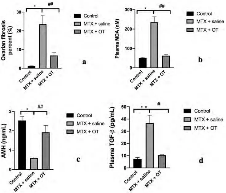
Plasma levels of MDA in the MTX + saline groups were sig-nificantly higher than in the control group (235.1 ± 28.5 nM vs. 51.3 ± 2.6 nM, respectively; p = 0.0084). Addition-ally, plasma levels of MDA were lower in the MTX + OT group than in the MTX + saline groups at a highly sig-nificant level (62.5 ± 5.1 nM vs. 235.1 ± 28.5 nM, respec-tively; p = 0.000036). Lipid peroxidation was significantly enhanced in the OT-added groups compared to the saline groups.
AMH levels in the MTX + saline groups were signifi-cantly lower than in the control group (0.6 ± 0.04 ng/mL vs. 2.53 ± 0.21 ng/mL, respectively; p = 0.0084). Administration of OT prevented the decrease of AMH levels compared to the saline groups (1.92 ± 0.35 ng/mL vs. 0.6 ± 0.04 ng/mL, respectively; p = 0.000036).
Plasma TGF-β levels were significantly higher in the MTX + saline groups than in the control group (36.8 ± 6.3 pg/mL vs. 7.4 ± 1.1 pg/mL, respectively;
p = 0.00069). However, OT treatment significantly reduced
TGF-β levels compared to the saline groups (36.8 ± 6.3 pg/ mL vs. 10.4 ± 0.7 pg/mL, respectively; p = 0.0044).
Plasma levels of MDA, AMH, and TGF-β in the study groups are listed in Table 1. Additionally, fibrosis percent-ages of ovary and plasma levels of MDA, AMH, and TGF-β are presented in Fig. 1.
Histopathological evaluation of ovaries
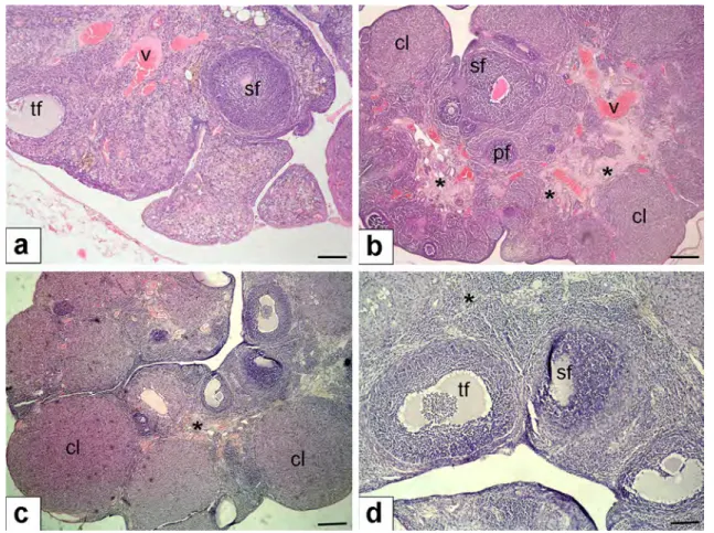
Histological examination of ovaries in the control group, the primary and developing follicles, corpus luteum, and ovarian stromal areas were found to be normal (Fig. 2a). Signifi-cant ovarian morphological alterations in view of stromal fibrosis were observed in the MTX + saline groups compared to the control group (Fig. 2b) (23.5 ± 4.8% vs. 1.2 ± 0.1%, respectively; p = 0.0084). When comparing stromal fibrosis in ovarian stroma between the MTX + OT and MTX + saline groups, a highly significant reduction was observed in the MTX + OT group (Fig. 2c, d) ( 6.8 ± 1.5% vs. 23.5 ± 4.8%, respectively; p = 0.000036).
Discussion
Cancer is a major public health problem all over the world. In recent decades, developed screening methods, increased awareness of cancer among women, and advanced radiodi-agnostic techniques have enabled clinicians to detect cancer at an early age and an early stage. Thus, a substantial number of cancer survivors are diagnosed at a younger age and then subject to cytotoxic chemotherapy alone or in addition to
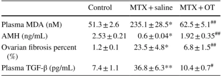
Table 1 Quantitative comparison of plasma MDA, AMH, TGF-β, and ovarian fibrosis percent
Values are expressed as mean ± SEM
OT oxytocin, MTX methotrexate, MDA malondialdehyde, AMH
anti-Müllerian hormone, TGF transforming growth factor
*p = 0.0084, MTX + saline group compared with control group **p = 0.00069, MTX + saline group compared with control group ## p = 0.000036, MTX + OT group compared with MTX + saline group # p = 0.0044, MTX + OT group compared with MTX + saline group
Control MTX + saline MTX + OT Plasma MDA (nM) 51.3 ± 2.6 235.1 ± 28.5* 62.5 ± 5.1## AMH (ng/mL) 2.53 ± 0.21 0.6 ± 0.04* 1.92 ± 0.35## Ovarian fibrosis percent
(%) 1.2 ± 0.1 23.5 ± 4.8* 6.8 ± 1.5 ## Plasma TGF-β (pg/mL) 7.4 ± 1.1 36.8 ± 6.3** 10.4 ± 0.7#
surgery [16]. The growing number of young cancer survivors are not only those with breast cancer; there are also gyneco-logic cancer survivors such as those with endometrial cancer (stage I A and grade 2) who wish to preserve their fertility during cancer treatment protocols [17]. There are some dev-astating challenges at this point, such as loss of ovarian fol-licular activity, toxicity to oocytes, fertility decline, POI, and menopause. Because of the finite number of oocytes, they are extremely sensitive to cytotoxic drugs. As a result, one of the main goals of physicians is the protection of the ovarian reserve and prevention of infertility in young women under-going chemotherapy. The main goal of the current study is to clarify the underlying mechanisms of MTX-related
ovarian toxicity and to investigate the likely ameliorative and protective role of OT on cytotoxic and fibrotic effects of MTX on the ovaries. In the present study, according to our results, MTX, a chemotherapeutic agent, impaired the ovarian follicular activity, as declining plasma AMH level and increasing plasma TGF-β via fibrosis. Moreover, MTX also increased the plasma MDA levels, a lipid peroxidation marker, compared to the control group. The protective and ameliorative effect of OT on ovaries in the MTX-induced gonadotoxicity model was clearly demonstrated in our study by reducing plasma TGF-β and MDA levels. Also, ovarian stromal fibrosis was significantly decreased in the MTX–OT group compared to the saline group. In addition, plasma
Fig. 1 a–d Bar graphics represent the alterations of fibrosis percent of
ovary, plasma MDA, AMH, and TGF-β levels in all groups. Results were presented as mean ± SEM. *p = 0.0084, MTX + saline group compared with control group. **p = 0.00069, MTX + saline group
compared with control group. ##p = 0.000036, MTX + OT group com-pared with MTX + saline group. #p = 0.0044, MTX + OT group com-pared with MTX + saline group
3
::r
E
2-
en
C -* 30I
300 • Control-c:J
MTX+saline :I!: C MTX+OTa
• ControlCl
MTX + saline MTX+OTC
-
<
C :I!: ftlE
Ill ftla:
:r
.§
a,a..
-200 100 50 40C9-
30 LL~
20ca
E
f/) 10 CVa:
0 * ##I
* * # • ControlCl
MTX + saline MTX+OTb
• Controlc::J
MTX
+ saline MTX+OTd
AMH levels were markedly higher in the MTX–OT group than in the saline group.
MTX—an inhibitor of dihydrofolate reductase commonly used in acute leukaemias; lymphomas; a number of solid tumors, including breast cancer, head and neck cancer, and bladder cancer; and some other malignant diseases—acts through nucleotide depletion, which composes DNA. With its other mechanisms of action, MTX is used in rheumato-logical diseases, such as rheumotoid arthritis, ankylosing spondilitis, and psoriasis. In gynecology, it is frequently used to decay and devitalize syncytiotrophoblasts for the management of clinically stable patients with non-ruptured ectopic pregnancy [18, 19]. Like other cytotoxic chemother-apeutics, MTX is not selective for cancerous cells; therefore, it has deleterious effects on ovarian follicles, gastrointesti-nal mucosa, bone marrow, and hair follicles, which have a high cellular proliferative index. Furthermore, because of its limited therapeutic index, MTX toxicity has been reported in various organs [20]. Armagan et al. showed that a single dose of MTX in rats caused extension of oxidative stress
in the testes [21]. Our study clearly showed that plasma MDA levels were decreased at a high statistically significant level in the OT group. The fibrosis effect of MTX has been reported in the literature in different organs, such as the liver, lung, and skin [22, 23]. The exact mechanisms underlying the fibrotic effect of MTX on several organs remain unclear. It is speculated that the damage of MTX may associated with oxidative stress through accumulating cell membrane oxidation markers, activation of proinflammatory cytokines, and triggering plasma TGF-β [24]. The current study dem-onstrated that OT attenuated the ovarian fibrosis induced by MTX in the rat ovaries. Additionally, OT improved ovarian fibrosis by reducing the TGF-β levels.
AMH, also called Mullerian inhibiting substance (MIS), is a member of the TGF-β family of glycoprotein differ-entiation factors that include inhibin and activin. AMH is secreted by the ovarian granulosa cells of the preantral and small growing follicles and substantially acts on a number of female reproductive functions. The levels of AMH are recognized as a more reliable marker of ovarian reserve
Fig. 2 Histopathological examination of ovaries. a Control group of ovaries with normal morphology (hematoxylin and eosin, × 10 mag-nification); b MTX + saline group with stromal fibrosis of the ovary (denoted with asterisk) (hematoxylin and eosin, × 10X magnifica-tion); c MTX + OT group demonstrating significant decrease of stro-mal fibrosis of the ovarian tissue (denoted with asterisk) (hematoxylin
and eosin, × 10 magnification); d MTX + OT group, reduction of stro-mal fibrosis appears to be better in higher magnification (hematoxylin and eosin, × 40 magnification). Scale bars indicate 225 µm for a–c; scale bar indicates 125 µm for d. pf primary follicle, sf secondary fol-licle, tf tertiary folfol-licle, cl corpus luteum, v vasculary
status compared to other tests, such as FSH, inhibin B, and estradiol. AMH is not a variable hormone during the ovar-ian–menstrual cycle like other gonadotropins. Its plasma level is strongly linked with the size of the follicle pool in the ovary [25]. Therefore, AMH is currently a gold stand-ard marker for ovarian reserve. AMH is also a promising marker for women who received chemotherapy. Hence, it was used in our study on MTX-induced ovarian toxicity in an experimental model. Recently, Erbas et al. reported that serum AMH levels were lower in the cisplatin-applied group than in the saline-applied group in a cisplatin-induced ovar-ian damage model in rats [14]. In another study, Karapinar et al. observed that the mean level of AMH was significantly lower in the MTX group than in the control group in experi-mentally induced ovarian injury by MTX [26]. In agree-ment with the literature, we observed that plasma AMH levels were significantly lower in the MTX–saline groups compared to the control groups. Moreover, with OT added to MTX-subjected rats, the plasma levels of AMH did not decrease as in the saline groups. Thus, OT prevented fol-licle loss in ovary and plasma AMH depletion from MTX toxicity. Besides, we observed a marked increase of AMH levels in the OT-treated group compared to the saline-treated group. This protective mechanism can be attribute to OT’s numerous features established in several other clinical and experimental studies [27].
OT is an essential drug of physicians practicing in obstet-rics. It is also used in different areas in experimental and clinical medicine. OT is a peptide hormone and has anti-oxidant and anti-inflammatory properties. OT prevents lipid peroxidation on the cell membrane by scavenging the free oxygen radicals. Moreover, effects of OT upon reducing neu-trophil accumulation and plasma TNF-α have been shown in the literature [28]. In a randomized, placebo-controlled clinical study, Clodi et al. reported that OT decreases the levels of proinflammatory cytokines, such as TNF-α, IL-6, monocyte chemoattractant protein 1 (MCP-1), and vascular endothelial growth factor (VEGF), in lipopolysaccharide-induced inflammatory response and endotoxemia [29]. Our study showed that OT has cytoprotective effects against MTX-induced ovarian injury and toxicity by decreasing plasma MDA and TGF-β levels at a highly significant level. Furthermore, OT significantly reduced fibrosis of the ovary compared to the MTX + saline groups. In addition, OT also improved the deleterious effects of a single dose of MTX on ovarian follicles. Thus, we observed plasma AMH levels at a significantly higher level in the OT group compared to the saline group.
Currently, several fertility preservation strategies have been implemented in young women with cancers. These include germ cell and embryo cryopreservation, ovarian tis-sue cryopreservation, and ovarian transposition [30]. How-ever, each type of technique should include counselling with
the patient or couples individually prior to starting adequate treatment for the sake of fertility and protection of ovar-ian function. Non-interventional, non-invasive methods such as gonadotropin-releasing hormone analogues (GnRH agonists) during cytotoxic chemotherapy have been utilized to protect women’s fertility [31]. However, the benefits of GnRH agonists in the context of protecting the ovary from cytotoxic drugs remain uncertain. A lack of high-power, high-numbered randomized controlled studies is an obsta-cle to obsta-clear decisions and directions for the use of GnRH agonists in this population. In another study, Gerber et al. reported uncertainty of resumption of ovarian functions during chemotherapy by GnRH agonists [32]. Del Mastro et al. demonstrated the temporary ovarian suppression and reduced occurrence of cytotoxic chemotherapy-induced early menopause during chemotherapy in young women with early-stage breast cancer [33]. Despite its beneficial effects on the ovary, GnRH agonists should not replace oocyte or embryo cryopreservation as the established modalities for fertility preservation [34]. To address this lack of clarity with regards to whether medical therapy can protect ovaries from cytotoxic effects of chemotherapeutics, we designed the current model to explore the role of OT.
To our knowledge, this is the first study that experimen-tally investigated the protective, alleviative, and antifibrotic effect of OT on MTX-induced ovarian cytotoxicity. Our results might open new directions for the protection of the ovary from chemotherapy-induced damage. In addition, there are many different areas of usage including gestational trophoblastic disease, rheumatologic diseases, and ectopic pregnancy of MTX in medicine. According to our results, OT can be implemented in clinical practice to alleviate the adverse, toxic, and fibrotic effects of MTX on the ovary and for the purpose of protection from ovarian insufficiency. Additional large-scale clinical studies will help support these results. Moreover, with the aid of new developments and new investigations, the mechanisms underlying the impacts of cytotoxic drugs on the ovary will be better understood.
On the other hand, there are some limitations that need to be considered in our study. A main limitation was the lack of evaluation of apoptotic markers and specific markers of oxidative stress and antioxidant enzymes in this experimen-tal model. The number of rats included in the subgroups was small; therefore, a larger sample is necessary to increase the power of our study. Furthermore, we did not apply dif-ferent and repeated dosage protocols of MTX and OT; so, large-scale experimental and clinical studies are required to determine the exact dosage and long-term effects of MTX and OT on the ovaries.
In conclusion, our findings suggest that OT can alleviate the deleterious, fibrotic, and cytotoxic effects of MTX on rat ovaries. These beneficial effects of OT might be related to its antioxidant capacities. Besides its antioxidant effects, OT
ameliorates oxidative stress injuries on ovarian follicles and supresses plasma TGF-β levels. As a consequence, harmful effects of MTX for women desiring further fertility may be reduced by OT.
Author contributions IH: data analysis and interpretation, writing manuscript; GO, AME: data collection, project development; GY: histological analysis; OA: data collection, statistical analysis; OE: performed experiment, supervising.
Compliance with ethical standards
Conflict of interest All the authors declare that there is no conflict of
interest.
Ethical approval The present study was approved by the Committee for Animal Research of Ege University, Izmir, Turkey. All procedures performed in studies involving animals were in accordance with the recommendations in the Guide for the Care and Use of Laboratory Animals of the National Institutes of Health.
Informed consent The informed consent is not applicable for the cur-rent study.
References
1. Bray F, Ferlay J, Soerjomataram I, Siegel RL, Torre LA, Jemal A (2018) Global cancer statistics 2018: GLOBOCAN estimates of incidence and mortality worldwide for 36 cancers in 185 coun-tries. CA Cancer J Clin 68:394–424
2. Elgindy EA, El-Haieg DO, Khorshid OM, El I, Abdeldawad M, Sallam HN, Abou-Setta AM (2013) Gonadotrophin suppression to prevent chemotherapy-induced ovarian damage: a randomized controlled trial. Obstet Gynecol 121:78–86
3. Roness H, Kalich-Philosoph L, Meirow D (2014) Prevention of chemotherapy-induced ovarian damage: possible roles for hormo-nal and non-hormohormo-nal attenuating agents. Hum Reprod Update 20:759–774
4. Kasum M, Beketic-Oreskovic L, Peddi PF, Oreskovic S, Johnson RH (2014) Fertility after breast cancer treatment. Eur J Obstet Gynecol Reprod Biol 173:13–18
5. Balachandren N, Davies M (2017) Fertility, ovarian reserve and cancer. Maturitas 105:64–68
6. Ganz PA, Land SR, Geyer CE Jr et al (2011) Menstrual history and quality-of-life outcomes in women with node-positive breast cancer treated with adjuvant therapy on the NSABP B-30 trial. J Clin Oncol 29:1110–1116
7. Cosgrove CM, Salani R (2019) Ovarian effects of radiation and cytotoxic chemotherapy damage. Best Pract Res Clin Obstet Gynaecol 55:37–48
8. Khan ZA, Tripathi R, Mishra B (2012) Methotrexate: a detailed review on drug delivery and clinical aspects. Expert Opin Drug Deliv 9:151–169
9. Iwase A, Nakamura T, Nakahara T, Goto M, Kikkawa F (2015) Anti-Müllerian hormone and assessment of ovarian toxic treat-ment: a systematic narrative review. Reprod Sci 22:519–526 10. Pergialiotis V, Frountzas M, Prodromidou A, Prapa S, Perrea DN,
Vlachos GD (2016) Propranolol and oxytocin versus oxytocin alone for induction and augmentation of labor: a meta-analysis of randomized trials. Arch Gynecol Obstet 293:721–729
11. Ragy MM, Aziz NM (2017) Prevention od renal ischemia/reper-fusion-induced renal and hepatic injury in adult male Albino rats by oxytocin: role of nitric oxide. J Basic Clin Physiol Pharmacol 28:615–621
12. Alizadeh AM, Faghihi M, Khori V, Sohanaki H, Pourkhalili K, Mohammadghasemi F, Mohsenikia M (2012) Oxytocin protects cardiomyocytes from apoptosis induced by ischemia-reperfusion in rat heart: role of mitochondrial ATP-dependent potassium chan-nel and permeability transition pore. Peptides 36:71–77 13. Gökçe A, Oktar S, Koc A, Yonden Z (2011) Protective effects
of thymoquinone against methotrexate-induced testicular injury. Hum Exp Toxicol 30:897–903
14. Erbaş O, Akman L, Yavaşoğlu A, Terek MC, Akman T, Taskiran D (2014) Oxytocin improves follicular reserve in a cisplatin-induced gonadotoxicity model in rats. Biomed Res Int 2014:703691 15. Zeb A, Ullah F (2016) A simple spectrophotometric method for
the determination of thiobarbituric acid reactive substances in fried fast foods. J Anal Methods Chem 2016:9412767
16. Bedoschi G, Navarro PA, Oktay K (2016) Chemotherapy-induced damage to ovary: mechanisms and clinical impact. Future Oncol 12:2333–2344
17. Vitale SG, Rossetti D, Tropea A, Biondi A, Laganà AS (2017) Fertility sparing surgery for stage IA type I and G2 endometrial cancer in reproductive-aged patients: evidence-based approach and future perspectives. Updates Surg 69:29–34
18. Committee on Practice Bulletin-Gynecology (2018) ACOG prac-tice bulletin No. 191: tubal ectopic pregnancy. Obstet Gynecol 131:e65–e77
19. Hortu İ, Akman L, Akdemir A, Ergenoğlu M, Yeniel Ö, Şendağ F (2017) Management of ectopic pregnancy in unusual location: five-year experience in a single center. J Clin Exp Invest 8:90–95 20. Padmanabhan S, Triphathi DN, Vikram A, Ramarao P, Jena GB
(2009) Methotrexate-induced cytotoxicity and genotoxicity in germ cells of mice: intervention of folic and folinic acid. Mutat Res 673:43–52
21. Armagan A, Uzar E, Uz E, Yilmaz HR, Kutluhan S, Koyuncuoglu HR, Soyupek S, Cam H, Serel TA (2008) Caffeic acid phenethyl ester modulates methotrexate-induced oxidative stress in testes of rat. Hum Exp Toxicol 27:547–552
22. Delyon J, Ortonne N, Benayoun E, Moroch J, Wolkenstein P, Sbid-ian E, Chosidow O (2015) Low-dose methotrexate-induced skin toxicity: keratinocyte dystrophy as a histologic marker. J Am Acad Dermatol 73:484–490
23. Mohamed DI, Khairy E, Tawfeek SS, Habib EK, Fetouh MA (2019) Coenzyme Q10 attenuates lung and liver fibrosis via modu-lation of autophagy in methotrexate treated rat. Biomed Pharma-cother 109:892–901
24. Norona LM, Nguyen DG, Gerber DA, Presnell SC, Mosedale M, Watkins PB (2019) Bioprinted liver provides early insight into the role of Kupffer cells in TGF- β1 and methotrexate-induced fibrogenesis. PLoS ONE 14:e0208958
25. Taylor HS, Pal L, Seli E (2020) Speroff’s clinical gynecologic endocrinology and infertility. In: Taylor HS, Pal L, Seli E (eds), Amenorrhea, 9th edn. Wolters Kluwer, New York, pp 821–939 26. Soylu Karapinar O, Pinar N, Özcan O, Özgür T, Dolapçıoğlu
K (2017) Protective effect of alpha-lipoic acid in methotrexate-induced ovarian oxidative injury and decreased ovarian reserve in rats. Gynecol Endocrinol 33:653–659
27. Wang P, Wang SC, Yang H, Lv C, Jia S, Liu X, Wang X, Meng D, Qin D, Hui Z, Wang YF (2019) Therapeutic potential of oxytocin in atherosclerotic cardiovascular disease: mechanisms and signal-ling pathways. Front Neurosci 13:454
28. Al-Amran F, Shahkolahi M (2013) Oxytocin ameliorates the immediate myocardial injury in rat heart transplant through downregulation of neutrophil-dependent myocardial apoptosis. Transplant Proc 45:2506–2512
29. Clodi M, Vila G, Geyeregger R, Riedl M, Stulnig TM, Struck J, Luger TA, Luger A (2008) Oxytocin alleviates the neuroendocrine and cytokine response to bacterial endotoxin in healthy men. Am J Physiol Endocrinol Metab 295:E686–E691
30. La Rosa VL, Shah M, Kahramanoglu I et al (2019) Quality of life and fertility preservation counseling for women with gynecologi-cal cancer: an integrated psychologigynecologi-cal and clinigynecologi-cal perspective. J Psychosom Obstet Gynaecol 2:1–7
31. Turner NH, Partridge A, Sanna G, Di Leo A, Biganzoli L (2013) Utility of gonadotropin—releasing hormone agonists for fertility preservation in young breast cancer patients: the benefit remains uncertain. Ann Oncol 24:2224–2235
32. Gerber B, Ortmann O (2014) Prevention of Early Menopause Study (POEMS): is it possible to preserve ovarian function by gonadotropin releasing hormone analogs (GnRHa)? Arch Gynecol Obstet 290:1051–1053
33. Del Mastro L, Boni L, Michelotti A et al (2011) Effect of the gonadotropin-releasing hormone analogue triptorelin on the occurrence of chemotherapy-induced early menopause in pre-menopausal women with breast cancer: a randomized trial. JAMA 306:269–276
34. Ethics Committee of the American Society for Reproductive Med-icine (2018) Fertility preservation and reproduction in patients facing gonadotoxic therapies: an Ethics Committee opinion. Fertil Steril 110:380–386
Publisher’s Note Springer Nature remains neutral with regard to jurisdictional claims in published maps and institutional affiliations.