ABSTRACT
OBJECTIVES: Nigella sativa oil and thymoquinone were comparatively tested in vitro for their effects on human cancer cell lines (glioma,T98; prostate, LnCaP) as well as mouse embryonic fi broblast cell lines (3T3), and for the induction of apoptosis.
METHODS: Individual cell lines were treated with thymoquinone and N. sativa oil for 24 and 48 hr. Survival rate with MTT, apoptosis with fl ow cytometry and caspase-9 mRNA enzyme levels with RT-PCR were deter-mined in vitro.
RESULTS: Application of respective concentrations of N. sativa oil (excluding 100 μg/mL for 48 hr) did not change the number of tested cell lines, however, treatment with thymoquinone reduced the number of all cells signifi cantly. Thymoquinone also exerted its apoptosis inducing effect through the activation of caspase-9. CONCLUSION: Differing with the type of cancer cells, thymoquinone posseses a strong contentration and time dependent survival reducing effect on cancer cells via apoptosis (Fig. 6, Ref. 22). Text in PDF www.elis.sk. KEY WORDS: thymoquinone, in vitro, apoptosis, antiproliferative, anticancer.
1Anadolu University, Department of Health, Faculty of Open Education,
Eskisehir, Turkey, 2Eskisehir Osmangazi University Department of
Physi-ology, Faculty of Medicine, Eskisehir, Turkey, 3Trakya University, Faculty
of Pharmacy, Department of Pharmacognosy, Edirne, Turkey, and 4
An-adolu University, Faculty of Pharmacy, Department of Pharmacognosy, Eskisehir, Turkey
Address for correspondence: G. Kus, Anadolu University Open Educa-tion Faculty, Yunus Emre Campus 26480 Eskisehir, Turkey.
Introduction
The plague of this century, “cancer” is a group of complex pa-thologies and diseases characterized by “out of control” cell growth (1). During apoptosis, a variety of molecules with up-regulatory and down-regulatory properties have a dynamic interaction and can inhibit pro-apoptotic molecules or apoptotic factors. And can-cer is caused by uncontrolled cell proliferation or failure of cells to pass through the apoptotic cell death (2–4). As a consequence, compounds which may trigger cancer cell apoptosis may lead to promising future drugs for the treatment of cancer.
Nigella sativa, well known also as black cumin seed of the
Ranunculaceae is an annual plant, has a long history in traditional medicine with therapeutic effects including analgesia, antihy-pertensive, anti-eczema, diuretic, antimicrobial, gastrointestinal problems as well as in various cancer therapies (5).
Thymoquinone is one of the bioactive components of Nigella
sativa seed oil and has a wide range of biological and
pharmaco-logical activities including anti-cancer, anti-tumoral, anti-oxidant and anti-proliferative properties. In vitro and in vivo
pharmacologi-cal studies have been reported for their potential antidiabetic (6), neuroprotective (7), cardiovascular (8), gastroprotective, hepato-protective (9) activities among others (10). Since there has baeen no encountered study examining the possible role of thymoqui-none on glioma (T98), prostate cancer cells (LnCaP) and mouse embryonic fi broblast cell line (3T3), we tried to reveal its role on these cell lines in vitro.
Materials and methods
General
Thymoquinone was obtained from commercial sources in high purity (> 98 %, Sigma-Aldrich) whereas Nigella sativa oil was obtained from local producer (> 10 % thymoquinone, AweCemre, Tokat, Turkey). Both samples were dissolved individually in di-methyl sulfoxide (DMSO, Sigma) and diluted further with Dul-becco’s modifi ed Eagle’s medium (DMEM, Sigma) to obtain the required fi nal concentrations for the assays listed below in detail.
Cell culture
T98, LnCaP and 3T3 cells were obtained from the American Type Culture Collection (ATCC, USA) and grown in complete medium containing DMEM supplemented with 10 % fetal calf se-rum (FCS, Sigma) and 1 % penicillin (10000 unit)-streptomycine (10mg/ml) solution (Sigma) in a humidifi ed atmosphere of 95 %
O2 and 5 % CO2 in air at 37 °C. After confl uence achieved more
than 95 %, the cells were detached with 0.25 % trypsin-EDTA (Sigma), centrifuged at 1200 rpm, 4 °C for 5 min and counted with
a cell counter (CEDEX, Roche). The cells were then transferred to microplates for cell survival studies and to fl asks for apoptosis and caspase measurements.
Test groups
Test groups were assigned as follows: control (complete me-dium only); dimethly sulfoxide (DMSO) (complete meme-dium with a fi nal concentration of 0.1 % DMSO, solvent); thymoquinone (treated with 1, 5, 10, 25 and 50 μM for 24 or 48 hr) and N. sativa oil (0.1, 1, 5, 10 and100 μg/mL for 24 and 48 hr).
Cell survival
The effects of thymoquinone and Nigella sativa oil on cell survival were determined by 3-[4,5-dimethylthiazol-2yl]-diphenyl tetrazolium bromide (MTT, Sigma) colorimetric assay (11). MTT test was applied and cell survival was determined by measuring the formazan absorbance at 550 nm with a microplate reader (BioTek; Winooski,VT). Since the number of cells in each well is proportion-ate to the absorbance of the soluble formazan, the optical density read from the drug-treated wells was converted to the percentage of live cells versus control using the following formula:
Absorbance of treated cells in each well x 100 / the mean ab-sorbance of control cells.
Flow cytometric analysis
To determine cell death type (necrosis or apoptosis) of cells, we used fl ow cytometric analysis with the 25 and 50 μM thymo-quinone doses that diminish cell survival by more than fi fty per-cent. Apoptotic cells were determined by staining with fl uores-cein isothiocyanate (FITC)-labeled Annexin V (Invitrogen) and subsequent fl ow cytometric analysis. Alive cells are negative for both PI and Annexin V, early apoptotic cells are PI negative but Annexin V positive while dead/late apoptotic cells are positive for both PI and Annexin V.
RNA extraction and determination of caspase-9 mRNA levels
The expression of caspase-9 mRNA was examined in control and thymoquinone treated cells. The mRNA levels of caspase-9 in relation to the housekeeping gene was determined by qT-PCR with TaqMan probes. RT-+PCR data were collected using the Roche lightcycler nano system.
Statistical analysis
All results are the mean of at least three independent assays and the p value less than 0.05 was considered to be signifi cant for MTT results. Data were expressed as the mean percent frac-tion of control ± standart error of mean. Statistical signifi cance ascertained by one way analysis of variance followed by Tukey’s multiple comparison test. The apoptotic results were depicted as
Fig. 1. Treatment of T98 cells with Nigella sativa oil for 24 and 48 hr.
Fig. 3. The infl uence of Thymoquinone on LnCaP cells survival after 24 or 48 h.
Fig. 2. Effects of Thymoquinone and DMSO (vehicle) on 3T3 cell sur-vival during 24 or 48 h treatments. * p < 0.05, ** p < 0.01, *** p < 0.001.
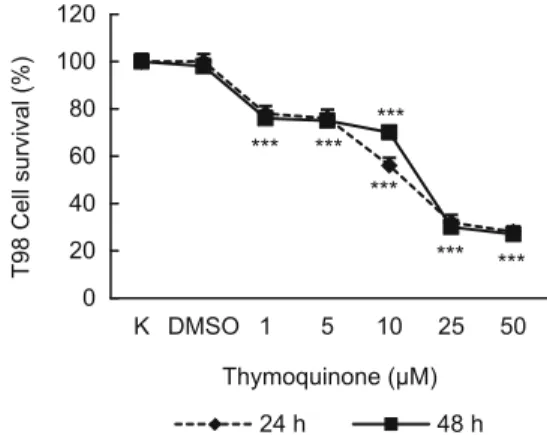
Fig. 4. The activity of 1, 5, 10, 25 and 50 μM thymoquinone on cell survival of T-98 cells by using MTT after 24 or 48 h.
percentage of cells. RT-PCR results were calculated by GraphPad software program. Transcript data were expressed relative to the control standart deviation.
Results
Cell viability assays
Treatment of the cells with DMSO did not cause any signifi -cant change in cell viability in pilot studies. Viability of T98, Ln-CaP and 3T3 cells was not changed after treatment with Nigella
sativa oil for 24 hr. Culturing of cells with 100 μg/mL of Nigella sativa oil for 48 hr reduced the number of only T98 cells by 20 %
(Fig. 1). However, treatment of cells with 1, 5, 10, 25 and 50 μM thymoquinone reduced the number of cells by 98, 95, 93, 82 (p < 0.5) and 75 % (p < 0.01) for 3T3 cells (Fig. 2); 97, 96, 90, 64 (p < 0.01) and 26 (p < 0.001) for LnCaP cells (Fig. 3) and 78, 76, 56, 32 and 28 (p < 0.001) for T98 cells (Fig. 4) for 24 hr, respective-ly. For 48 hr these rates were 95, 94, 93, 75 (p < 0.01) and 68 % (p < 0.001) for 3T3 cells (Fig. 2); 95, 88, 77, 60 and 18 (p < 0.001) % for LnCaP cells (Fig. 3) and 76, 75, 70, 30 and 27 (p < 0.001) % for T98 cells (Fig. 4) for 48 hr.
Flow cytometric analysis and gene expression
To further investigate the underlying mechanism of reduc-tion of cell survival detected with MTT assay, we examined the
apoptotic effect of the thymoquinone on cancer cells using fl ow cytometric analysis with 25 and 50 μM thymoquinone that de-creases cell survival by more than 50 %. As seen in Figs 5 and 6, time and concentration dependent apoptosis of LnCap cells, not T98 cells, was detected. The analysis after 24 hr treatment with 25 and 50 μM thymoquinone demonstrated that 2 % and 23 % of the LnCaP cells underwent early apoptosis, whereas 3 and 76 % of the cancer cells underwent early apoptosis after 48 hr treatment. Caspase-9 mRNA level was increased in LnCaP cellline treated with 25 and 50 μMthymoquinone (p>0.05) whereas notchanged in T98 cell line.
Discussion
There are increasing number of scientifi c studies on the rela-tionship between thymoquinone and cancer. In vitro and in vivo pharmacological studies have been reported for their potential an-titumor, anticancer (12), antidiabetic (13), cardiovascular activity (8), gastroprotective, anti-infl ammatory, hepatoprotective activity (9) and pulmonary activity (14).
Several other studies demonstrated that thymoquinone, one of the most active components in Nigella sativa seed, inhibits the proliferation of various cancer cell lines. Thymoquinone dimin-ished cell survival and induced apoptosis of canine osteocarcinoma cells, human breast adenocarcinoma (MCF7) and human ovarian
Fig. 5. T-98G cells, treated or non-treated with 25 or 50 μM Thymoquinone for 24 (A) or 48 (B) h, then stained with FITC Annexin V apop-tosis assay kit with PI (Invitrogen). Lower left sections of all the Figure, AnnexinV/PI (−), living cells; lower right sections of all the Figure, AnnexinV (+)/PI (−), early apoptotic cells; upper left sections of all the fi güre, Annexin (−)/PI (+), necrotic cells; upper right sections of all the Figure, Annexin (+)/PI (+), late stages of apoptosis and secondary necrosis. Results of only one independent experiment out of 3 is pointed.
adenocarcinoma (BG-1) (15). Similarly, thymoquinone has been shown to inhibit cell proliferation in cultured cells derived from myeloblastic leukemia cells, fi brosarcoma cells, laryngeal neoplas-tic cells, pancreaneoplas-tic cells and human colon cancer cells (16, 17, 18, 19, 20). El-Mahdy et al. showed that thymoquinone induced apoptosi s is p53-independent and occurs through the activation of caspase 3, 8 and 9 (21). In contrast, normal cells and primitive mouse keratinocytes are resistant to the apoptotic and antiprolifera-tive effects of thymoquinone and their lack of signifi cant changes in morphology and proliferation confi rms the selectivity of this compound for cancer cells (15, 22).
Accordi ng to our results, N. sativa oil did not affect the survival of cancer cells and 3T3 cells, however thymoquinone diminishes survival rate and induces apoptosis of only LnCaP cells in vitro and it possibly this effect through caspase-9. However, thymo-quinone did not show the same effects on T98 and 3T3 cells. Our study shows thymoquinone has strong dose, time and cancer type dependent effects through decreasing cell survival and inducing apoptosis in prostate cancer cells. Although much work is needed, our data provides evidence of potential implications for the appli-cation of thymoquinone as a novel possible antiproliferative drug against prostate cancer.
References
1. Clark HP, Carson WF, Kavanagh PV, Ho CPH, Shen P, Zagoria RJ. Staging and current treatment of hepatocellular carcinoma. Radi Graph-ics, 2005; 25: 3–23.
2. Salseven GS, Dixit VM. Caspase activation: The induced proximity model. Proc Natl Acad Sci USA 1999; 96: 10964–10967.
3. Huerta S, Gaulet EJ, Haerta-Yepez S, Livingston EH. Screening and detection of apoptosis. J Surg Res 2007; 1: 143–156.
4. Herr I, Debatin KM. Cellular stress response and apoptosis in cancer therapy. Blood 2001; 98: 2603–2614
5. Ahmad A, Husain A, Mujeeb M et al. A review on therapeutic poten-tial of Nigella sativa: A miracle herb. Asian Pacif J Tropic Biomed 2013; 3 (5): 337–352.
6. Mathur ML, Gaur J, Sharma R, Haldiya KR. Antidiabetic proper-ties of a spice plant Nigella sativa. J Endocrinol Metabol 2011; 1 (1): 1–8. 7. Ezz HSA, Khadrawy, YA, Noor NA. The neuroprotective effect of curcumin and Nigella sativa oil against oxidative stress in the pilocar-pine model of epilepsy: a comparison with valproate. Neurochem Res 2011; 36 (11): 2195.
8. Tasawar Z. Siraj Z, Ahmad N, Lashari MH. The effects of Nigella sativa (Kalonji) on lipid profi le in patients with stable coronary artery dis-ease in Multan, Pakistan. Pak J Nutr 2011; 10 (2): 162–167.
9. Al-Suhaimi EA. Hepatoprotective and immunological functions of Nigella sativa seed oil against hypervitaminosis A in adult male rats. Int J Vitam Nutr Res 2012; 82 (4): 288–297.
10. Gholamnezhad, Z., Havakhah, S., Boskabady, M. H. Preclinical and clinical effects of Nigella sativa and its constituent, thymoquinone: A review. J Ethnopharmacol 2016; 190: 372–386.
11. Mossmann T. Rapid colorimetric assay of cellular growth and sur-vival: Application to proliferation and cytotoxicity assay. J Immun Method 1983; 65: 55–63.
The antitumor activity of thymoquinone and thymohydroquinone in vitro and in vivo. Exp Oncol 2006; 28 (3): 220–224.
17. Womack K, Anderson M, Tucci M, Hamadain E, Benghuzzi H. Evaluation of biofl avonoids as potential chemotherapeutic agents. Biomed Sci Instrum 2006; 42: 464–469.
22. Gali-Muhtasib HU, Kheir WGA, Kheir LA, Darwiche N, Crooks PA. Molecular pathway for thymoquinone-induced cell-cycle arrest and apoptosis in neoplastic keratinocytes. Anti-cancer Drugs 2004; 15 (4): 389–399.
Received December 29, 2017. Accepted February 5, 2018.