Journal of Turkish Sleep Medicine 2019;1:37-42
Introduction
Obstructive Sleep Apnea (OSA) is common in the general population. Recurrent episodes of the collabing upper airway during sleep, and the resulting intermittent hypoxia, are
important features of OSA. The reported estimated prevalence of OSA in adults is 3~7% for males and 2~5% for females (1). During OSA, intermittent cycles of falling oxygen saturation and reoxygenation, and rises in pharyngeal resistance and esophageal pressure, microarousal, and hypercapnia occur. Ad dress for Cor res pon den ce/Ya z›fl ma Ad re si: Aynur Yılmaz Avcı MD, Başkent University Faculty of Medicine, Department of Neurology, Ankara, Turkey
Phone: +90 242 510 25 25 E-mail: [email protected] ORCID-ID: orcid.org/0000-0001-9004-9382 Re cei ved/Ge liş Ta ri hi: 23.12.2018 Ac cep ted/Ka bul Ta ri hi: 15.05.2019
©Copyright 2018 by Turkish Sleep Medicine Society / Journal of Turkish Sleep Medicine published by Galenos Publishing House.
Amaç: Periferik nöropati için risk faktörlerinden birinin kronik hipoksi
olduğu bilinmektedir. Bununla birlikte, aralıklı hipoksinin periferal sinirler üzerindeki etkisi tam olarak anlaşılamamıştır. Bu çalışmada Obstrüktif Uyku Apnesi (OSA) hastalarında aralıklı hipoksi ve periferik sinir fonksiyonu arasındaki ilişki değerlendirildi.
Gereç ve Yöntem: Bu retrospektif çalışmaya polisomnografi (PSG) ve
elektronöromiyografi uygulanmış 86 hasta alındı. Periferik sinirleri ve akciğer fonksiyonlarını etkileyen hastalıkları olan katılımcılar çalışma dışı bırakıldı. Hipoksi parametreleri PSG çalışmasından elde edildi. Tüm hastaların alt ekstremite motor ve duyu siniri iletim çalışmaları değerlendirildi.
Bulgular: OSA’lı hastalarda kontrollere göre peroneal sinir distal motor
latansı ve sural duyusal sinir aksiyon potansiyeli amplitüdü düşük ve hızı anlamlı olarak yavaştı (p<0,001, p<0,04, p<0,001, sırasıyla). Yaş ve vücut kitle indeksi için ayarlama yapıldıktan sonra, sonuçlar anlamlı kaldı (p<0,001, p<0,01, p<0,001, sırasıyla). Sinir iletim sonuçları hipoksi parametreleri ile anlamlı olarak korele idi. Karıştırıcı faktörler ayarlandıktan sonra, lojistik regresyon analizleri, hipoksi parametrelerinin sinir iletimi sonuçlarıyla bağımsız olarak ilişkili olduğunu gösterdi.
Sonuç: OSA ve aralıklı hipoksi, hem motor hem de duyusal sinir iletimini
etkileyebilmekte, bu da subklinik sensorimotor periferik nöropatinin OSA ile ilişkili olduğunu düşündürmektedir. OSA ve ilgili aralıklı hipoksi, aksonal ve demiyelinizan nöropatinin bir nedeni olabilir.
Anahtar Kelimeler: Uyku Apne sendromu, uykuda solunum bozukluğu,
elektromiyografi, hipoksi, nöropati
Öz Introduction: Chronic hypoxia is known to be one of the risk factors
for peripheral neuropathy. However, the effect of intermittent hypoxia on peripheral nerves is not fully understood. This study evaluated the relation between intermittent hypoxia and peripheral nerve function in Obstructive Sleep Apnea (OSA) patients.
Materials and Methods: In this retrospective study, 86 patients who
underwent polysomnography (PSG) and electroneuromyography were enrolled. Participants with diseases affecting peripheral nerves and lung function were excluded from the study. Hypoxia parameters were obtained from the PSG study. Lower extremity motor and sensory nerve conduction studies of all patients were evaluated.
Results: In patients with OSA, peroneal nerve distal motor latency and
sural sensory nerve action potential amplitude was low and velocity was significantly slower than controls (p<0.001, p<0.04, p<0.001, respectively). After adjustment for age and body mass index, the results remained significantly (p<0.001, p<0.01, p<0.001, respectively). The nerve conduction results were significantly correlated with the hypoxia parameters. After adjustment for confounding factors, logistic regression analyses revealed that hypoxia parameters were independently associated with nerve conduction results.
Conclusion: OSA and intermittent hypoxia may affect both motor and
sensory nerve conduction, which suggests that subclinical sensorimotor peripheral neuropathy is associated with OSA. The related intermittent hypoxia and OSA may be a cause of axonal and demyelinating neuropathies.
Keywords: Sleep Apnea syndrome, sleep-disordered breathing,
electromyography, hypoxia, neuropathy
Abstract
Başkent University Faculty of Medicine, Department of Neurology, Ankara, Turkey *Başkent University Faculty of Medicine, Department of Otolaryngology, Ankara, Turkey
Aynur Yılmaz Avcı, Suat Avcı*
Obstrüktif Uyku Apne’de Hipoksiye Bağlı Periferal Sinir Fonksiyonu Değişiklikleri
Peripheral Nerve Function Changes Due to Hypoxia in
Obstructive Sleep Apnea
The chronic cycles of desaturation-reoxygenation during intermittent hypoxia induce oxidative stress, producing an inflammatory response and reactive oxygen species, which are responsible for OSA-related complications, such as metabolic, neurocognitive, and cardiovascular disorders (2,3).
In patients with chronic obstructive pulmonary disease (COPD) neurophysiological changes have been found in in 90% of the cases. The reported high incidence is thought to be related to hypoxia (4,5). Chronic hypoxia induces peripheral polyneuropathy and this form of nerve damage is associated with the severity and duration of hypoxia (6-8). Prevalence of polyneuropathy due to chronic hypoxia have been reported in a similar range of 28% to 70% in patients with OSA and COPD (5,9-11).
Previous studies have reported that peripheral nerve sensory amplitudes are impaired in OSA patients, but the relationship with hypoxemia has not been fully elucidated (9,12,13). We hypothesized that patients with severe OSA and hypoxia show significantly impaired peripheral nerve conduction compared to controls. In this study, patients were subgrouped according to hypoxia parameters and the apnea-hypopnea index (AHI). Following this we determined whether the severities of OSA and hypoxia were associated with lower extremity motor and sensory nerve conduction abnormalities.
Materials and Methods
Patient’s Characteristics
This retrospective study was performed at the neurology outpatient clinic of university hospital. Subjects who underwent polysomnography (PSG) at our sleep disturbance center (accredited by the Turkish Sleep Association) and who underwent nerve conduction studies (NCSs) between October 2008 and January 2018 were selected. In the patients’ medical histories, the presence of vascular risk factors and systemic diseases were recorded. Exclusion criteria were patients aged under 18 years, presence of any radiculopathy, traumatic neuropathy, Central Sleep Apnea syndrome, narcolepsy, COPD, bronchial asthma, chronic alcohol intake, hypertension, coronary heart disease, hyperlipidemia, diabetes mellitus, goiter, chronic renal failure, liver failure, malignancy, or use of neurotoxic drugs.
After applying the exclusion criteria, 86 of 237 selected patients were included. Using the AHI, the participants were divided into a control group (AHI ≤5) (n=40) and OSA group (AHI >5) (n=46). Age, sex, body mass index (BMI), Epworth Sleepiness Scale were measured. Hypoxia parameters including the lowest oxygen saturation (min SaO2), total sleep duration with oxygen saturation <90% (ST90), percentage of cumulative time with oxygen saturation <90% (CT90), 3% oxygen desaturation index (ODI), and sleep time were calculated and recorded from the patients’ PSG records. The participants were further subdivided according to ST90, CT90, min SaO2, and ODI.
This study was exempted from the requirement for ethics committee approval from the institutional review board of our university due to its retrospective nature.
Polysomnography
All participants underwent PSG using a computerized PSG device in the sleep laboratory (E series, 44 channels; Compumedics, Victoria, NSW, Australia). The following parameters were
documented during the PSG study (16 channels): four-channel electroencephalogram, electrooculogram, submental and leg electromyogram, electrocardiogram, nasal airflow using a nasal pressure cannula, airflow at the nose and mouth (thermistors), chest and abdominal respiratory movements, oxygen saturation (pulse oximetry), snoring microphone, and body position. All studies were interpreted by a sleep specialist (pulmonologist) who was blinded to the participants’ characteristics. Apnea, hypopneaand sleep staging were defined and performed according to standards of the American Academy of Sleep Medicine criteria (14). The ST90 was recorded in minutes, and min SaO2 and CT90 were recorded as percentages. The min SaO2 value was the lowest oxygen saturation during sleep.
Nerve Conduction Studies
NCSs of bilateral tibial, peroneal, and sural nerves were performed using an electromyography device (Neuropack M1 MEB 2002; Nihon Kohden, Tokyo, Japan). Standardized techniques and procedures were used as described in the guidelines of electrodynamic medicine (15). Filter settings were 3 Hz-10 kHz for the motor NCSs and 20 Hz-2 kHz for the sensory NCSs. During the testing procedures, the room temperature was maintained at an average of 25 °C and skin temperature was maintained at >33 °C in all participants.
Statistical Analysis
The statistical data were evaluated using IBM SPSS Statistics for Windows (ver. 20.0; IBM Corp, Armonk, NY, USA). To test the homogeneity of variances, which is a prerequisite of parametric tests, Levene’s test was used. The assumption of normality was tested via the Shapiro-Wilk test. To compare differences between the characteristics of patients with OSA and controls, the Student’s t-test was used when the parametric test prerequisites were met, and the Mann-Whitney U test was used when such prerequisites were not met. The chi-square test was used to determine the relationships between two discrete variables. Values were determined through the Monte Carlo simulation method when the expected sources were less than 20% to include such sources in analyses. Age and BMI were determined as covariates (to be excluded), and groups were compared by a covariance analysis (Table 1). The relationship between two continuous variables was assessed by Pearson’s correlation analyses and Spearmans correlation analyses when the parametric test prerequisites were not met (Table 2). Binary logistic regression analyses were used to reveal the model for the relationship between the independent and dependent variables (Table 3). A p value <0.05 was set for level of significance.
Results
Characteristics of the subjects with OSA and the controls are described in Table 1. Significant differences in age, sex, BMI, the AHI, and the hypoxia parameters were observed among both groups (Table 1). In OSA subjects, peroneal nerve distal motor latency was prolonged and sural Sensory nerve action potential (SNAP) amplitude and velocity were significantly decreased (Table 1). After adjusting for age and BMI, the differences in peroneal nerve motor latency, sural SNAP amplitude, and sural nerve conduction velocity (NCV) of the two groups remained significant (Table 1). Univariate analyses revealed a significant mild to moderate correlation
Table 1. Comparison of the characteristics of the study participants with Obstructive Sleep Apnea and control subjects. Age- and body mass index-adjusted analyses
Control Obstructive Sleep Apnea
p Adjust BMI and Age ||
n=40 n=46 p
Age (years) 44.4±13.9 54.7±12.4 0.001† –
Sex Female 22 (55%) 12 (26.1%) 0.006§ –
Male 18 (45%) 34 (73.9%)
Epworth Sleepiness Scale 6.7±6.3 10.8±0.5 0.001‡ 0.001
Body mass index (kg/m2) 25.8±3.4 32.3±5.3 0.001‡ –
Apnea-hypopnea index (events/h) 1.5±1.3 23.3±20.6 0.001† 0.001
Total sleep time (min) 447.1±22.9 440.6±40.5 NS‡ NS
Oxygen desaturation index (%) 3.1±1.4 4.9±1.6 0.001‡ 0.001
ST90 (min) 1.3±3.3 23.3±47.5 0.001‡ 0.001
CT90 (%) 0.3±0.7 5.6±12.2 0.01‡ 0.01
Minimum oxygen saturation (%) 90.7±4.7 81.2±7.8 0.001‡ 0.001
Tibial motor nerve latency (ms) 4.2±0.7 4.3±0.91 0.001‡ 0.001
Tibial CMAP amplitude (mV) 15.4±6.2 11.8±4.5 0.040‡ 0.01
Tibial motor nerve velocity (m/s) 48.1±4.03 45.1±8.3 NS‡ NS
Peroneal motor nerve latency (ms) 3.9±0.5 4.2±0.7 0.001‡ 0.001
Peroneal CMAP amplitude (mV) 10.3±4.3 6.9±2.9 NS‡ NS
Peroneal motor nerve velocity (m/s) 50.9±4.9 49.3±5.4 NS‡ NS
Sural nerve latency (ms) 2.3±0.3 2.2±0.3 0.01‡ 0.01
Sural SNAP amplitude (mV) 19.5±9.04 14.2±8.3 0.04‡ 0.01
Sural nerve velocity (m/s) 55.01±6.2 50.6±12.2 0.001† 0.001
CMAP: Compound muscle action potential, CT90: Percentage of cumulative sleep time with oxygen saturation <90%, SNAP: Sensory nerve action potential,
ST90: Total sleep time with oxygen saturation <90%
*Data are means ± standard deviations, numbers of subjects (%)
†NS: nNt significant (p>0.05)
†Mann-Whitney U Test
‡Student’s t-test
§Fisher Exact chi-square test ||Covariance analysis
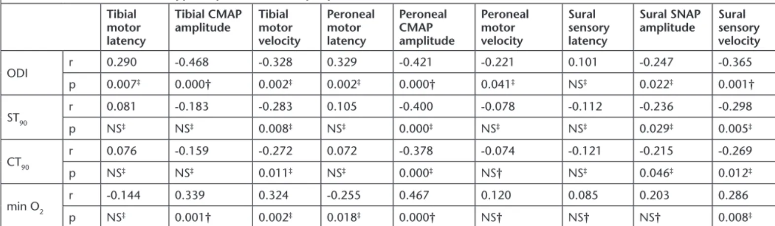
Table 2. Correlation between hypoxia parameters and peripheral nerves Tibial
motor latency
Tibial CMAP
amplitude Tibial motor velocity Peroneal motor latency Peroneal CMAP amplitude Peroneal motor velocity Sural sensory latency Sural SNAP
amplitude Sural sensory velocity ODI r 0.290 -0.468 -0.328 0.329 -0.421 -0.221 0.101 -0.247 -0.365 p 0.007‡ 0.000† 0.002‡ 0.002‡ 0.000† 0.041‡ NS‡ 0.022‡ 0.001† ST90 r 0.081 -0.183 -0.283 0.105 -0.400 -0.078 -0.112 -0.236 -0.298 p NS‡ NS‡ 0.008‡ NS‡ 0.000‡ NS‡ NS‡ 0.029‡ 0.005‡ CT90 r 0.076 -0.159 -0.272 0.072 -0.378 -0.074 -0.121 -0.215 -0.269 p NS‡ NS‡ 0.011‡ NS‡ 0.000‡ NS† NS‡ 0.046‡ 0.012‡ min O2 r -0.144 0.339 0.324 -0.255 0.467 0.120 0.085 0.203 0.286 p NS‡ 0.001† 0.002‡ 0.018‡ 0.000† NS† NS† NS† 0.008‡
CMAP: Compound muscle action potential, CT90: Percentage of cumulative sleep time with oxygen saturation <90%, min O2: Lowest oxygen saturation,
ODI: Oxygen desaturation index, SNAP: Sensory nerve action potential, ST90: Total sleep time with oxygen saturation <90% †NS: Not significant (p>0.05) (n=86)
†Pearson correlation
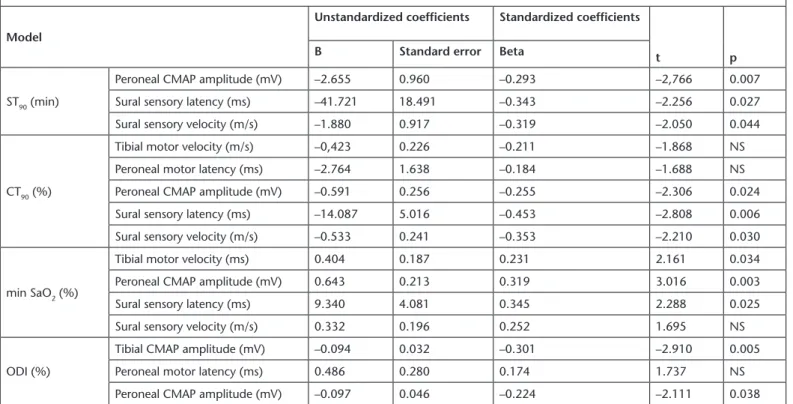
between the NCS results and the hypoxia parameters (Table 2). After adjusting for confounding factors, multiple logistic regression analyses revealed that prolonged sural sensory latency [odds ratio OR: 0.018, 95% confidence interval (CI): 0.001-0.550, p=0.021] and slowing of sural conduction velocity (OR: 0.816, 95% CI: 0.982-0.031, p=0.031) were independently associated with OSA. Furthermore, logistic regression analyses revealed that the ST90, CT90, min SaO2, and ODI were independently affecting the NCSs (Table 3).
Discussion
This study suggested that OSA and intermittent hypoxia have significant effects on motor and sensory peripheric nerve function. The peroneal nerve distal latency was prolonged, sural SNAP amplitude reduced, and sural nerve velocity decreased in the OSA group (Table 1). NCSs results showed a significant correlation with the hypoxia (Table 2). The hypoxia parameters were independent factors associated with nerve conduction abnormalities (Table 3). This subclinical peripheral neuropathy is therefore related to severe OSA and intermittent hypoxia seems to cause axonal and demyelinating neuropathy.
Our study shows a relationship among axonal neuropathy, hypoxia parameters and nerve conduction in OSA patients, which is consistent with previous reports (9,10,13). These studies also reported that subjects with severe OSA, min SaO2 ≤80%, and ST90 have lower amplitude for sensory and mixed nerve
action potentials, and that sural nerve velocity is slower in OSA patients (8,9). Similar to our study, hypoxia-associated axonal degeneration and less frequent demyelination were reported in COPD subjects in another study (16). These pathophysiological changes in nerve damage are more functional than structural because sleep apnea treatment revealed that the impaired nerve function was partially reversible (10,17).
In contrast to our study, Evlice et al. (12) described a decrease in tibial CMAP amplitude and slower tibial NCV in the OSA group, but they found no correlation among the severity of OSA, min SaO2, and neuropathy. The patients with OSA were older compared to our study and the authors only used the min SaO2 saturation as a hypoxia parameter (12).
The etiology of hypoxia-related diseases is complex. The episodic upper airway obstruction in patients with OSA results in hypoxia and hypercapnia. Hypoxia causes oxidative stress, sympathetic activation, metabolic dysfunction, systemic inflammation, endothelial dysfunction, and arterial disease together with pathological changes in morphology (2,18,19). Electrophysiological abnormalities are reported in approximately 95% of subjects with COPD associated with hypoxia (4,5). The pathophysiology of the nerve conduction abnormalities in patients with OSA is not exactly known. There are several possible explanations for the association between neuropathy of the peripheral nerve and OSA. Oxidative stress plays an important role in hypoxia-induced neuropathy. Endoneurial
Table 3. Factors affecting peripheral nerves in Obstructive Sleep Apnea Model
Unstandardized coefficients Standardized coefficients
t p
B Standard error Beta
ST90 (min)
Peroneal CMAP amplitude (mV) –2.655 0.960 –0.293 –2,766 0.007
Sural sensory latency (ms) –41.721 18.491 –0.343 –2.256 0.027
Sural sensory velocity (m/s) –1.880 0.917 –0.319 –2.050 0.044
CT90 (%)
Tibial motor velocity (m/s) –0,423 0.226 –0.211 –1.868 NS
Peroneal motor latency (ms) –2.764 1.638 –0.184 –1.688 NS
Peroneal CMAP amplitude (mV) –0.591 0.256 –0.255 –2.306 0.024
Sural sensory latency (ms) –14.087 5.016 –0.453 –2.808 0.006
Sural sensory velocity (m/s) –0.533 0.241 –0.353 –2.210 0.030
min SaO2 (%)
Tibial motor velocity (ms) 0.404 0.187 0.231 2.161 0.034
Peroneal CMAP amplitude (mV) 0.643 0.213 0.319 3.016 0.003
Sural sensory latency (ms) 9.340 4.081 0.345 2.288 0.025
Sural sensory velocity (m/s) 0.332 0.196 0.252 1.695 NS
ODI (%)
Tibial CMAP amplitude (mV) –0.094 0.032 –0.301 –2.910 0.005
Peroneal motor latency (ms) 0.486 0.280 0.174 1.737 NS
Peroneal CMAP amplitude (mV) –0.097 0.046 –0.224 –2.111 0.038
CT90: Percentage of cumulative sleep time with oxygen saturation <90%, min SaO2: Lowest oxygen saturation, ODI: Oxygen desaturation index, ST90: Total sleep time
with oxygen saturation <90%, CMAP: Compound muscle action potantial
†NS: Not significant (p>0.05)
hypoxia causes increased intercapillary distance resulting from nerve edema (7,20). The nerve capillary basement membrane thickens and endothelial cell hyperplasia and hypertrophy occur in hypoxic neuropathy. These microvascular angiopathic changes inhibit transport of nutrients and oxygen, altering nerve function and structure and predispose the patient to narrowing of the capillary lumen and vascular occlusion (6,21). In addition, the high-frequency intermittent hypoxia associated with OSA is characterized by a reoxygenation and hypoxemia cycle similar to ischemia-reperfusion injury and generates oxidative stress and reactive oxygen species. During severe hypoxemia, the ischemia and reperfusion sequence leads to the development of tissue acidosis, while intracellular sodium and calcium accumulation lead to sarcolemma damage (22,23). Furthermore, an excitotoxicity mechanism mediated by excitatory amino acids has been reported in hypoxic central nervous system damage. This excitotoxicity is thought to be related to oxidative stress-induced energy metabolism, leading to neurodegeneration (24,25). Last, hypoxic nerves develop resistance to ischemic conduction block (RIBC) due to reduced energy requirement and an increased efficiency of anaerobic glycolysis (26). RIBC is thought to be partially reversible with treatment (10). Chronic severe intermittent hypoxia exposure often results in RIBC, leading to axonopathies. RIBC is a result of an adaptive mechanism that depends on a critical threshold of nocturnal oxygen desaturation and microvascular disorders (10,26). The combination of changes in OSA associated hypoxia results in a high susceptibility to peripheral nerve injury. The strength of our study is that we have excluded patients with risk factors that cause neuropathy and evaluated detailed hypoxia parameters. Nevertheless, there are limitations of this work included its retrospective design and single-center analysis. Hyperinsulinemia combined with insulin resistance states, such as obesity, may contribute to nerve dysfunction (6). In our study, we did not exclude subjects with BMI ≥30 kg/ m2. But when we adjusted for age and BMI, the results were
persistent. Although our study excluded subjects with diabetes, hyperinsulinemia and insulin resistance were not evaluated. Another limitation is that we did not include upper extremity NCS, only lower extremity NCS.
In conclusion, our study revealed that OSA and associated hypoxia affect both motor and sensory nerve conduction, revealing that subclinical peripheral neuropathy is related to OSA. The severity of OSA and severe intermittent hypoxia may cause axonal and demyelinating neuropathy. Future studies comparing the results of our nerve conduction study with oxidative stress and hypercapnia are necessary to provide better insight into the pathophysiology of neuropathy in OSA. These studies should be multicentered, and prospective with a larger OSA patient population.
Acknowledgments
The author thanks Mustafa Agah Tekindal for performing the statistical analysis (PhD, Associate Professor, Facult of Medicine, Department of Biostatistics), Hakan Gökahmetoğlu (technician) for helping collecting data, Hüseyin Lakadamyalı for evaluating the all the polysomnographs (from the department of Pulmonology, Associate Professor), and Hatice Gönüllü (M.D. and MSc) for English editing.
Ethics
Ethics Committee Approval: This study was deemed to be
exempt from the requirement for informed consent by the Institutional Review Board of University due to its retrospective design. All procedures performed in studies involving human participants were in accordance with the ethical standards of the institutional and/or national research committee and with the 1964 Helsinki Declaration and its later amendments or comparable ethical standards.
Informed Consent: Oral informed consent was taken from all
the participants.
Peer-review: Internally peer-reviewed. Authorship Contributions
Desing: A.Y.A, S.A., Data Collection or Processing: A.Y.A., S.A., Analysis or Interpretation: A.Y.A., Literature Search: A.Y.A., Writing: A.Y.A.
Conflict of Interest: The authors declare that they have no
conflict of interest.
Financial Disclosure: The authors declare that this study
received no financial support.
References
1. Punjabi NM. The epidemiology of adult obstructive sleep apnea. Proc Am ThoracSoc 2008;5:136-43.
2. Sforza E, Roche F. Chronic intermittent hypoxia and obstructive sleep apnea: An experimental and clinical approach. Hypoxia 2016;4:99-108.
3. Beaudin AE, Waltz X, Hanly PJ, Poulin MJ. Impact of obstructive sleep apnoea and intermittent hypoxia on cardiovascular and cerebrovascular regulation. Exp Physiol 2017;102:743-63.
4. Kayacan O, Beder S, Deda G, Karnak D. Neurophysiological changes in COPD patients with chronic respiratory insufficiency. Acta Neurol Belg 2001;101:160-5.
5. Valli G, Barbieri S, Sergi P, Fayoumi Z, Berardinelli P. Evidence of motor neuron involvement in chronic respiratory insufficiency. J Neurol Neurosurg Psychiatr 1984;47:1117-21.
6. Malik RA, Masson EA, Sharma AK, Lye RH, Ah-See AK, Compton AM, Tomlinson DR, Hanley SP, Boulton AJM. Hypoxic neuropathy: Relevance to human diabetic neuropathy. Diabetologia 1990;33:311-8.
7. Low PA, Nukada H, Schmelzer JD, Tuck RR, Dyck PJ. Endoneurial oxygen tension and radial topography in nerve edema. Brain Res 1985;341:147-54.
8. Tapan ÖO, Tapan U, Kılıç US. Obstruktf uyku apne sendromlu hastalarda hipoksinin göz üzerine etkileri. JTSM 2018;5:62-6. 9. Ludemann P, Dziewas R, Soros P, Happe S, Frese A. Axonal
polyneuropathy in obstructive sleep apnoea. J NeurolNeurosurg Psychiatry 2001;70:685-7.
10. Mayer P, Dematteis M, Pepin JL, Wuyam B, Veale D, Vila A, Levy P. Peripheral neuropathy in sleep apnea. Am J Respir Crit Care Med 1999;159:213-9.
11. Agrawal D, Vohra R, Gupta P, Sood S. Subclinical peripheral neuropathy in stable middle-aged patients with chronic obstructive pulmonary disease. Singapore Med 2007;48:887-94.
12. Evlice A, Ugurel B, Baklan B, Oztura I. Neuropathy and dysautonomia in patients with obstructive sleep apnea syndrome. Arch Neuropsychiatr 2015;52:24-8.
13. Mihalji M, Lusic L, Dogas Z. The AASM manual for the scoring of sleep and associated events: Rules, Reduced evoked motor and sensory potential amplitudes in obstructive sleep apnea patients. J Sleep Res 2016;25:287-95.
14. Berry RB, Brooks R, Gamaldo CE, Harding SM, Lloyd RM, Marcus CL, Vaughn BV. American Academy of Sleep terminology and technical specifications, version 2.2. Darien IL: American Academy of sleep medicine. J Clin Sleep Med 2015;11:1253-4.
15. American Association of Electrodiagnostic Medicine. Guidelines in electrodiagnostic medicine. Muscle Nerve 1992;15:229-53. 16. Vila A, Reymond F, Paramelle B, Stoebner P, Quvrard-Hernandez AM,
Muller P, Pollak P. Neuropathies and chronic respiratory insufficiency: Electrophysiologic study. Rev Electroencephalgr Neurophysiol Clin 1986;15:331-40.
17. Dziewas R, Schilling M, Engel P, Boentert M, Hor H, Okegwo A, Ludemann P, Ringelstein EB, Young P. Treatment for obstructive sleep apnoea: Effect on peripheral nerve function. J Neurol Neurosurg Psychiatry 2007;78:295-7.
18. Song D, Fang G, Greenberg H, Liu SF. Chronic intermittent hypoxia exposure-induced atherosclerosis: A brief review. Immunol Res 2015;63:121-30.
19. Deng F, Wang S, Xu R, Yu W, Wang X, Zhang L. Endothelial microvesicles in hypoxic hypoxia disease. J Cell Mol Med 2018;22:3708-18.
20. Frolkis VV, Tanin SA, Gorban YN. Age-related changes in axonal transport. Exp Gerontol 1997;32:441-50.
21. Malik RA, Veves A, Masson EA, Sharma AK, Ah-See AK, Schady W, Lye RH, Boulton AJM. Endoneurial capillary abnormalities in mild human diabetic neuropathy. J Neurol Neurosurg Psychiatry 1992;55:557-61. 22. Iida H, Schmelzer JD, Schmeihel AM, Wang Y, Low PA. Peripheral
nerve ischemia: Reperfusion injury and fiber degeneration. Exp Neurol 2003;184:997-1002.
23. Wang Y, Schmelzer JD, Schmeichel A, Iida H, Low PA. Ischemia-reperfusion injury of peripheral nerve in experimental diabetic neuropathy. J Neurol Sci 2004;227:101-7.
24. Bondy SC, Le Bel CP. The relationship between excitotoxicity and oxidative stress in the central nervous system. Free Radic Biol Med 1993;14:633-42.
25. Ludolph AC, Riepe M, Ulrich K. Excitotoxicity, energy metabolism and neurodegeneration. J Inherit Metab Dis 1993;16:716-23. 26. Hampton KK, Alani SM, Wilson JI, Price DE. Resistance to ischaemic
conduction failure in chronic hypoxemia and diabetes. J Neurol Neurosurg Psychiatry 1989;52:1303-5.