A retrospective study on traumatic diaphragmatic hernias in cats
Omer BESALTI1, Zeynep PEKCAN2, Murat CALISKAN1, Z. Gamze AYKUT1
1Ankara University, Faculty of Veterinary Medicine, Department of Surgery, Ankara, Turkey 2Kirikkale University, Faculty of
Veterinary Medicine, Department of Surgery, Kirikkale, Turkey
Summary: The purpose of the study was to evaluate the clinical and surgical findings of traumatic diaphragmatic hernia in 52 cats and to determine the association with mortality rate. The medical and surgical records of fifty two cats that underwent surgical repair for traumatic diaphragmatic hernia were reviewed retrospectively. Survival rate was 82.7% (43/52) in cats. The most frequently herniated organ was liver (81%). This was followed by small intestines (67%), stomach (48%), omentum (38.5%), spleen (25%), pancreas (13.5%) and large intestines (8%) respectively. Thirty-one cases had acute and 21 cases had chronic diaphragmatic hernia and mortality rate was 16.1% and 19% respectively. Although the majority of the rupture was in the right side, death was not recorded in these cases. The mortality rates according to the rupture sites were 57% in central, 24% in ventral and 19% in the left side. On the other hand, it was recorded that the mortality rate was associated with the amount of organ herniation. The more excessive amount of organ herniation resulted in higher mortality rate. It was concluded that mortality rate could be related to the location of hernia, amount of herniated organs and time from trauma to the surgery.
Key words: Cat, diaphragm, hernia, rupture localization
Kedilerde travmatik diyafram fıtıkları üzerine geriye yönelik çalışma
Özet: Bu çalışmanın amacı kedilerde travmatik diyafram fıtıklarının klinik ve cerrahi bulguları ile mortalite oranı arasındaki ilişkiyi sunmaktır. Çalışmanın materyalini oluşturan 52 travmatik diyafram fıtıklı kedinin klinik ve operatif bulguları retrospektif olarak değerlendirildi. Kedilerdeki sağ kalım oranı %82.7 (43/52) olarak saptandı. Göğüs boşluğu içine en sık fıtıklaşan organın karaciğer olduğu (81%), bunu sırası ile ince bağırsaklar (%67), mide (%48), omentum (%38.5), dalak (%25), pankreas (%13.5) ve kalın bağırsakların (%8) izlediği belirlendi. Otuz bir olgunun akut, 21 olgunun ise kronik fazda olduğu ve mortalite oranlarının sırası ile %16.1 ve %19 olduğu ortaya konuldu. Diyafram üzerindeki rupturun en çok sağ tarafta görülmesine rağmen bu olguların hiçbirinde mortalite olmaması dikkati çekti. Yırtığın yerine göre mortalite oranı sırasıyla merkez %57, ventral %24 ve sol %19 olarak belirlendi. Ayrıca mortalite oranı ile fıtıklaşan organlar arasında ilişki tespit edildi; buna göre birden fazla organın fıtıklaşması ile ölüm oranının arttığı gözlendi. Sonuç olarak mortalite oranı ile fıtık yeri, fıtıklaşan organ sayısı, travma ve operasyon zamanı arasında ilişki olabileceği kanısına varıldı.
Anahtar sözcükler: Diyafram, fıtık, kedi, yırtık lokalizasyonu
Introduction
Traumatic diaphragmatic hernia is a protrusion of the abdominal viscera to the thoracic cavity through the disrupted diaphragm. Approximately 85% of the diaphragmatic hernias are of traumatic origin in cats (2, 4). The loss of diaphragmatic line and cardiac silhouette, appearance of the abdominal organs, intestinal loops or gas in the thorax or thin abdomen are characteristic radiographic signs (5, 8, 12). Almost all abdominal structures have been reported to protrude into the thoracic cavity; liver, stomach, small intestines, spleen, omentum, pancreas are the commonly herniated organs but on rare occasion the kidney have been reported to be in the herniated organs (3, 6, 8, 10, 14). Perioperative mortality rates ranging from 8-18% have been reported in the researches (7, 10).
The objective of this study is to determine the relationships between the clinical signs, duration of hernia, localization of the disruption, contents of hernia, concurrent disorders and mortality rates in cats.
Materials and Methods
The medical and surgical records of fifty two cats that underwent surgical repair of traumaticall diaphragmatic hernia at Department of Surgery Faculty of Veterinary Medicine Ankara University between May 2002 and April 2010 were reviewed. Data retrieved included age, sex, clinical signs and the duration of hernia which was presumed as the time of the traumatic event or onset of clinical signs and the time of admission. The nature of trauma, clinical signs, and location of diaphragmatic rupture, herniated organs, concurrent disorders, postoperative
complications and survival rate (at least 10 days after surgery) were detailed.
Clinical and physical examination findings at the time of admission were recorded in all 52 cats. The cases admitted for surgery in less than or including 14 days were accepted as acute, and more than 14 days were chronic. Abdominal and thoracic radiography was taken for the radiographic diagnosis. In debatable cases celiography or barium contrast radiography was performed.
Cephalosporin (20 mg/kg IV, Sefazol, Mustafa Nevzat, Turkey) and dexamethasone (0.5 mg/kg IV) was administered to prevent infection and reexpansion injury of lungs respectively. General anesthesia was induced with propofol (4-6 mg/kg, IV, Diprivan, AstraZeneca, UK), and the trachea was intubated and anesthesia was maintained with isoflurane (Forane, Abbott, UK) in oxygen. The cats were placed in dorsal recumbency with the head positioned toward the top of the table and the table was tilted at a 30 to 40 degree to facilitate gravitation of abdominal viscera from the thorax. All cats breathed 16-18 times/min, 0.09 ml/kg tidal volume with 18 cmH2O positive pressure ventilation with an
automatic ventilator and received an infusion of 10 ml/kg/hr Lactated Ringer’s solution during surgery. Surgical herniorrhapy was carried out via a ventral midline celiotomy approach to the diaphragm in all cases. Hernia was repaired with monoflament nonabsorbable suture material in pattern of simple continuous or simple interrupted form (Prolene 3/0, Ethicon, Johnson & Johnson, Brussels, Belgium). After repairing the hernia, thoracocentesis were carried out with a three ways cannula placed through the diaphragm and the air within the thorax was aspirated with an injector to achieve negative pressure. Analgesia was provided with 2 mg/kg ketoprofen postoperatively for 3 days.
Results
Fifty two cats that were examined met the inclusion criteria in this study. Except for two Angora cats, all of the subjects were domestic short hair cats. Thirty cats were male and twenty two cats were female. Mean age was 22.4 months, varying from 3 months to 5 years.
Data regarding cause of trauma was available for 26 of 52 (50 %) cats, and the etiology of trauma was traffic
accidents in 17 cats, fall from the height in 7 cats and dog attacks in 2 cats. Even though the causes of trauma for the remaining 26 cats were unknown it was speculated as traumatic because they were either missing for a period of time and found, or recently adopted.
The most common clinical and physical examination findings were dyspnea in 31 cats (59.6%), anorexia in 8 cats (15.4%), open mouth breathing in 8 cats (15.4%), abdominal respiration in 5 cats (9.6%), exercise intolerance in 5 cats (9.6%), tachypnea in 1 cat (1.9%), coughing in 1 cat (1.9%) and vomiting in 1 cat (1.9%).
Radiographic diagnosis was carried out by plain radiography in 42 cats. Cranial displacement of the liver, displacement of the stomach and/or intestinal loops into the thorax and loss of diaphragmatic line were the radiographic signs in these cases. Additional imaging tests were needed to confirm the diagnosis in 10 cases. Positive contrast celiography was performed in 8 cats. In addition to celiography, thoracic ultrasonography was applied in two cases with pleural effusion. Indirect gastrointestinal radiography with barium was applied in the other 2 cases.
Concomitant disorders were identified on clinical and physical examination and /or radiography or during surgery in 16 of 52 cats. Five of 16 cats with femur fractures, two with coxofemoral luxations, 2 with pelvic fractures, 2 with vertebral body fractures and paraplegia, 1 with a tibial fracture, 1 with a rib fracture, 1 with a pericardial effusion and abscess formation on the cheek, 1 with a traumatic abdominal hernia, and 1 with an open wound in the pelvic region were observed. One cat was pregnant before the surgery and ovariohysterectomy was performed as the owners’ request during the same surgery. The elapsed time from the trauma to the diagnosis ranged from one day to one year (mean 54 days, median 7 days). Diagnosis of the hernia was achieved within 7 days of trauma in 24 cats, between 7 and 14 days in 7 cats and longer than 14 days (chronic) in 21 cats.
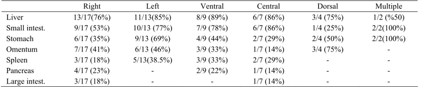
The location of disruption in the diaphragm was in the right-side in 17 cats, in the left-side in 13 cats, in ventral (over the sternum) in 9 cats and central (over the esophageal, vena caval and /or aortic hiatus) in 7 cats, in dorsal (under the vertebrae) in 4 cats and multiple in 2 cats.
Table 1: Herniated organs and their percentage according to the localization. Tablo1: Fıtıklaşan organlar ve lokalizasyona göre fıtıklaşma yüzdeleri.
Right Left Ventral Central Dorsal Multiple
Liver 13/17(76%) 11/13(85%) 8/9 (89%) 6/7 (86%) 3/4 (75%) 1/2 (%50) Small intest. 9/17 (53%) 10/13 (77%) 7/9 (78%) 6/7 (86%) 1/4 (25%) 2/2(100%) Stomach 6/17 (35%) 9/13 (69%) 4/9 (44%) 2/7 (29%) 2/4 (50%) 2/2(100%) Omentum 7/17 (41%) 6/13 (46%) 3/9 (33%) 1/7 (14%) 3/4 (75%) - Spleen 3/17 (18%) 5/13(38.5%) 3/9 (33%) 2/7 (29%) - - Pancreas 4/17 (23%) - 2/9 (22%) 1/7 (14%) - - Large intest. 3/17 (18%) - - 1/7 (14%) - -
The most frequently herniated organs were liver (42/52; 81%) in all localizations. This was followed by small intestines (35/52; 67%), stomach (25/52; 48%), omentum (20/52; 38.5%), spleen (13/52; 25%), pancreas (7/52; 13.5%) and large intestines (4/52; 8%), respectively. The herniated organs according to their side and their percentage were given in table 1.
In 6 cases, the rupture in the hernia had to be enlarged to pull the liver backwards, because of liver congestion and enlargement. In all cases the hernia was repaired by median celiotomy without sternotomy. Adhesions between the liver and diaphragm or thoracic viscera were recorded in 11 cases and these adhesions were separated by gentle dissection at the expense of diaphragm. After hernial contents replaced, atalectesia in lung lobe was recorded in 13 cases especially in caudal lung lobes and 3 of them died.
All deaths occurred intraoperatively (4/9) or at the postoperative early period (5/9, up to 5 hours) because of cardio or cardiopulmonary arrest. Among 9 dead cases, 3 of them had adhesions between the liver and thoracic viscera, and 3 had atelectasia within the lung lobes.
Forty three out of 52 cats (82.7%) were discharged alive. In one cat, frothy fluid accumulated within the endotracheal tube after the operation. The fluid was aspirated when it accumulated. Furosemide (4 mg/kg, IV) was administered. The oxygen supplementation was continued via mask after extubation. As the respiratory function deteriorated, the cat was reintubated, the fluids were aspirated and positive pressure ventilation was initiated. However, the cardiopulmonary arrest was eventually followed by death.
One cat was euthanized 10 days after the surgery because of paraplegia resulting from the same traumatic incident that caused traumatic diaphragmatic hernia. Although there were no clinical signs related to the respiratory disorders, this cat showed no neurological improvement and on owner’s request euthanized.
Among 9 dead cases, the major cause was the amount of excessive organ herniation. Except for one case; the others had at least two different herniated organs. In all dead patients, liver was exclusively herniated contents. In addition to liver, 1 with small intestines, 2 with stomach and small intestines, 1 with omentum and small intestines, 1 with spleen and small intestines, 2 with spleen, stomach and small intestines and 1 with stomach, spleen, omentum, pancreas and small intestine were noted.
Regardless of the organ herniation, among 9 dead cases the common point was localization of the rupture. Although the majority of the disruption was in the right side in our study, the deaths were not recorded among 17 right sided herniated cases. Considering the localization
of the disruption, among 7 cases with centrally located hernia, 4 (57%) of them died. The other localization and death ratio was 2 deaths in 9 cases (22%) with ventrally located hernia, 2 deaths in 13 cases (15%) with left-sided located hernia and 1 death in 2 cases (50%) with multiple side hernia.
The elapsed time from trauma to surgery varied from 1 day and 1 year for dead cases. Among 9 dead cases, 5 had acute diaphragmatic hernia and the others were chronic. Forty three cats were discharged from the hospital. None developed evidence of recurrence in 10 days of period postoperatively.
Discussion and conclusion
In our study, the location of hernia in diaphragm seems to be related to the mortality rate besides the chronicity. Especially in cases with centrally located hernia the mortality rate is the highest when compared to the other sites.
As in other studies (1, 5, 9, 10, 13), the most commonly reported sex was male in our study. Liver was the most frequently herniated organ and this was followed by small intestines, stomach, omentum, spleen, pancreas and large intestines (3, 5, 10, 17).
The location of hernia in this study was parallel to the several reports (1, 5, 8), as the majority of patients had right sided hernia. However some reports assumed that the liver has a protective effect on the right side of the diaphragm and could serve as a barrier for herniation (13). Except Wilson et al. (1971), there was no report that focused on the location of the hernia. In the authors’ knowledge, this is the first report to evaluate the relationship between exact location of hernia and mortality rate. As a result of our study, the higher percentage of liver herniation can be speculated that the liver which is the adjacent massive organ to the diaphragm cannot play its role in prevention of hernia, in contrast to this hypothesis it can potentialize the effect of trauma. On the contrary, tubular structures like intestines and stomach can absorb the shock much during trauma.
Herniated organs were associated with the anatomical position of the organ and the location of the disruption. Liver was the predominantly herniated organ as several authors stated (5, 10). However, there were some disagreement between the reports. Some authors (5) mentioned stomach was the predominant herniated organ in the left side; however small intestine was prominent in the left side in our study. Although the stomach is the closest organ to the diaphragm on the left side, it was the third predominant organ within the herniated organs on the left side. This can be explained by intestines have more mobility compared with the stomach.
In our study, interval from trauma to surgery varied from one day to one year. Some authors assumed 2 weeks for chronicity and they reported that the mortality rate for the chronic diaphragmatic hernia was higher because of complications such as pneumothorax, complications during the breakdown of the adhesions, liver lobe torsion or necrosis, lung lobe torsion or strangulation of the intestines within the hernial ring (9). In our study among 21 chronic diaphragmatic hernia cases, 5 died. Two of five had liver congestion, one had liver adhesion to the collapsed lung and it was freed without problem. The only common point with them was liver herniation. Neither liver nor lung lobe torsion or necrosis was seen in all death series. All cats that died had herniation of liver, whereas 35 of 43 cats that survived had liver herniations. Although all dead cases included liver herniations in the study of Schmiedt et al (2003), reported that the contents of hernia were not significantly associated with the mortality. It was similar to our study that all dead patients had liver herniations. Additionally in our study, amount of organ herniations had an important role on the mortality rate, because except for one case, all had more than two organ herniations.
Removal of the abdominal organs from thorax to the abdomen rarely caused difficulty. However some authors (9) combined median sternotomy with celiotomy to improve visualization and division of the adhesions. Stokhof (1986) performed either lateral thoracotomy or median sternotomy upon midline laparotomy for diaphragmatic repair. This author reported that if the location of the rupture was well-defined, thoracic approach provided best view of the diaphragm. As the thoracotomy and sternotomy was more painful operations, they could cause significant hypoventilation and were not recommended for diaphragmatic hernioraphy (4). In these case series, ventral median celiotomy provided adequate exposure for the diaphragmatic rupture. In this study, liver could not be replaced through the rupture because of the congestion and swelling in 6 cases and the hernia was enlarged as some authors stressed (15). Afterwards exposure was apparent and replacement of the liver or division of adhesions was applied easily.
Although the majority of the disruption was in the right side in our study, death was not recorded among 17 right sided herniated cases. This can be explained by location of the heart and the development of heart failure after the replacement of the herniated organs on the left side during or after the hernioraphy operation.
The diagnosis of diaphragmatic hernia was especially done by survey thoracic radiography. The presence of pleural effusion, pleural mass or herniations of relatively
small volumes of abdominal structures can make the diagnosis difficult. In debatable cases celiography, barium contrast radiography or ultrasonography can be used (9, 12, 16). Celiography was the most frequently utilized supplemental imaging technique in our study because of its quick application. However in 2 of 8 celiography performed cases definitive diagnosis could not be performed, and it was carried out by ultrasonography.
Schmiedt (2003) reported that mortality rate was greater in cats that had concurrent injuries than those did not. However in our study, similar to findings of Kraje et al (2000), the mortality rate was not different from the cats with or without concurrent injuries. Among 52 cats, 16 of them had concurrent injuries and only 3 of them died.
The cause of pulmonary edema is rapid re-expansion of the chronic collapsed lung and symptoms developed within a few hours after lung re-expansion (2, 10, 11). Positive pressure ventilation after replacement of the abdominal organs and aspiration of the air from the thorax via a three way cannula can cause rapid reexpansion of the lungs. It was advised to aspirate the air over a 12 hour period by use of an underwater seal or multiple aspirations. After pulmonary edema developed, it was suggested to apply hypertonic colloid solutions, diuretics and dobutamine (11). Oxygen radical scavengers can be administered to prevent pulmonary edema (2, 11). The circulation of liver is also disrupted in cats with hepatic herniation. In these case series, dexamethasone was used for attenuating reexpansion injury of lungs, and massive release of the toxins into the circulation because of hepatic strangulation or vascular compromise. The air was aspirated from the thorax at once through the diaphragm just after the repair. However, the pulmonary edema occurred in one cat and not recovered despite of all efforts.
In conclusion, the location of hernia, amount of herniated organs and time from trauma to surgery are all responsible for the perioperative mortality rate.
References
1. Bellenger JR, Hunt GB, Goldsmid SE, Pearson MRB (1996): Outcomes of thoracic surgery in dogs and cats. Aust Vet J, 74: 25-30.
2. Fossum TW (2002): Traumatic Diaphragmatic Hernia. In: TW Fossum (Ed), Small Animal Surgery, 2nd ed.,
Mosby, Missouri, pp795-798
3. Gibson TWG, Brisson BA, Sears W (2005): Perioperative survival rates after surgery for diaphragmatic hernia in dogs and cats: 92 cases (1990-2002). J Am Vet Med Assoc, 227: 105-109.
4. Hunt GB, Johnson KA (2003): Diaphragmatic, pericardial, and hiatal hernia. In: D Slatter (ed.) Textbook
of Small Animal Surgery, 3rd ed., Saunders, Philadelphia, pp 471-487.
5. Hyun C (2004): Radiographic diagnosis of diaphragmatic hernia: Review of 60 cases in dogs and cats. J Vet Sci, 5: 157-162.
6. Katic N, Bartolomeus E, Bohler A, Dupre G (2007): Traumatic diaphragmatic rupture in a cat with partial kidney displacement into the thorax. J Small Anim Pract,
48: 705-708.
7. Kraje BJ, Kraje AC, Rohrbach BW, Anderson KA, Marks SL, Macintire DK (2000): Intrathoracic and
concurrent orthpedic injury associated with traumatic rib fracture in cats: 75 cases (1980-1998). J Am Vet Med Assoc, 216: 51-54.
8. Marlof A, Kraft S, Lowry J, Pelsue D, Veir J (2002): Radiographic diagnosis- Right kidney herniation in a cat. Vet Radiol Ultrasound, 43: 237-240.
9. Minihan AC, Berg J, Evans KL (2004): Chronic diaphragmatic hernia in 34 dogs and 16 cats. J Am Anim Hosp Assoc, 40: 51-63.
10. Schmiedt CW, Karen MT, Stevenson M (2003): Traumatic diaphragmatic hernia in cats: 34 cases (1991-2001). J Am Vet Med Assoc, 222: 1237-1240.
11. Stampley AR, Waldron DR (1993): Reexpansion pulmonary edema after surgery to repair a diaphragmatic hernia in a cat. J Am Vet Med Assoc, 203: 1699-1701.
12. Stickle RL (1984): Positive-contrast celiography (peritoneography) for the diagnosis of diaphragmatic hernia in dogs and cats. J Am Vet Med Assoc, 185: 295-298. 13. Stokhof AA (1986): Diagnosis and treatment of acquired
diaphragmatic hernia by thoracotomy in 49 dogs and 72 cats. Vet Q, 8: 177-183.
14. Stork CK, Hamaide AJ, Schwedes C, Clercx CM, Snaps FR, Balligand MH (2003): Hemiurothorax
following diaphragmatic hernia and kidney prolapse in a cat. J Feline Med Surg, 5: 91-96.
15. Walker RG, Hall LW (1965): Rupture of the diaphragm: Report of 32 cases in dogs and cats. Vet Rec, 77: 830-837. 16. White JD, Tisdall PL, Norris JM, Malik R (2003):
Diaphragmatic hernia in a cat mimicking a pulmonary mass. J Feline Med Surg, 5: 197-201.
17. Wilson GP, Newton CD, Burt JK (1971): A review of 116 diaphragmatic hernias in dogs and cats. J Am Vet Med Assoc, 159: 1142-1145.
Geliş tarihi: 26.04.2010 / Kabul tarihi: 24.09.2010
Address for Correspondance:
Omer Besalti, Ankara University, Faculty of Veterinary Medicine,
Department of Surgery, Diskapi, 06110, Ankara, Turkey Telephone number: +90535 2840546