Ankara Üniversitesi Tıp Fakültesi Mecmuası 2008, 61(2)
Araştırma Makalesi / Research Article
Utility Of The Serum C - Reactive Protein And Procalcitonin For
Detection Of Occult Bacterial Infection In 3-36 Month Old Children
Üç-36 Ay Arası Çocuklarda Gizli Bakteriyel Enfeksiyonları Saptamada C-reaktif Proteinin Ve Prokalsitoninin KullanıIabilirliği
Ayhan Abacı
1, Mehmet Ali Öktem
2, Edip Ünal
1, Mehmet Atilla Türkmen
11Dokuz Eylül Üniversitesi Tıp Fakültesi, Çocuk Sağlığı ve Hastalıkları, Anabilim Dalı
2Dokuz Eylül Üniversitesi, Tıp Fakültesi, Klinik Mikrobiyoloji, Anabilim Dalı
Received: 12.09.2008 • Accepted: 08.12.2008 Corresponding author
Uzm. Dr. Ayhan Abacı
Dokuz Eylül Üniversitesi Tıp Fakültesi Çocuk Sağlığı ve Hastalıkları Anabilim Dalı Phone : (505) 477 66 04 E-mail address : [email protected]
Aim: To assess the utility of serum C-reactive protein (CRP) and Procalcitonin (PCT) as a screening
method for occult bacterial infection (OBI) in 3-36 month old children.
Materials and Methods: Febrile children, who were admitted to emergency department with
ages ranging from 3 to 36 months, temperatures > 38.3°C, and clinically undetectable source of fever, were enrolled in this study. Sex, age, degree of fever, Yale Observation Scale (YOS) score, antibiotic treatment and hospitalization were recorded at the time of the initial evaluation. Rou-tine urine analysis, white blood cell count (WBC) count, CRP and PCT determination tests were performed at the initial evaluation. Viral serology was done and patients having specific IgM posi-tivity during the acute phase and/or a four-fold increase in specific IgG titer during convalescence phase were considered to be positive. A chest radiograph, blood culture and urine cultures were taken from all patients. The patients were divided into two groups: OBI group and viral infection group. These two groups were compared for each predictor.
Results: There were 10 patients with OBI, six with urinary tract infection two with pneumonia,
one with bacterial meningitis and one with bacteremia and 16 patients with viral infection, (five Adenovirus, five Respiratory syncytial virus, four Enterovirus, one Parainfluenza type 1, one Epste-in-Barr virus. Regarding the CRP values, there was no statistical difference between OBI and viral infection groups (respectively, median 44.84 mg/L and 23.3 mg/L, p=0.097), but PCT values were significantly higher in OBI group (respectively, median 2.38 ng/mL and 0.28 ng/mL, p=0.014).
Conclusion: This study showed that PCT is superior than CRP in detecting OBI and could be used
for screening purposes in pediatric emergency departments while elevated levels might be found in some viral infections.
Key Words: Occult bacterial infection, C-reactive protein, Procalcitonin
Amaç: Üç–36 ay arası gizli bakteriyel enfeksiyon riski taşıyan hastaların saptanmasında C-reaktif
proteinin (CRP) ve Prokalsitoninin (PCT) kullanılabilirliğini saptamak.
Gereç ve Yöntem: Yaşları 3-36 ay arası olan, 38.3oC üzerinde ateş yüksekliği şikayeti ile acil
servi-se başvuran ve ateş odağı saptanamayan hastalar çalışmaya alındı. Başvuru anında, cinsiyetleri, yaşları, ateş yüksekliğinin derecesi, Yale Gözlem Ölçeği (YGÖ) skoru, hastaneye yatma ve antibi-yotik başlanma durumları kaydedildi. Başvuru anında, rutin idrar analizi, beyaz küre sayıları, CRP ve PCT’nin düzeylerinin ölçümü için testler yapıldı. Viral serolojik inceleme yapıldı ve akut faz dö-nemindeki spesifik IgM pozitifliği ve/veya konvalasan fazda spesifik IgG düzeylerinde 4 katlık artış pozitif olarak kabul edildi. Tüm hastalardan kan kültürü, idrar kültürü yapıldı ve akciğer grafileri çekildi. Hastalar gizli bakteriyal enfeksiyon ve viral enfeksiyon grubu olmak üzere ikiye ayrıldı. İki grup verileri her bir belirleyici açısından karşılaştırıldı.
Bulgular: Gizli bakteriyel enfeksiyonlu 10 hastada, altı idrar yolu enfeksiyonu, iki bronkopnömoni,
bir menenjit, bir bakteriyemili hasta saptanırken, viral enfeksiyon saptanan 16 hastada, beş Ade-novirus, beş Respiratory syncytial virus, dört Enterovirus, bir Parainfluenza Tip1 ve bir Epstein-Barr virus enfeksiyonlu hasta saptandı. Gizli bakteriyel enfeksiyon grubu, viral enfeksiyon grubu ile CRP değerleri açısından karşılaştırıldığında istatistiksel olarak anlamlı fark saptanmazken (sırasıyla, or-tanca 44.84 mg/L ve 23.3 mg/L, p=0.097), PCT değerleri istatistiksel olarak anlamlı saptandı (sıra-sıyla, ortanca 2.38 ng/mL, 0.28 ng/mL, p=0.014).
Sonuç: Bu çalışma, çocuk acil bölümlerinde PCT’nin gizli bakteriyel enfeksiyonları saptamada
CRP’ye göre daha üstün bir belirleyici olduğunu ve tarama amaçlı kullanılabileceğini, ancak bazı viral enfeksiyonlarda yüksek saptanabileceğini göstermiştir.
Febrile infants and children frequ-ently present to primary care and emergency physicians. The majo-rity of these children are younger than 3 years. Most have an appa-rent source of infection (ie, a viral respiratory infection, acute otitis media, or enteritis). However, 20% of febrile children have fever wit-hout source of infection after his-tory and physical examination and occult bacterial infections (OBI) (urinary infection, pneumonia, oc-cult bacteremia, early bacterial me-ningitis, etc.) develop in 11.3% of them (1-4).
The most important feature of Pro-calcitonin (PCT) is that it increases in bacterial infections, it does not increase or slightly increases in vi-ral and local infections and it dec-reases in a very short time upon a successful antibiotic treatment (5-13). It has been found out that PCT starts to increase in the first 2-3 hours, reaches the peak level in 6-8 hours, stays high for 24 hours and it has an half-life of approxi-mately 25-30 hours because of its rapid kinetic effect (12-14). The kinetic of serum C-reactive prote-in (CRP) is slower than that of PCT and to increase within 4 – 6 h after onset of inflammation. It then do-ubles every 8 h and peaks at app-roximately 36 – 50 h (15). Due to this feature of PCT, the monitoring of the patient is more reliable and the early differential diagnosis of bacterial and viral infections is pos-sible (16,17).
The objective of this study was to in-vestigate the utility of PCT and CRP and to compare them in the diffe-rential diagnosis of OBI and viral infections in the patient group of 3-36 month old children who are admitted to pediatric emergency services for high fever complaints with fever without a focus despite medical history and physical exa-mination.
Materials and Methods
Children aged 3 to 36 months of age consulting the Pediatric Emergency Department of the Dokuz Eylul Uni-versity, Faculty of Medicine (Izmir/ Turkey) with an axillary temperatu-re ≥ 38.3°C and without localizing signs of infection in their history or at physical examination were pros-pectively enrolled. Each infant was examined by a pediatric resident who took a complete history, perfor-med a physical examination, recor-ded the degree of fever and deter-mined a clinical score according to Yale Observation Scale (YOS) scores (18).
Routine urine analysis, white blood cell (WBC) count and CRP determi-nation were performed at the initial evaluation. Chest radiography was taken for all study patients regard-less of respiratory symptoms. Pyuria was defined as ≥5 white blood
cells/high-power field in a centrifu-ged urine sample. Urine for culture was obtained by urethral catheteri-zation using standard sterile tech-nique from all the patients having pyuria and the patients under the age of 12 months without pyuria. Blood culture, viral serology panel and 2 mL blood were taken from all patients for the study of PCT levels. Blood taken was centrifuged for vi-ral serology and PCT examinations for 10-15 minutes in 3000 rpm and then serums were separated and stored in - 70oC. At the initial
admis-sion, lumber puncture was done in all patients with poor general con-dition (fever with convulsion, slee-piness, toxic appearance etc.). Pa-tients with good general condition were followed clinically.
The exclusion criteria for potential study subjects were: having high fe-ver for more than a week with cli-nical localization symptoms; antibi-otic treatment within 48 hours prior
to admission to the hospital; vacci-nation in the days before the study, which may have caused the febrile syndrome; surgery performed wit-hin 7 days before the start of the study; any chronic pathology that could alter CRP values; a history of prior urinary tract infection, patho-logy involving malformation of the kidney or of the urinary tract and vesicoureteral reflux.
Patients with more than >104 CFU/mL
pathogenic bacteria in urine cultu-res obtained by catheter (19), having a reproduction in blood and cereb-rospinal fluid (CSF) cultures, having infiltration in chest radiography and having negative test results for viral serology formed the OBI group. Patients with positive Immunoglo-bulin M (IgM) upon examination of viral serology and/or having a four-fold increase of antibody in Immu-noglobulin G (IgG) titer of control serum taken in the second week and having no positive bacteriologic cultures formed the viral infection group.
Phone numbers of patients in both groups whose general condition se-emed good were taken for monito-ring future complications and data about the prognosis was collected by getting into touch with the pati-ents on 1st, 2nd, 3rd, 7th and 15th days. Physical examinations and la-boratory analysis for etiologic diag-nosis of patients with high fever and with new symptoms or diagnosis were repeated. Antibiotic treatment was started in both patients with oc-cult bacterial infections and patients who have been recalled for reexami-nation and found having an infecti-on focus.
Informed consent was obtained from parents or guardians and the study was approved by board of ethics.
Radiological Evaluation
The diagnosis of “occult pneumonia” was based on a radiologic diagnosis of lobar infiltrate or pneumonia in a patient with no abnormalities noted on physical examination.
Evaluation of Blood Cultures
Blood culture samples were taken af-ter systemic examination of patients and before decreasing the tempera-ture. The area was cleaned by po-vidon iodine and 3 mL blood was taken after waiting for 30 seconds. The blood taken was cultured in BACTEC which is blood culture medium and evaluated by BACTEC 9240 device (Becton Dickinson Ca-nada U.S.A).
Organisms which reproduce within the first 48 hours were passaged to their mediums like bloody agar and chocolate agar. The samples were examined in terms of newly repro-duced colonies after incubation of samples at 35-37oC for 24 hours.
Samples having more than one dif-ferent colony were determined as contaminated.
Serologic Diagnosis of Viral Infections
Adenovirus, Enterovirus, Parainfluenza type 1,2,3, Epstein-Barr virus (EBV), Respiratory syncytial virus (RSV), Influenza A and B viruses were in-vestigated in serums of the patients by using Respiscreen Clinikit (Orge-nium Helsinki, Finland), an indirect commercial EIA kit. Patients having specific IgM positivity during the acute phase and/or a four-fold inc-rease in specific IgG titer during convalescence phase were included in the viral infection group. The sen-sitivity and specificity values of viral serologic markers were all above 89% (range, 89.2-95%) (according to manufacturer’s catalog: http:// www.orgenium.com/Orgenium%20 Catalog %202008.pdf).
White Blood Cell Count and Measu-rement of CRP and PCT Levels Number of white blood cells was
co-unted by STKS coulter LH 750 de-vice (Coulter Electronics, Miami, Florida). Plasma concentrations of CRP were measured by means of particle-enhanced immunolometry with the Behring nephe-lometer using N Latex CRP mono reagent (Dade Behring Marburg GmbH, Germany). An immunolu-minometric assay for the measure-ment of PCT serum concentration was performed with LUMItest PCT kit (Brahms Diagnostica GmbH, Berlin, Germany). The cut-off valu-es for determination of OBI were accepted as ≥15 000 cell/mm3 for WBC, ≥40 mg/L for CRP, ≥0.5 ng/ mL for PCT (13,19).
Statistical Analysis
Statistical analysis was made by using SPSS Software 11.0. Compari-son between group averages was made by non-parametric tests. To this aim, Mann-Whitney U-test was used for comparison between the averages of two groups. Data were given as median (range). Spear-man correlation analysis was used to determine the correlation bet-ween the two groups. The
deter-mined value of p<0.05 was consi-dered significant.
Results
A total of 87 patients participated in the study and 10 patients were de-termined as having OBI and 16 pa-tients were determined as having a viral infection (Figure 1).
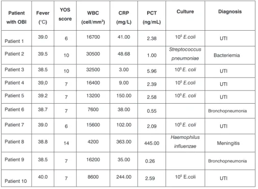
There were six patients with urinary tract infection, two with bronc-hopneumonia, one with meningi-tis, one with bacteremia among 10 patients with OBI, there were five patients with Adenovirus, five with Respiratory syncytial virus (RSV), four with Enterovirus, one with Parainfluenza type 1 and one with Ebstein–Barr virus (EBV) infection among 16 patients with viral infec-tion (Table 1 and 2). Escherichia coli was responsible for most of the urinary tract infections, Strep-tococcus pneumoniae was isola-ted in the bacteremia patient and Haemophilus influenzae was iso-lated in CSF culture in the patient with meningitis.
Both groups were compared in terms of average ages, genders, YOS scores, antibiotic treatment, and hospitalization, degree of fever, WBC count, PCT and CRP Figure 1. Flow chart of the present study
* Four fold IgG antibody increment of control serum
** Did not give permission us for taking control serum because their children had already ben recovered
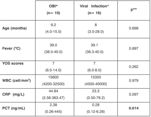
levels. Regarding the CRP values, there was no statistical difference between occult bacterial infection and viral infection groups (me-dian 44.84 mg/L [range 2.56 to 363.47], 23.3 mg/L [range 0.50 to 79.2], p=0.097), but PCT values were significantly higher in OBI group (median 2.38 ng/mL [range 0.26 to 445], 0.28 ng/mL [range 0.12 to 6.28], p=0.014) (Table 3). There was no statistically signifi-cant difference between the two groups with regard to YOS scores, application ratio of antibiotic tre-atment, degree of fever and WBC count (p>0.05). There was a sta-tistically high hospitalization ratio in OBI group than viral infection group (62.5%, 32.5%, p=0.03). It was determined that all patients in OBI group and 81.3% of those in viral infection group had antibi-otic treatment. There was no sta-tistically significant difference bet-ween the two groups with regard to treatment (p=0.262).
While there was a positive correlati-on between PCT levels and YOS score and CRP levels (r=0.778, p=0.001; r=0.759, p=0.001, res-pectively) determining OBI, there was no significant correlation bet-ween PCT and WBC count, degree of fever (p > 0.05).
Procalcitonin level was found to be more than 0.5ng/mL for 90% of pa-tients in the OBI group and 37.5% of the viral infection group. Ove-rall, two patients were diagnosed ashaving Adenovirus, one patient as having EBV, two patients as ha-ving Enterovirus, two patients as having RSV who were among the patients with viral infection and who have high PCT values. CRP was found to be greater than 40 mg/L in 60% of OBI cases and in 30% of viral infection cases. While the number of patients with CRP below 40 mg/L was 4 (40%), PCT level of one of the patients with OBI was found to be below 0.5 ng/mL.
Discussion
The management of febrile young children without apparent source of infection and the use of accura-te diagnostic laboratory methods remains controversial. In 1993, Baraff published a consensus pra-ctice guideline for management of febrile infants aged 3-36 months and two options were suggested for febrile nontoxic-appearing in-fants: obtaining a blood culture and giving empirical antimicrobial therapy (ceftriaxone) or obtaining a blood culture and giving empi-rical antimicrobial therapy in case of a WBC count equal to or gre-ater than 15000 cell/mm3 (20). In
spite of the fact that a consensus has not been reached on the eva-luation and management of fever in young infants without an obvi-ous focus of infection, most would agree that no single laboratory test has been shown to reliably iden-tify young infants at high or low risk of having bacterial illness. Many inflammatory determinants such as Interleukin (IL)-1, IL-6, tu-mor necrosis factor-, fibronectin, -integrin, CRP have been studied up to now for evaluation and true diagnosis of children between 3-36 months with fever and with fe-ver without a focus (2,6,7). Howe-ver, the developed clinical scoring methods and assaying methods of many inflammatory determinants are neither useful nor cheap and simple and they take a long time to obtain the results. With the identification of reliable determi-nants providing the diagnostic ac-curacy of OBI, the use of antibioti-cs will be limited, development of bacterial resistance to antibiotics and complications will be reduced and unnecessary hospitalization for the administration of parental antibiotics in viral infections will be prevented.
It has been reported that the risk of
UTI: Urinary Tract Infection, YOS: Yale observation scale, CRP: C-reactive protein, PCT: Procalcitonin,
OBI: Occult Bacterial Infection, WBC: White blood cell
Table 1: Clinical and laboratory finding of patients with occult bacterial infection
Patient with OBI Fever (°C) YOS score WBC (cell/mm3) CRP (mg/L) PCT (ng/mL) Culture Diagnosis Patient 1 39.0 6 16700 41.00 2.38 10 5 E.coli UTI Patient 2 39.5 10 30500 48.68 1.00 Streptococcus pneumoniae Bacteriemia Patient 3 38.5 10 32500 3.00 5.96 105 E. coli UTI Patient 4 39,0 7 16400 9.00 2.39 105 E.coli UTI Patient 5 39.2 7 13200 150.00 2.58 105 E. coli UTI Patient 6 38.7 7 7600 38.00 0.55 Bronchopneumonia Patient 7 39.0 6 15600 102.00 2.09 105 E. coli UTI Patient 8 38.8 14 4200 363.00 445.00 Haemophilus influenzae Meningitis Patient 9 38.5 7 16200 35.00 0.26 Bronchopneumonia Patient 10 40.0 7 8600 244.00 2.59 10 5 E.coli UTI
bacteremia is 6% in patients with a WBC count of 15000 cell/mm3 or greater, and this risk decreases to 0.7% in those patients with a WBC count of 15000 cell/mm3 or below
(21). However, studies in the past have indicated that WBC count is not a reliable indicator of severe bacterial infection (SBI) in febri-le infants (1,16,22). Pulliam et al. examined a febrile pediatric po-pulation with SBI and found that WBC count was not an ideal indi-cator than CRP (1). Although, it is suggested that the blood cultures should be taken from patients whose number of white blood cells is 15000 cell/mm3 and above
and their empiric antibiotic treat-ments should be started (23). In our study, it has been determined that 30% of patients whose num-ber of WBC is above 15000 cell/ mm3 and 81.3% of the patients
who had viral infection have had unnecessary antibiotic treatment. It was reported that the CRP levels
do not increase until 12 h after the onset of fever (1,24,25). In ad-dition, CRP levels can be elevated in minor or viral infections and do not always enable confirmati-on of the severity of an infecticonfirmati-on, especially in the first 12 h of the process (16). In our study, it was
ascertained that 4 of the patients in the OBI group were not detec-ted when the cut-off value for CRP was taken as ≥40 mg/L. Pulliam et al. determined that CRP level of 3 patients, who had serious bacteri-al infection with a fever lasting less than 9 hours, was below 7 mg/L (1). Nevertheless, the duration of degree of fever of our patients was not recorded. Therefore, it has been thought CRP is an inadequa-te deinadequa-terminant in identifying pati-ents with OBI in early stage. It has been reported that the serum CRP is not specific and sensitive eno-ugh for bacterial infections since it can remain at low concentrations in bacterial infections and can inc-rease significantly in viral infecti-ons (15,25,26).
Recently, it has been emphasized that PCT is a more specific and sensiti-ve method than CRP in the early diagnosis of patients having risk of bacterial infection and in the diffe-rentiation of viral and bacterial in-fections (6,7,9,25,27). It is known that the serum PCT level low in the healthy people (<0.5 ng/mL) and rises slightly in viral infections (13,16,22). This increase often correlates with the severity of the disease and can increase nearly a thousand-fold in bacterial infecti-ons (22,28-30). In 2003, Galetto-Lacour et al. have reported that PCT was the single most sensitive test (31). Fernandez et al. evalua-ted the usefulness of PCT as well as CRP as predictors of bacterial infections in febrile children aged 1 to 36 months. In this study, the authors found PCT to be the best predictor of bacterial infection. Using a calculated optimum cutoff as 0.53 ng/mL the authors noted a sensitivity of 65.5% and a speci-ficity of 94.3% (16). In our study, the sensitivity and specificity valu-es were not calculated because of small sample population.
Patient with Viral Infections Fever (°C) YOS score WBC (cell/ /mm3) CRP (mg/L) PCT
(ng/mL) Culture infectious agent Determined
Patient 1 39.5 7 15600 60.0 2.53 - Enterovirus Patient 2 39.2 6 16800 35.7 1.59 - Adenovirus Patient 3 39.5 7 7600 0.50 0.28 - RSV Patient 4 38.3 6 10900 13.0 0.20 - Adenovirus Patient 5 38.5 8 15100 24.0 0.12 - Enterovirus Patient 6 38.5 6 5800 1.50 0.93 - RSV Patient 7 39.0 7 14000 5.50 0.20 - Enterovirus Patient 8 40.0 8 40000 34.4 6.28 - Epstein-Barr virus Patient 9 38.5 6 4500 1.45 0.17 - Enterovirus Patient 10 39.0 7 13200 9.31 0.38 - Adenovirus Patient 11 39.5 6 29500 22.6 0.13 - Adenovirus Patient 12 39.0 8 30600 75.0 0.28 - RSV Patient 13 39.8 7 7200 4.99 0.21 - Parainfluenza Type 1 Patient 14 40.0 6 17400 47.7 5.75 - Adenovirus (Bronchopneumonia) Patient 15 39.2 6 22100 79.2 1.10 - RSV Patient 16 38.5 8 16200 34.9 0.24 - RSV (Bronchopneumonia) YOS: Yale Observation Scale, CRP: C-reactive protein, PCT: Procalcitonin, WBC: White blood cell, RSV: Respiratory Syncytial Virus
During the first 12 hours of the fever CRP level does not reach the peak level and most of the children with fever are generally admitted to emergency services in the first 12 hours. Therefore, CRP levels do not provide sufficient informa-tion about the seriousness of the infection (1,24). In our study, it has been found that PCT level of 3 cases, which have been diagno-sed of OBI, have been calculated above 0.5ng/mL although their CRP levels have been below 40 mg/L. The duration of the fever before the admission of the pati-ents to the emergency department was not recorded. However these results indicated that this durati-on should have been less than 12 hours in 3 patients diagnosed with OBI and who had low CRP levels. Many studies have compared the OBI
groups with control groups witho-ut infection or with patient groups who are thought to have a pro-bable viral infection. Lacour et al. compared the patients in the OBI
group with the patients who were thought to have clinically probable viral infection which was not con-firmed with viral serological evalu-ation (7). Fernandez et al conduc-ted a prospective, observational and multi-centered study (among patients aged 1 to 36 months) in which invasive bacterial infection groups were compared with the viral infection groups of which the diagnoses were confirmed with serologic viral evaluation (16). Therefore, a significant difference of
our study from the others is the comparison of the patients with OBI (3-36 month age) with the patients with viral infection who-se diagnowho-ses have been confirmed with serological methods similar to those in the study of Fernandez et al. (16).
It was reported that bacterial pne-umonia cannot be differentiated from viral pneumonia on the basis of a patient’s characteristics, routi-ne laboratory tests, or chest
radi-ographic findings (32,33). White blood cell count or serum CRP le-vels sometimes help to differentia-te between bacdifferentia-terial or viral infec-tion (1,3). In a prospective study carried out by Toikka et al. which aimed the differential diagnosis of viral and bacterial pneumonia, the levels of CRP, PCT and IL-6 were investigated in the sera of patients aged 1 month to 17 years (2.6 ye-ars average). Although there was no statistically significant diffe-rence between the PCT and CRP values of the patients, the patients with specific pneumonic infiltra-tion of bacterial origin which was confirmed with chest radiography had higher PCT and CRP values than the patients with viral pneu-monias (34). In the same study, a viral agent was identified in only 18% of the patients who had chan-ges in their specific whole chest radiography, while it was determi-ned that the agent was a virus in 45% of the patients who had mo-derate radiologic changes (34). In our study, viral agents (Adenovirus (patient 14), RSV (patient 16) Tab-le 2) was identified in only 2 of the patients while there were mo-derate radiologic changes in the whole chest radiography of 3 pa-tients who were thought to have pneumonia. It has been found out that PCT level was low in one of the 2 patients in the OBI group in which bronchopneumonia was diagnosed. The PCT level of this patient was low and although the viral serologic examination of this patient was negative, the patient was thought as having viral bronc-hopneumonia as a result of the low CRP and PCT levels and mo-derate infiltrations in the whole chest radiography.
Appenzellera et al. have reported that CRP may increase in adeno-viral infections (35).Kawasaki et al. have demonstrated that there might be some increases in the
OBI* (n= 10) Viral Infection* (n= 16) p** Age (months) 9.2 (4.0-15.0) 8 (3.0-28.0) 0.698 Fever (oC) 39.0 (38.5-40.0) 39.1 (38.3-40.0) 0.897 YOS scores 7 (6.0-14.0) 7 (6.0-8.0) 0.262 WBC (cell/mm3) 15600 (4200-32500) 15350 (4500-40000) 0.979 CRP (mg/L) 44.84 (2.56-363.47) 23.3 (0.50-79.2) 0.097 PCT (ng/mL) 2.38 (0.26-445) 0.28 (0.12-6.28) 0.014
* Data are given as median (range) ** Mann-Whitney U-test
Table 3: Comparing the laboratory and clinical features of occult bacterial infection and viral
levels of CRP and IL-6 which are inflammatory determinants in res-piratory diseases caused by ade-noviruses (36). Ruuskanen et al. have found that increases in the levels of CRP may be seen in ade-novirus and influenza infections similar to invasive bacterial infec-tions (37). In a different study, it has been shown that PCT did not increase in severe acute respira-tory syndrome (SARS) infection of viral etiology (38). Korczowski et al. have found that PCT could be increased in 7% of the gastroente-ritis related to rotavirus infection (39). In this study, a wide viral se-rologic study has been carried out for the first time and it has been showed that the level of the PCT could be increased in viral infecti-ons. In our study, it has been iden-tified that in 6 of the 16 patients (37.5%) the PCT level was above 0.5ng/mL. The reason for the high
level of PCT in viral infections might be the fact that some viral infections that are strong immune stimulants (Adenovirus, RSV, EBV, Enterovirus) may increase the le-vel of PCT.
In our study the first limiting factor was the small size of the study sample and the second limiting fa-ctor was that some viral pathogens (rhinovirus, coronavirus etc.) whi-ch are commonly encountered in this age group were not sought. A recent study has shown that low
PCT level cannot be used to exclu-de OBI in this population and sug-gested a combination of PCT, CRP and WBC count may be more use-ful in predicting OBI (11). Howe-ver, we found that PCT is a better determinant than CRP in OBI di-agnosis. Measuring PCT levels in fever without focus could reduce
the number of hospitalizations and unnecessary antibiotic usage, hospital stays and finally the cost. Finally;
1. High PCT levels were also found in viral infections such as Adeno-virus, EBV, EnteroAdeno-virus, RSV, which show clinical symptoms of bacteri-al infections. However, this study showed that studies with larger patient populations and studies to find out in which viral infections PCT is more elevated are needed. 2. Although PCT may increase in
some viral infections, it still shows that PCT level is currently the most reliable and rapid method for the differentiation of viral infections from bacterial infections and for distinguishing occult bacterial in-fections according to CRP and the white blood cell count.
REFERENCES
1. Pulliam PN, Attia MW, Cronan KM. C-reactive protein in febrile children 1 to 36 months of age with clinically undetectable serious bacterial infec-tion. Pediatrics 2001;108:1275-1279. 2. Kuppermann N. Occult bacteremia
in young febrile children. Pediatr Clin North Am 1999;46:1073-1109. 3. Isaacman DJ, Burke BL. Utility of the
serum C-reactive protein for detec-tion of occult bacterial infecdetec-tion in children. Arch Pediatr Adolesc Med 2002;156:905-909.
4. Finkelstein JA, Christiansen CL, Platt R. Fever in pediatric primary care: occurrence, management, and out-comes. Pediatrics 2000;105:260-266. 5. Braithwaite S. Procalcitonin: new in-sights on regulation and origin. Crit Care Med 2000;28:586-588.
6. Hatherill M, Tibby SM, Sykes K,et al. Diagnostic markers of infection: comparison of procalcitonin with C reactive protein and leucocyte count. Arch Dis Child 1999;81:417-421. 7. Lacour AG, Gervaix A, Zamora SA,
et al. Procalcitonin, IL-6, IL-8, IL-1 receptor antagonist and C-reactive protein as identificators of serious bacterial infections in children with fever without localising signs. Eur J Pediatr 2001;160:95-100.
8. Nijsten MW, Olinga P, The TH, et al. Procalcitonin behaves as a fast responding acute phase protein in vivo and in vitro. Crit Care Med 2000;28:458-461.
9. Schwarz S, Bertram M, Schwab S, et al. Serum procalcitonin levels in bac-terial and abacbac-terial meningitis. Crit Care Med 2000;28:1828-1832. 10. Smolkin V, Koren A, Raz R, et al.
Pro-calcitonin as a marker of acute pye-lonephritis in infants and children. Pediatr Nephrol 2002;17:409-412. 11. Thayyil S, Shenoy M, Hamaluba M,
et al. Is procalcitonin useful in ear-ly diagnosis of serious bacterial in-fections in children? Acta Paediatr 2005;94:155-158.
12. Vincent JL. Procalcitonin: THE marker of sepsis? Crit Care Med 2000;28:1226-1228.
13. Whicher J, Bienvenu J, Monneret G. Procalcitonin as an acute phase mar-ker. Ann Clin Biochem 2001;38:483-493.
14. Rush S.S. Procalcitonin--Mar-ker or mediator? Crit Care Med 1998;26:977-978.
15. Jaye DL, Waites KB. Clinical applica-tions of C-reactive protein in pediat-rics. Pediatr Infect Dis J 1997;16:735-746.
16. Fernandez LA, Luaces CC, Garcia JJ, et al. Procalcitonin in pediatric emer-gency departments for the early diag-nosis of invasive bacterial infections in febrile infants: results of a multi-center study and utility of a rapid qu-alitative test for this marker. Pediatr Infect Dis J 2003;22:895-903. 17. Gendrel D, Raymond J, Coste J, et
al. Comparison of procalcitonin with C-reactive protein, interleukin 6 and interferon-alpha for differentiation of bacterial vs. viral infections. Pedi-atr Infect Dis J 1999;18:875-881. 18. McCarthy PL, Sharpe MR, Spiesel SZ,
et al. Observation scales to identify serious illness in febrile children.
Pe-diatrics 1982;70:802-809.
19. Alper BS, Curry SH. Urinary tract in-fection in children. Am Fam Physici-an 2005;72:2483-2488.
20. Baraff LJ. Management of fever wit-hout source in infants and children. Ann Emerg Med 2000;36:602-614. 21. Kuppermann N, Fleisher GR, Jaffe
DM. Predictors of occult pneumococ-cal bacteremia in young febrile child-ren. Ann Emerg Med 1998;31:679-687.
22. Hsiao AL, Baker MD. Fever in the new millennium: a review of recent studies of markers of serious bacteri-al infection in febrile children. Curr Opin Pediatr 2005;17:56-61.
23. Baraff LJ, Bass JW, Fleisher GR,et al. Practice guideline for the manage-ment of infants and children 0 to 36 months of age with fever without source. Agency for Health Care Po-licy and Research. Ann Emerg Med 1993;22:1198-1210.
24. Pratt A, Attia MW. Duration of fever and markers of serious bacterial in-fection in young febrile children. Pe-diatr Int 2007;49:31-35.
25. van Rossum AM, Wulkan RW, Oudes-luys-Murphy AM. Procalcitonin as an early marker of infection in neona-tes and children. Lancet Infect Dis
2004;4:620-630.
26. Peltola H, Jaakkola M. C-reactive pro-tein in early detection of bacteremic versus viral infections in immuno-competent and compromised child-ren. J Pediatr 1988;113:641- 646. 27. Simon L, Gauvin F, Amre DK, et al.
Serum procalcitonin and C-reactive protein levels as markers of bacte-rial infection: a systematic review and meta-analysis. Clin Infect Dis 2004;39:206-217.
28. Assicot M, Gendrel D, Carsin H, et al. High serum procalcitonin concen-trations in patients with sepsis and infection. Lancet 1993;341:515-518. 29. Muller B, Becker KL. Procalcitonin:
how a hormone became a marker and mediator of sepsis. Swiss Med Wkly 2001;131:595-602.
30. Whang KT, Steinwald PM, White JC, et al. Serum calcitonin precur-sors in sepsis and systemic inflam-mation. J Clin Endocrinol Metab 1998;83:3296-3301.
31. Galetto-Lacour A, Zamora SA, Ger-vaix A. Bedside procalcitonin and C-reactive protein tests in children with fever without localizing signs of infection seen in a referral center. Pediatrics 2003;112:1054-1060. 32. Turner RB, Lande AE, Chase P, et al.
Pneumonia in pediatric outpatients: cause and clinical manifestations. J Pediatr 1987;111:194-200.
33. Bettenay FA, de Campo JF, McCros-sin DB. Differentiating bacterial from viral pneumonias in children. Pediatr Radiol 1988;18:453-454.
34. Toikka P, Irjala K, Juven T, et al. Serum procalcitonin, C-reactive protein and interleukin-6 for distinguishing bac-terial and viral pneumonia in child-ren. Pediatr Infect Dis J 2000;19:598-602.
35. Appenzeller C, Ammann RA, Dup-penthaler A, et al. Serum C-reac-tive protein in children with ade-novirus infection. Swiss Med Wkly 2002;132:345-350.
36. Kawasaki Y, Hosoya M, Katayose M, et al. Correlation between serum interleukin 6 and C-reactive protein concentrations in patients with ade-noviral respiratory infection. Pediatr Infect Dis J 2002;21:370-374. 37. Ruuskanen O, Putto A, Sarkkinen
H, et al. C-reactive protein in res-piratory virus infections. J Pediatr 1985;107:97-100.
38. Chua AP, Lee KH. Procalcitonin in severe acute respiratory syndrome (SARS). J Infect 2004;48:303-306.