Antimicrobial susceptibility and serotype distribution of Listeria
monocytogenes isolates obtained from raw milk cheese samples sold in
Nigde
*Fulden KARADAL1, Yeliz YILDIRIM2
1 University of Niğde, Bor Vocational School, Niğde; 2University of Erciyes, Faculty of Veterinary Medicine, Department of Food Hygiene and Technology, Kayseri, Turkey.
Summary: This study was designed to evaluate public health risks in respect to listeriosis due to consumption of local raw milk cheese sold in villages and public bazaars of Nigde. In addition, this study aims to contribute to the treatment process of listeriosis by determining serotype distribution and antimicrobial susceptibility profiles of the isolates. In the study, two of cheese (1 %) samples of total 200 raw milk cheese samples (100 white cheeses and 100 tulum cheeses) were found contamined with L.
monocytogenes. Serotype distribution of the isolates were determined as 1/2a (white cheese isolate) and 4a (tulum cheese isolate).
Finally, antimicrobial resistance profiles of the isolates against 11 antibiotics (tetracycline, ciprofloxacin, gentamicin, ampicillin, erythromycin, trimethoprim, vancomycin, chloramphenicol, nalidixic acid, penicillin G and sulphanilamide) were determined and the MIC values were specified by a microdilution method. Both of the isolates were found to be resistant to nalidixic acid but sensitive to the others analysed. As a result, although low L. monocytogenes prevalance rates were detected in this study, it is a public health concern to isolate serotype 1/2a which is secondly incriminated serotype to cause human listeriosis. In addition, it is important in terms of public health to continue surveillance of emerging antimicrobial resistance among L. monocytogenes isolated especially from foods of animal origin though incidence of antibiotic resistance in L. monocytogenes isolates is rare.
Keywords: Antimicrobial susceptibility, cheese, Listeria monocytogenes, microdilution, serotype distribution.
Niğde’de satışa sunulan çiğ sütten yapılmış peynir örneklerinden elde edilen Listeria monocytogenes izolatlarının serotip dağılımının ve antimikrobiyel duyarlılık profilinin belirlenmesi
Özet: Bu çalışma, Niğde ili köy ve pazarlarında tüketime sunulan çiğ sütten yapılmış peynirlerin listeriozis açısından taşıdığı halk sağlığı risklerini değerlendirmek ve peynirlerden elde edilen izolatların serotip dağılımını ve antimikrobiyel duyarlılık profillerini belirleyerek tedavi sürecine katkıda bulunmak amacıyla planlanmıştır. Çalışma sonucunda 200 çiğ sütten yapılmış peynir örneğinden (100 beyaz peynir ve 100 tulum peyniri) ikisinin (% 1) L. monocytogenes ile kontamine olduğu bulunmuştur. L.
monocytogenes izolatlarının serotip dağılımı 1/2a (beyaz peynir izolatı) ve 4a (tulum peyniri izolatı) olarak belirlenmiştir. Son olarak
izolatların 11 antibiyotiğe (tetrasiklin, siprofloksasin, gentamisin, ampisilin, eritromisin, trimetoprim, vankomisin, kloramfenikol, nalidiksik asit, penisilin G ve sülfonilamid) karşı direnç profilleri belirlenmiş ve MIC değerleri mikrodilüsyon metodu ile ortaya konmuştur. İzolatların her ikisinin de nalidiksik asite dirençli, diğer antibiyotiklere duyarlı olduğu tespit edilmiştir. Sonuç olarak incelenen örneklerde L. monocytogenes prevalansının çok düşük düzeylerde bulunmasına karşın insan listeriozisinden ikinci sırada sorumlu tutulan 1/2a serotipinin belirlenmiş olması halk sağlığı açısından risk unsuru olarak değerlendirilmiştir. Ayrıca, L.
monocytogenes izolatları arasındaki antimikrobiyel dirençlilik nadir olsa da özellikle hayvansal gıdalardan elde edilen L. monocytogenes izolatlarının antimikrobiyel direnç profillerinin sürekli gözetim altında tutulması halk sağlığı açısından önem
arzetmektedir.
Anahtar sözcükler: Antimikrobiyel duyarlılık, Listeria monocytogenes, mikrodilüsyon, peynir, serotip dağılımı.
Introduction
Listeria monocytogenes is a intracellular foodborne pathogen that can cause listeriosis in humans and animals (14). As a facultative intracelluler pathogen, L. monocytogenes is remarkably tolerant to external stress such as extreme pH, low temperature and osmolarity, oxidative stress, carbon starvation etc… (23). A large
variety of foods, particularly milk, cheeses and ready-to eat products have been implicated as vehicles of listeriosis (15).
L. monocytogenes isolates are divided in to at least 4 lineages (I, II, III, and IV) and 12 common serotype. L. monocytogenes isolates from different lineages differ in their virulence characteristics (18). Ninety six percent of
the human cases of listeriosis are reported to be caused only by three serotypes among the 12 serotypes that are 1/2a, 1/2b and 4b. The other serotypes (especially 4a) are reported to be found mostly in foods and animals but rarely responsible for human L. monocytogenes infections (12).
The use of antimicrobials at subtherapeutic levels in food-producing animals results in increased antibiotic resistance and entrance of resistant pathogens through the food chain (7). L. monocytogenes is usually susceptible to antibiotics that are active against Gram-positive bacteria (24). Antimicrobial resistance of L. monocytogenes has been conscientiously traced since the first emergence of acquired resistance in L. monocytogenes isolates in France in 1988 (20). Other resistant isolates to one or more antibiotics obtained from food and human listeriosis have been reported by recent studies (26, 27).
The aim of this study was to determine the presence, serotype distribution and antimicrobial susceptibility of L. monocytogenes in raw milk cheese sold at retail in Nigde. Developing the effective risk management strategies and enriching the data on the MIC (Minimum Inhibitory Concentration) value of the antimicrobials for the strains isolated from raw milk cheeses are also within the frame of this study.
Materials and Methods
In this study total of 200 cheese samples (100 white cheeses and 100 tulum cheeses) were collected from open-air bazaars in Nigde during April-June 2011. All samples were placed in sterile bags, numbered and transported to the laboratory inside cold portable insulated boxes and processed within 3 hours of collection.
The reference strain L. monocytogenes (RSKK 472, Refik Saydam Hıfzıssıhha Institute, Ankara, Turkey) was used as a positive control.
Conventional culture method: Samples were examined in accordance with International Standardization Organization (ISO) procedure (8). Briefly, 25 g cheese samples were pre-enriched in 225 ml half Frazer Broth (Merck Frazer Broth, Germany) at 30°C for 24 h. After enrichment, 0.1 ml pre-enriched sample was inoculated in 10 ml full Fraser broth (Merck Frazer Broth, Germany) and incubated at 37°C for 24–48 h. A loopfull of enriched sample was streaked onto Oxford Listeria Selective Agar (Merck, Germany) agar plates which contained polymyxin, acriflavin, lithium chloride, ceftazidime, esculin and mannitol. After 48 hours, suspected colonies were purified and further identified as L. monocytogenes by Microbact 12L Listeria identification system (Oxoid, MB1128A).
DNA extraction: Total genomic DNA was extracted from control and test strains using a commercial DNA
extraction kit (Axygen, Bioscience, USA) as described by the manufacturer. All the extracts were stored at -20ºC until they are used the PCR procedure.
Polimerase Chain Reaction: All L. monocytogenes isolates identified by phenotypic tests were confirmed by PCR using species specific primers. Primers were composed of hly F [5`-CCT AAG ACG CCA ATC GAA-3`] and hly R [5`-TAG TTC TAC ATC ACC TGA GAC AGA-3`] were used for the amplification of 840 bp region of the hly gene (3, 19). PCR reaction was performed in a reaction mixture of 25 μl final volume containing; 5 mM template DNA, 5 μl 10XPCR buffer A (Vivantis, 500mM KCl, 100mM Tris HCl and 1% TritonTM X-100), 1 U Taq polymerase (Vivantis), 0,1mM
dNTP mix (Vivantis), 1.5 mM MgCl2 (Vivantis) and 0.2
µM of each primer (28). PCR amplification was performed with an initial denaturation of 94 °C for 4 min, followed by 30 cycles, each consisting of 94°C for 40 s, 62 °C for 40 s and 72 °C for 40 s. The final extension was applied 5 min at 72 °C (Techne TC-512, USA) (39).
All amplification products were detected by agarose gel (1.5%) electrophoresis performed at 100 V for 50 min (EC250-90, Thermo, USA). The gels were stained with 0.5 μl/ml ethidium bromide and inspected visually under a UV transilluminator (Vilber Lourmat, Marne La Vallee, France).
Serotyping: Serotyping was performed by commercially available serotyping kit (Denka Seiken Co., Tokyo, Japan) following the manufacturer’s instructions:
Determination of the O-antigen was carried out with heat-inactivated bacteria using the slide agglutination method. Bacterial suspensions were prepared in sodium chloride (0.2% w/v) to adjust the cell concentration to about 10 mg/ml from the isolates grown in BHI (Brain Heart Infusion Agar, Merck, Germany). Then bacterial suspensions were heated 121°C 30 min and they were centrifuged at 3.000 rpm during 20 min and resuspended with small amount of sodium chloride (0.2%w/v). Suspensions placed on the slide after dropping each of I/II antiserum, V/VI antiserum and physiological salinewater (30µl) as a control. Positive agglutination pattern was observed on the mix of antiserum and bacterial suspension slide in 1 minute. Positive evaluated isolates with antiserum I/II were tested with I and IV antiserums. Isolates agglutinated with V/VI antiserums were tested with VI, VII and IX antiserums.
Determination of the H-antigen was carried out using the test tube method with the bacteria cultured in semi-liquid BHI media (Brain Heart Infusion Broth, Merck, Germany and %0.2 Agar No.2 LAB M). Bacterial cultures were placed into Craigie’s tubes included semi-liquid BHI and passaged 3 times. Then they were inoculated on BHI agar and incubated overnigth at 30°C. Bacterial suspension was prepared
adding 1v/v physiological salinewater with formaline. Two drops of HA, HB, HC and HD antiserums and 0.5 ml bacterial suspension was added four sterile tubes. The fifth tube that did not include antiserum were used as control. Each tube were incubated water bath 1 h at 52°C after mixed for homogenization. At the end of the incubation, agglutination was assessed visually in possitive tubes.
Antimicrobial Susceptibility Testing: Antimicrobial resistance profiles of the isolates against 11 antibiotics (tetracycline HCl (Sigma, Lot#110M1693V), ciprofloxacin (Sigma, Lot#BCBC7322V), gentamicin (Fluka, Lot#SZBA050XV), ampicillin (Duchefa, Lot#006701.05), erythromycin (Sigma, Lot#090M1715V), trimethoprim
(Sigma, Pcode: 100946080), vancomycin HCl (Multicell, Lot#400138010), chloramphenicol (Sigma, Lot#100M 0061V), nalidixic acid (Sigma, Lot#020M1562V), penicillin G (Sigma, Lot# 071M074V) and sulphanilamide (Sigma, Lot#STBB4751V) frequently used for both animal and human treatments were determined and the MIC values were specified by broth microdilution method as recommended by the Clinical and Laboratory Standards Institute (CLSI) (5). Antimicrobial stock solutions were prepared by distilled water (for ciprofloxacin, gentamicin, tetracycline HCl, penicillin G, vancomycin HCl and trimethoprim), 95% ethanol (for chloramphenicol and erythromycin), phosphate buffer pH 8.0 (for ampicilin), distilled water and 1mol/l sodium hydroxide (for
Table 1. Serotype distribution of L. monocytogenes isolates. Tablo 1. L. monocytogenes izolatlarının serotip dağılımı.
Antiserums of O Antigen Antiserums of H Antigen
Serotype I/II V/VI I IV VI VII VIII IX HA HAB HC HD
4a
Tulum cheese isolate (-) a (+) b (-) (-) (-) (+) (-) (+) (+) (+) (+) (-) 1/2a
White cheese isolate (+) (-) (-) (-) (-) (-) (-) (-) (+) (+) (-) (-) a Agglutination was observed.
b Agglutination wasn’t observed.
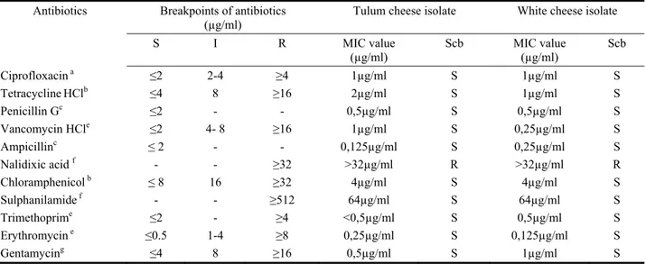
Table 2. The antimicrobial susceptibility profiles, MIC values and breakpoints of the L. monocytogenes isolates and S. aureus ATCC 25923 to 11 antibiotics.
Tablo 2. L. monocytogenes izolatlarının ve S. aureus ATCC 25923’ün kırılma noktaları, MIC değerleri ve antimikrobiyel duyarlılık profilleri.
Antibiotics Breakpoints of antibiotics
(µg/ml) Tulum cheese isolate White cheese isolate
S I R MIC value (µg/ml) Scb MIC value (µg/ml) Scb Ciprofloxacin a ≤2 2-4 ≥4 1µg/ml S 1µg/ml S TetracyclineHClb ≤4 8 ≥16 2µg/ml S 1µg/ml S Penicillin Gc ≤2 - - 0,5µg/ml S 0,5µg/ml S Vancomycin HCle ≤2 4- 8 ≥16 1µg/ml S 0,25µg/ml S Ampicillinc ≤ 2 - - 0,125µg/ml S 0,25µg/ml S Nalidixic acid f - - ≥32 >32µg/ml R >32µg/ml R Chloramphenicol b ≤ 8 16 ≥32 4µg/ml S 4µg/ml S Sulphanilamide f - - ≥512 64µg/ml S 64µg/ml S Trimethoprime ≤2 - ≥4 <0,5µg/ml S 0,5µg/ml S Erythromycin e ≤0.5 1-4 ≥8 0,25µg/ml S 0,125µg/ml S Gentamycing ≤4 8 ≥16 0,5µg/ml S 1µg/ml S
a Reported by Ruiz-Bolivar et al., (22).
bDetermined by CLSI for all microorganisms except Streptococcus spp (5). c Determined by CLSI for Listeria spp (5).
d For S. aureus determined by CLSI (5). e For Staphylococcus spp. determined by CLSI. f Reported by Zhang et al., (27).
g Determined by CLSI (5). S: Sensitive
I: Intermediate R: Resistance
nalidixic acid) and distilled water and 2.5mol/l sodium hydroxide (for sulphanilamide). Concentrations of antimicrobial agents ranged from 0.0625 to 4 μg/ml for ciprofloxacin, 0.125 to 8 μg/ml for ampicillin, penicillin G and erythromycin, 0.25 to 16 μg/ml for gentamicin, tetracycline HCl, trimethoprim and vancomycin HCl, 1 to 32 μg/ml for nalidixic acid and chloramphenicol and 8 to 512 μg/ml for sulphanilamide. Fifty μl of initial concentration of the eleven antimicrobial agents were added first and eighth wells of 96-well sterile U microdilution tray (Eren Kimya, Turkey) with 50 μl Cation-adjusted Mueller Hinton broth (CAMHB; GLB, Turkey) supplemented with 5% v/v lysed horse blood. Serial twofold dilutions were used since seventh wells and last 50 μl diluent was threw out. Eighth wells of 96-well sterile U microdilution tray was used for negative control. Bacterial inoculum preparation was emulsified in 0.45% saline solution to the equivalent 0.5 McFarland turbidity standard and 50 μl of bacterial suspension were added to each well. The final inoculum contained 5x10⁵
CFU/ml bacteria. MIC’s were determined after the inoculated microdilution trays were incubated at 35 °C for 16 to 20 h. The resistance, sensitive and intermediate breakpoints of the antibiotics for L. monocytogenes are mentioned in Table 2. All treatments included S.aureus ATCC 25923 as negative control.
Results
Two L. monocytogenes isolates were recovered, representing 1% of total samples (n=200). The pathogen occured in 1% (1/100) of white cheese and 1% (1/100) of tulum cheese samples. According to Denka Seiken Antisera tests, white cheese isolate found to belong to serotype 1/2a while tulum cheese isolate to serotype 4a (As shown in Table 1).
Table 2 resumes the antimicrobial susceptibility profiles, MIC values and the breakpoints of L. monocytogenes isolates for 11 antibiotics. Each of L. monocytogenes isolates were found susceptible to tetracycline HCl, ciprofloxacin, gentamicin, ampicillin, erythromycin, trimethoprim, vancomycin HCl, chloramphenicol, penicillin G and sulphanilamide and were resistant to nalidixic acid. S.aureus ATCC 25923 were susceptible to 11 antibiotics except nalidixic acid and penicillin G.
Discussion and Conclusion
The prevalence of L. monocytogenes in cheese samples from some provinces in Turkey were previously reported; in Nigde as 6% (2); in Kayseri as 17.2 % (11) and in Bolu as 9.2% (1). Our results are comperatively lower (1%) when compared to other studies. This variation in isolation rate among different provinces might be due to the differences in cheese processing environment, human activity, farm management, agriculture
application (silage feeding), type of cheese samples, sampling seasons and isolation methods etc…. (16).
In different studies reported around the world, isolation percentages of L. monocytogenes from soft and semi-hard cheeses were 15% in Iran (21) and 40% in Greece (6). Manfreda et al., (13) reported low L. monocytogenes contamination rates (2.1%) from soft cheeses which is in agreement with our results whereas several authors reported no L. monocytogenes in cheese samples (4, 9).
Lineage II isolates ( 1/2a, 1/2c and 3a serotypes) are more frequently isolated from foods and food environments compared to lineage I isolates (12, 18). One of the isolates obtained from cheese samples in this study belonged to serotype 1/2a which is in agreement with previous studies in several countries (10, 17). The occurance of 1/2a serotype in local cheese samples that are commonly consumed in this region of Turkey is an important public health concern as this serotype is one of the the most frequently incriminated serotype associated with human listeriosis.
Lineage III isolates (4a, 4b and 4c serotypes) have rarely been isolated from foods or processing plant and retail environments (18). It is suggested that lineage III isolates may be better adopted to the animal production environment than the food-processing environment (25). The isolate obtained from tulum cheese in this study belonged to 4a serotype which is in agreement with Zhang et al., (27) who isolated 4a serotype from other food categories. This result is thought to indicate that contamination is derived from environment or goat skin in which tulum cheese is stuffed.
Another approach to different prevalence of lineages among food and food related samples may be due to the more resistant appearance of lineage II isolates to bacteriocins than lineage I isolates which could be a selective advantage for lineage II isolates in food samples and enrichments containing bacteriocins (25). The notable diversity in the pathogenicity among L. monocytogenes serotypes necessitates the development of rapid and accurate laboratory procedures that readily distinguish virulent from avirulent serotypes. This information has critical importance for the effective control and prevention of listeriosis in addition to provide data on prevalence and distribution of virulent and avirulent L. monocytogenes strains in foods of animal origin.
In the present study MIC values were specified by a broth microdilution method. Among the other antimicrobial susceptibility tests, broth microdilution method is used as gold standard for L. monocytogenes (5).
In this study, both of the L. monocytogenes isolates showed natural resistance to nalidixic acid that is used as a selective agent during isolation of this bacterium. This
result is in aggrement with Zhang et al., (27). In this study, no isolate was resistant to ampicillin, gentamicin, trimethoprim, tetracycline, ciprofloxacin, erythromycin, vancomycin, chloramphenicol, penicillin G and sulphanilamide.
The standard therapy for listeriosis is ampicillin or penicillin G combined with an aminoglycoside, such as streptomycin or gentamicin (26). In this study, MIC value of penicillin G, ampicillin and gentamicin were detected 0.5 μg/ml, 0.125-0.25 µg/ml and 0.5-1 µg/ml respectively which were similar to those reported by other authors (13, 10, 15). On the other hand MIC values for penicilin G was slightly higher than those reported by Zhang et al., (27) as 4 µg/ml. Penicillin G, ampicillin and gentamicin resistance of L. monocytogenes are mentioned in several studies (1, 21, 17, 26).
The MIC ranges obtained in this study for trimethoprim and sulphanilamide were 0.5 µg/ml and 64 µg/ml which were similar to those noted for food L. monocytogenes isolates (10, 27). Trimethoprim and sulphonamide resistant L. monocytogenes isolated from various food were reported in recent studies (17, 26). This resistance is important as trimethoprim-sulfamethoxazole treatment has been successfully used for listeriosis patients with beta-lactams allergy.
Both of isolates showed susceptibility to tetracycline with MIC value of 1µg/ml (white cheese isolate) and 2µg/ml (tulum cheese isolate). Similar MIC values for tetracycline were also reported by several authors (6, 10, 15). On the other hand tetracycline resistance among L. monocytogenes isolates has been recently observed by some researchers (1, 10, 17, 26, 27). High prevelance of tetracycline resistance in food isolates may be partly due to the frequent use of tetracycline in animal production (26).
In the present study both isolates showed high sensitivity to ciprofloxacin, chloramphenicol, erythromycin, and vancomycin. MICs for ciprofloxacin, chloramphenicol, erythromycin and vancomycin were determined as 1µg/ml, 4 µg/ml, 0.125- 0.25 µg/ml and 0.25- 1 µg/ml and µg/ml respectively. Similar MIC values were also noticed by some authors (6, 10, 15). Resistance to these antimicrobials have been previously documented at different levels (17, 26, 27).
The results of this study suggest that the overall incidence of L. monocytogenes isolated from commonly consumed cheese types in Nigde is low. However, traditionally produced raw milk cheese types can potentially be the source of L. monocytogenes serotype 1/2a capable of causing human listeriosis. In addition, althought L. monocytogenes isolates obtained from cheeses have shown uniform susceptibility to antibiotics in this study, it is important to continued surveillance of MIC value for various antibiotics of L. monocytogenes isolates to ensure effective treatment of human listeriosis.
References
1. Arslan S, Ozdemir F (2008). Prevalence and
antimicrobial resistance of Listeria spp. in homemade white cheese. Food Control 19: 360– 363.
2. Bagcı C, Cinar M (2005). Isolation and idenlifiealion of
Listeria monocytogenes from white pickled cheese consumed in Niğde region. Vet Bil Derg 21: 69- 74.
3. Border P, Howard J, Plastow G, Siggens K (1990).
Detection of Listeria species and Listeria monocytogenes using polymerase chain reaction. Lett Appl Microbiol 11:
158- 162.
4. Buyukoruk S, Goksoy EO (2011). Investigation the
presence of Listeria spp. from village cheese in Aydın province. Uludag Univ J Fac Vet Med 30: 9- 12.
5. CLSI (2008). Development of In vitro Susceptibility Testing Criteria and Quality Control Parameters for Veterinary Antimicrobial Agents—Third Edition: Approved Guideline M37-A3., 28, 8. and Informational Supplement M31- A2, 2004; 24, 17. Clinical and Laboratory Standards Institute.
6. Filiousis G, Johansson A, Frey J, Perreten V (2009).
Prevalence, genetic diversity and antimicrobial susceptibility of Listeria monocytogenes isolated from open-air food markets in Greece. Food Control 20: 314– 317.
7. Gyles CL (2008). Antimicrobial resistance in selected
bacteria from poultry. Anim Health Res Rev 9: 149- 158.
8. ISO (2004). Horizontal Method for Detection of Listeria
monocytogenes International Standardization Organization
ISO NORM, 11290-1-/A1.
9. Jajali M, Abedi D (2008). Prevalence of Listeria species
in food products in Isfahan, Iran. Int J Food Microbiol
122: 336–340.
10. Korsak D, Borek A, Daniluk S, Grabowska A, Pappelbaum K (2012). Antimicrobial susceptibilities of
Listeria monocytogenes strains isolated from food and food processing environment in Poland. Int J Food
Microbiol 158: 203– 208.
11. Kum E, Yildirim Y, Ertas N (2011). Detection of Listeria
monocytogenes in cheese samples retailed in Kayseri by
classical cultural technique. J Fac Vet Med Univ Erciyes 8: 105 -109.
12. Liu D, Lawrence M, Wiedmann M, Gorski L, Mandrell RE, Ainsworth AJ, Austin FW (2006) Listeria
monocytogenes subgroups IIIA, IIIB, and IIIC delineate genetically distinct populations with varied pathogenic potential. J Clin Microbiol 44: 4229– 4233.
13. Manfreda G, De Cesare A, Stella S, Cozzi M, Cantoni C (2005). Occurrence and ribotypes of Listeria
monocytogenes in Gorgonzola cheeses. Int J Food
Microbiol 102: 287– 293.
14. McLauchlin J, Mitchell RT, Smerdon WJ, Jewell K (2004). Listeria monocytogenes and listeriosis: a review of
hazard characterisation for use in microbiological risk assessment of foods. Int J Food Microbiol 92: 15– 33.
15. Navratilova P, Schlegelova J, Sustackova A (2004).
Prevalence of Listeria monocytogenes in milk, meat and food stuff of animal origin and the phenotype of antibiotic resistance of isolated strains. Vet Med Czech 49: 243–252.
16. Nightingale KK, Schukken YH, Nightingale CR, Fortes ED, Ho AJ, Her Z, Grohn YT, McDonough PL, Wiedmann M (2004). Ecology and transmission of
Listeria monocytogenes infecting ruminants and in the farm environment. Appl Environ Microbiol 70: 4458–
4467.
17. O’Connor L, O’Leary M, Leonard N, Godinho M, O'Reilly C, Coffey L, Egan J, O'Mahony R (2010). The
characterization of Listeria spp. isolated from food products and the food-processing environment. Lett Appl
Microbiol 51: 490– 498.
18. Orsi RH, Bakker CH, Wiedmann M (2011). Listeria
monocytogenes lineages: Genomics, evolution, ecology, and phenotypic characteristics. Int J Med Microbiol 301:
79- 96.
19. Pourjafar M, Badiei K, Oryan A, Tabatabei M, Ghane M, Ahmadi N (2010). Clinico-pathological, bacteriological
and PCR findings of Ovine Listeriosis: an emerging disease in southern Iran. Global Vet 5: 226- 232.
20. Poyart-Salmeron C, Carlier C, Trieu-Cuot P, Courtieu AL, Courvalin P (1990). Transferable plasmid-mediated
antibiotic resistance in Listeria monocytogenes. Lancet
335: 1422–1426.
21. Rahimi E, Ameri M, Momtaz H (2010). Prevalence and
antimicrobial resistance of Listeria species isolated from milk and dairy products in Iran. Food Control 21: 1448-
1452.
22. Ruiz-Bolivar Z, Neuque-Rico MC, Poutou-Pinales RA, Carrascal-Camaco AK, Mattar S (2011). Antimicrobial
susceptibility of Listeria monocytogenes isolated from different cities in Colombia. Foodborne Pathog Dis 8: 913-
919.
23. Sleator RD, Francis GA, O’Beirne D, Gahan CGM, Hill C (2003). Betaine and carnitine uptake systems in
Listeria monocytogenes affect growth and survival in foods and during infection. J Appl Microbiol 95: 839- 846.
24. Troxler R, von Gravenitz A, Funke G, Wiedemann B, Stock I (2000). Natural antibiotic susceptibility of Listeria
species: L. grayi, L. innocua, L. ivanovii, L. monocytogenes, L. seeligerii and L. welshimerii strains. Clin Microbiol
Infec 6: 525– 535.
25. Ward TJ, Gorski L, Borucki MK, Mandrell RE, Hutchins J, Pupedis K (2004). Intraspecific phylogeny and lineage group identification based on the prfA virulence gene cluster of Listeria monocytogenes. J Bacteriol 186: 4994–5002.
26. Yan H, Neogi SB, Mo Z, Guan W, Shen Z, Zhang S, Li L, Yamasaki S, Shi L, Zhong N (2010). Prevalence and
characterization of antimicrobial resistance of foodborne Listeria monocytogenes isolates in Hebei province of Northern China, 2005–2007. Int J Food Microbiol 144:
310- 316.
27. Zhang Y, Yeh E, Hall G, Cripe J, Bhagwat AA, Meng J (2007). Characterization of Listeria monocytogenes isolated
from retail foods. Int J Food Microbiol 113: 47- 53.
28. Zhang D, Zhang L, Wang D, Suo B, Shi X (2010). A
PCR method for the detection of Listeria monocytogenes based on a novel target sequence identified by comparative genomic analysis. J Food Safety 30: 832–847.
29. Zeng H, Zhang X, Sun Z, Fang W (2006). Multiplex
PCR identification of Listeria monocytogenes isolates from milk and milk-processing environments. J Sci Food Agric
86: 367– 371.
Geliş tarihi: 15.01.2014 / Kabul tarihi: 31.03.2014
Address for correspondence:
Yrd. Doç. Dr. Fulden Karadal
Niğde Üniversitesi, Bor Meslek Yüksekokulu, Bor, NİĞDE.