ABSTRACT
394
Erciyes Med J 2019; 41(4): 394–7 • DOI: 10.14744/etd.2019.98470 ORIGINAL ARTICLE – OPEN ACCESSThis work is licensed under a Creative Commons Attribution-NonCommercial 4.0 International License.
Emine İnce , Semire Serin Ezer , Abdülkerim Temiz , Hasan Özkan Gezer , Akgün Hiçsönmez
Post-necrotizing Enterocolitis Stricture:
Misdiagnosis of this Complication Results in Greater
Infant Mortality
Objective: Intestinal stricture following necrotizing enterocolitis (NEC) is often misdiagnosed as recurrent functional con-stipation, enteritis, and malnutrition, and it increases the rates of morbidity and mortality in infants. Although a number of studies have focused on the potential etiologic factors leading to NEC, the information regarding the occurrence and diagnosis of post-NEC strictures is limited. The aim of this study was to evaluate the clinical presentation and diagnostic and surgical methods to treat NEC.
Materials and Methods: The medical records of infants who had undergone surgery for post-NEC strictures between Jan-uary 2005 and September 2018 were evaluated retrospectively in a single institution.
Results: This study included 38 infants (20 males, 18 females) with post-NEC stricture. Their histories revealed that they had been treated medically (20 of 38) or surgically (18 of 38) for NEC. Symptoms typical of intestinal obstruction (vomiting, abdominal distension, constipation, growth retardation, etc.) were present in the medically treated patients. The average time of onset of symptoms after the acute episode of NEC was 1.64±0.78 months. Contrast studies revealed strictures in the small intestine in 13 (65%) medically treated patients, while 13 (72.2%) surgically treated patients had strictures in the colon. Additionally, 2 of surgically treated patients presented with ileocolic fistulae. In 11 of 38 (28.9%) patients, the contrast studies were false-negative.
Conclusion: Post-NEC strictures may present with vague nutritional problems, causing the diagnosis to often be missed, which leads to high rates of morbidity and mortality in infants. Colon enemas, distal loopograms, and small bowel passage radiograms are useful in making a diagnosis, but a careful examination of the intestines for the presence of any other stric-tures should be done during the surgery.
Keywords: Intestinal stricture, necrotizing enterocolitis, neonatal surgery, post-NEC strictures
INTRODUCTION
Necrotizing enterocolitis (NEC) is a serious disorder manifesting with partial or complete ischemia of the intestines. The mortality ranges from 30% to 40% (1). In patients suffering from an acute NEC episode, a degree of obstruc-tion secondary to ischemic intestinal damage develops at rates of up to 40% during the recovery period (2–4). The critical period for stricture development is during the first 3 months following the remission of the initial acute NEC episode (2, 3). The incidence of intestinal stricture is higher in patients who have been treated surgi-cally (20%–43%), as compared to those who have been treated medisurgi-cally (15%–30%) (3–7). Post-NEC strictures may cause mortality or severe long-term morbidity due to intestinal obstruction, septicemia, and/or intestinal perforations (3–4). Although many studies have focused on the potential etiologic factors leading to NEC, studies regarding the development of post-NEC strictures after the acute episodes are limited (2–9). Therefore, the aim of this study was to evaluate post-NEC intestinal strictures, their presenting symptoms, the methods of making a diagnosis, and whether the initial treatment plays a role in their development.
MATERIALS and METHODS
Thirty-eight patients with post-NEC strictures, who were treated at our hospital between January 2005 and September 2018, were evaluated retrospectively. The data collection and review were performed in compliance with the principles of the ethics committee (project no: KA19/43) and the Declaration of Helsinki. The data col-lected and recorded for each patient included the gender, gestational age, extreme prematurity (<28 weeks), birth weight, extremely low birth weight, NEC history, treatment (medical or surgical), and initial surgical procedure (bowel resection, primary anastomosis, ostomy formation).
The clinical symptoms and the laboratory findings of the patients at the time of the first presentation (includ-ing C-reactive protein [CRP], hemoglobin [Hb], leucocytes, and platelet counts) were collected. Contrast studies
Cite this article as: İnce E, Ezer SS, Temiz A, Gezer HÖ, Hiçsönmez A. Post-necrotizing Enterocolitis Stricture: Misdiagnosis of this Complication Results in Greater Infant Mortality. Erciyes Med J 2019; 41(4): 394–7.
Departments of Pediatric Surgery, Başkent University Faculty of Medicine, Ankara, Turkey Submitted 03.05.2019 Accepted 26.08.2019 Available Online Date 06.11.2019 Correspondence
Emine İnce, Departments of Pediatric Surgery, Başkent University Faculty of Medicine, Ankara, Turkey Phone: +90 322 458 68 68-1000 e-mail: [email protected] ©Copyright 2019 by Erciyes University Faculty of Medicine - Available online at www.erciyesmedj.com
İnce et al. Post-Necrotizing Enterocolitis Strictures
Erciyes Med J 2019; 41(4): 394–7
395
performed (small bowel passage radiogram, contrast enema, distal loopogram), strictures detected on contrast studies, age at ostomy closure, strictures detected at further operation, mortality, and length of hospital stay were evaluated.
Statistical Analysis
This is a single-center, retrospective, cross-sectional, and descrip-tive study. Patients were not randomly selected and all patients had undergone surgery. Data were analyzed using descriptive statistical tests based on data distribution.
RESULTS
We included 20 (52.6%) males and 18 (47.4%) females with post-NEC strictures in this study. The demographic data and the lab-oratory test results for the patients at the time of admission are summarized in Table 1. During the acute NEC episode, 27 pa-tients were treated at another institution, while the remaining 11 were treated at our institution. In this study, the patients had been treated for the acute NEC episode either surgically or medically (18 [47.4%] and 20 [52.6%], respectively). In all surgically treated patients, an ostomy was performed (ileostomy: 15, colostomy: 3), and at the same time, primary perforation repair (4 of 18), seg-mental resection of the ileum (5 of 18), and segseg-mental resection of the colon (9 of 18) were also performed. Medically treated patients were admitted to our clinic after an average of 1.64±0.78 months post-NEC with symptoms of vomiting (40%), abdominal distension (95%), constipation (95%), and/or inability to gain weight (75%).
Two of these patients were septic. For each of the 20 patients treated medically at another institution, no medical information or hospital discharge report was available. However, when detailed medical histories of these patients were taken, it was learned that a majority of them had experienced an acute NEC episode previ-ously (18 of 20).
Contrast studies were performed on all patients, the results of which are summarized in Table 2. The contrast studies showed that in 6 (30%) patients treated medically, bowel stricture could not be excluded. In the 2 surgically treated patients, jejuno-colic fistulae were found simultaneously (Fig. 1).
Twenty patients who had been treated medically and presented with intestinal obstruction were made to undergo laparotomies. The patients’ intestines were evaluated with extreme care during the operation and suspicious areas were examined with saline solu-tion administrasolu-tion or under contrast fluoroscopy. In 11 patients, a stricture was detected in accordance with the contrast enema find-ings, following which resection with anastomosis was performed (Fig. 2). In 6 (30%) patients, strictures were not shown in the contrast enema, but were found in the cecum and terminal ileum
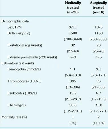
Table 1. Patients’ demographic data and laboratory test results
Medically Surgically treated treated (n=20) (n=18) Demographic data Sex, F/M 9/11 10/8 Birth weight (g) 1500 1150 (700–3440) (730–2800)
Gestational age (weeks) 32 28
(27–40) (25–40)
Extreme prematurity (<28 weeks) n=3 n=5 Laboratory test results
Hemoglobin (mmol/L) 9.1 9.1 (6.4–13.3) (6.8–17.1) Thrombocytes (109/L) 385 93 (13–904) (21–368) Leukocytes (109/L) 12.2 6.7 (2.1–28.7) (1.7–19.3) CRP (mg/L) 20.8 31.8 (1.2–270.1) (2.1–277.1) Mortality rate (%) 1 2 (5%) (11.1%)
Data are expressed as median (range) or as numbers unless specified otherwise. F: Female; M: Male; NEC: Necrotizing enterocolitis; CRP: C-reactive protein
Table 2. Localization of post-NEC strictures determined via contrast studies
Medically Surgically treated treated (n=20) (n=18) Distal ileum 2 – Hepatic flexure 3 – Splenic flexure 4 – Descending colon 2 – Sigmoid colon – 10
Two or more strictures are as in the colon 3 3 Low calibration of the intestines – 5 (27.7%) Stricture could not be shown 6 (30%) –
NEC: Necrotizing enterocolitis
b a
Figure 1. a, b. (a) A 3-month-old baby girl had a laparotomy for acute NEC episode and underwent ileostomy. Distal loopogram showed jejuno-colic fistula (descending colon: dashed arrow, small bowels: thick arrows). (b) Colon en-ema showed jejuno-colic fistula (descending colon: dashed arrow, small bowels: thick arrows)
İnce et al. Post-Necrotizing Enterocolitis Strictures
396
Erciyes Med J 2019; 41(4): 394–7during the operation. These patients underwent stricture resection and primary anastomosis. A 3rd stricture was found in the cecum or
terminal ileum in 3 patients with strictures in the transverse colon and splenic flexure. Since performing 3 anastomoses would be risky, stricture resection, primary anastomosis, and ileostomy were performed in these patients. The ileostomies in these patients were closed after 3 months.
Eighteen patients who had previously undergone an ostomy during the surgery to treat the acute NEC episode, underwent ostomy closure at 3.93±2.23 months after the first operation. In addi-tion to ostomy closure, 10 patients underwent stricture resecaddi-tion in the sigmoid colon, and 2 patients underwent resection of the splenic flexure with primary anastomosis. In 1 patient, a long-seg-ment stricture of the left colon with a total length of 10-12 cm was found. In this patient, approximately 15 cm of the colon was resected, primary anastomosis was performed, and the colostomy was closed 2 months later. In 2 patients with jejuno-colic fistulae, a fistula repair and resection of the stricture with anastomosis were performed initially, following which their ostomies were closed after 3 months. In 5 (27.7%) patients, who had presented with no strictures in the distal loopograms, cecal, and terminal ileac strictures were found during surgery. In addition to ostomy clo-sure, these patients underwent resection of the cecum and terminal ileum with ileocolic anastomosis.
Two patients who were septic at the time of admission died due to multi-organ failure in the early postoperative period. One patient died of ileus after a period of 3.5 months. The median length of hospital stay of the patients was 1 month (range: 1–3 months). The patients were followed up at intervals during a period of 1 year after the surgical interventions. During the follow-up period, the patients did not present with short bowel syndrome or long-term constipation. It was observed that the patients had a good nutritional status and favorable weight gain.
DISCUSSION
A missed diagnosis of a post-NEC intestinal stricture causes mortal-ity and life-threatening morbidmortal-ity due to sepsis or perforation of the
intestinal wall (2, 3, 6). The incidence of strictures was reported to be between 14% and 48% after an episode of NEC (1-3, 8). The critical period when stricture development may occur is the in the first 3 months following the remission of the initial acute episode (1, 2, 8). To avoid delays in the diagnosis and treatment, the staff of the neonatal intensive care unit, pediatricians, pediatric gastroen-terologists, and pediatric surgeons should be well-informed about the symptoms of post-NEC strictures (1-5, 8). These symptoms are often misdiagnosed as recurrent functional constipation, enteri-tis, and nutritional disorders. In our study, 18 of the patients with post-NEC strictures were medically managed at healthcare centers other than our own. Therefore, a detailed history of the previous NEC episode in the neonatal period could not be obtained. The symptoms occurred at an average of 1.64±0.78 months after the medically treated acute NEC episodes. The most common symp-toms at the time of admission were abdominal distension (95%), constipation (95%), vomiting (40%), an inability to put on weight (75%), and, in 2 patients, sepsis (10%).
Contrast studies are most commonly used in the diagnosis of post-NEC strictures. These studies may include contrast enemas, distal loopograms, or both. The sensitivity and specificity of the con-trast enema are 93% and 88%, respectively, and for the distal loopogram, they are 50% and 96%, respectively (10, 11). Despite its high sensitivity, the contrast enema may still miss strictures that are present. When a sufficient amount of contrast does not pass through the ileocecal valve, small bowel strictures can remain undi-agnosed (10). Contrast studies were performed in all the patients in our study. In the patients who had received medical treatment, the contrast enema showed either 1 (40%) or 2 (15%) areas of stricture at different levels of the colon. In 6 (30%) patients, the contrast enema was false-negative. In these patients, strictures in the ileo-cecal valve and cecum were detected during surgery. Small bowel passage radiograms of 2 patients demonstrated areas of stricture in the distal ileum.
More than 50% of surgically treated NEC patients have been re-ported to have asymptomatic post-NEC strictures distal to the os-tomy (2). Therefore, performing distal loopograms prior to osos-tomy
b c
a
Figure 2. a–c. (a) A 2-month-old baby boy was admitted with abdominal distension. Direct radiography showed dilated intestinal lobes. (b) Colon enema showed stricture in the splenic flexure of the colon (arrow). (c) Stricture was observed in the splenic flexure of the colon in surgery (arrow)
İnce et al. Post-Necrotizing Enterocolitis Strictures
Erciyes Med J 2019; 41(4): 394–7
397
closure is important to reduce the risk of overlooked strictures and to prevent unnecessary bowel dissection during surgery (2, 4). In the patients who had undergone an ostomy previously, distal loo-pograms revealed 1 (10 of 18), 2 (2 of 18), or 3 (1 of 18) strictures in 13 (72.2%) patients in our study. In addition, a jejuno-colic fistula was detected in 1 patient. The distal loopograms of 5 (27.7%) pa-tients showed low intestinal calibration. In these papa-tients, strictures in the cecum and terminal ileum were detected during surgery. Surgically treated patients are more likely to develop post-NEC strictures (20%–43%) as compared to patients treated medically (15%–30%) during the acute NEC episode (1, 6-10). It has been re-ported that the rate of post-NEC strictures in the surgically treated patients is as high as 80% (2). In our study, 18 patients had under-gone a surgical intervention. Of them, 8 had underunder-gone surgery at another institution and 10 underwent surgery in our clinic. In our clinic, the patients treated surgically during the acute NEC episode were those who were worsening clinically, or those who did not recover despite the medical treatment. The literature suggests that the potential to develop post-NEC strictures is higher in more se-vere forms of NEC (2, 3). Similarly, we consider the patients with very serious acute NEC episodes to be at a higher risk of develop-ing strictures.
Some authors have argued that contrast studies should be per-formed routinely in the medically treated infants with acute NEC, however, others claim that these tests, when routinely applied, pose a risk for unnecessary laparotomies in this patient group due to the high occurrence of false-positive results (3, 10, 11). In our clinical practice, we do not routinely perform contrast studies in the management of medically treated acute NEC patients unless there are symptoms that suggest a partial or complete obstruction. In our study, 2 patients were in sepsis at the time of admission to the hospital and they died in the early postoperative period. There-fore, we are of the opinion that the patients should be followed up at regular intervals over a mean period of 1 year after the acute NEC episode. In addition, the pediatricians, pediatric gastroen-terologists, and pediatric surgeons should be knowledgeable about the common symptoms of post-NEC stricture, and care should be exercised to prevent any delays in the diagnosis and treatment of at-risk patients.
The limitation of our study is that it is a retrospective study from a single institution with a relatively small sample size, and our sam-ple may not reflect the data on global post-NEC strictures, since it was performed only among patients receiving treatment in the pediatric surgery unit. This descriptive study was carried out only in cases of post-NEC stricture.
CONCLUSION
Post-NEC stricture symptoms may be frequently overlooked, as they are similar to the clinical presentation of nutritional problems, growth retardation, and chronic constipation. Therefore, pediatri-cians, pediatric gastroenterologists, and pediatric surgeons should
together follow patients who are being treated medically during the period of acute NEC, and then at regular intervals for an average of 1 year. A missed diagnosis may lead to mortality due to sepsis or perforation. Colon enemas, distal loopograms, and small bowel passage radiograms are useful for diagnosis, but a careful examina-tion of the intestines for the presence of any other strictures should be performed during the operation.
Ethics Committee Approval: This article was approved by the Ethics Committee of the Başkent University Faculty of Medicine (Date: 05/02/2019, No: KA19/43).
Informed Consent: Written informed consent was obtained from patients who participated in this study.
Peer-review: Externally peer-reviewed.
Author Contributions: Concept – Eİ; Design – Eİ, HÖG; Supervision – SSE, AT, AH; Resource – Eİ, HÖG, AH; Materials – Eİ; Data Collection and/or Processing – Eİ, HÖG, SSE; Analysis and/or Interpretation – AT; Literature Search – Eİ, HÖG, SSE; Writing – Eİ; Critical Reviews – AH, AT.
Conflict of Interest: The authors have no conflict of interest to declare.
Financial Disclosure: Supported by Başkent University Research Fund.
REFERENCES
1. Bellodas Sanchez J, Kadrofske M. Necrotizing enterocolitis. Neurogas-troenterol Motil 2019; 31(3): e13569. [CrossRef]
2. Heida FH, Loos MH, Stolwijk L, TeKiefte BJ, van den Ende SJ, On-land W, et al. Risk factors associated with postnecrotizing enterocolitis strictures in infants. J Pediatr Surg 2016; 51(7): 1126–30. [CrossRef] 3. Houben CH, Chan KW, Mou JW, Tam YH, Lee KH. Management of
Intestinal Strictures Post Conservative Treatment of Necrotizing Ente-rocolitis: The Long Term Outcome. J Neonatal Surg 2016; 5(3): 28. 4. Blackwood BP, Hunter CJ, Grabowski J. Variability in Antibiotic
Reg-imens for Surgical Necrotizing Enterocolitis Highlights the Need for New Guidelines. Surg Infect (Larchmt) 2017; 18(2): 215–20. [CrossRef] 5. Hong CR, Han SM, Jaksic T. Surgical considerations for neonates
with necrotizing enterocolitis. Semin Fetal Neonatal Med 2018; 23(6): 420–5. [CrossRef]
6. Bazacliu C, Neu J. Necrotizing Enterocolitis: Long Term Complica-tions. CurrPediatr Rev 2019; 15(2): 115–24. [CrossRef]
7. Robinson JR, Rellinger EJ, Hatch LD, Weitkamp JH, Speck KE, Danko M, et al. Surgical necrotizing enterocolitis. Semin Perinatol 2017; 41(1): 70–9. [CrossRef]
8. Rich BS, Dolgin SE. Necrotizing Enterocolitis. Pediatr Rev 2017; 38(12): 552–9. [CrossRef]
9. Zhang H, Chen J, Wang Y, Deng C, Li L, Guo C. Predictive factors and clinical practice profile for strictures post-necrotizing enteroclitis. Medicine (Baltimore) 2017; 96(10): e6273. [CrossRef]
10. Burnand KM, Zaparackaite I, Lahiri RP, Parsons G, Farrugia MK, Clarke SA, et al. The value of contrast studies in the evaluation of bowel strictures after necrotizing enterocolitis. Pediatr Surg Int 2016; 32(5): 465–70. [CrossRef]
11. Grant CN, Gorden JM, Anselmo DM. Routine contrast enema is not required for all infants prior to ostomy reversal: A 10-year single-center experience. J Pediatr Surg 2016; 51(7): 1138–41. [CrossRef]