Cukurova Medical Journal
Cukurova Med J 2016;41(Suppl 1):84-87 ÇUKUROVA ÜNİVERSİTESİ TIP FAKÜLTESİ DERGİSİ DOI: 10.17826/cutf.254645Yazışma Adresi/Address for Correspondence: Dr. Elif Karadeli, Baskent University, Faculty of Medicine, Department of Radiology, Adana, Turkey E-mail: [email protected]
Geliş tarihi/Received: 05.05.2015 Kabul tarihi/Accepted: 25.06.2015
OLGU SUNUMU/CASE REPORT
Radiologic findings mimicking acute abdomen in a patient with
colorectal cancer: are these side effects of FOLFIRI therapy?
Kolorektal kanserli hastada akut batını taklit eden radyolojik bulgular: FOLFIRI
tedavisinin yan etkileri mi?
Elif Karadeli
1, Gürcan Erbay
1, Zafer Koç
11Baskent University Faculty of Medicine, Department of Radiology, Adana, Turkey
Cukurova Medical Journal 2016;41(Suppl 1):84-87.
Abstract Öz
Bowel perforation is an emergency problem, it presents as an acute abdomen. Computurize tomography (CT) is important imaging modality used to evaluate patients with acute abdomen. CT shows anatomical detail and intestinal wall, evaluating secondary signs of bowel disease within the surrounding mesentery and detecting even small amounts of extraluminal air or oral contrast leakage into the peritoneal cavity. The aim of this article is to illustrate acute abdominal CT findings due to FOLFIRI therapy of a patient with colorectal cancer. Interestingly, this patient was an emergency case based on radiologic findings, but he felt good, had no emergency problem clinically.
Bağırsak perforasyonu, akut batını işaret eden tehlikeli bir problemdir. Bilgisayarlı tomografi (BT), akut batını olan hastaları değerlendirmede kullanılan önemli bir metoddur. BT, anotomik detayları ve bağırsak duvarını göstermekte, çevre mezenterde bağırsak hastalığı ile ilgili ikincil işaretleri değerlendirmekte ve peritonel kavitede oral kontrast sızıntıları ya da az miktarda ekstraluminal havayı saptamaktadır. Bu makalenin amacı, kolorektal kanserli hastada FOLFIRI tedavisine bağlı olarak akut batının BT bulgularını açıklamaktır. İlginç olarak, bu hastanın radyolojik bulgularının aciliyeti olmasına rağmen, hasta kendini iyi hissetmekte ve klinik olarak acil bir problemi bulunmamaktadır.
Key words: Acute abdomen, colorectal cancer, FOLFIRI,
computerized tomography. Anahtar kelimeler: Akut batın, kolorektal kanser, FOLFIRI, bilgisayarlı tomografi.
INTRODUCTION
Bowel perforation is an emergency problem, it presents as an acute abdomen. Computurize tomography (CT) is important imaging modality used to evaluate patients with acute abdomen. CT shows anatomical detail and intestinal wall, evaluating secondary signs of bowel disease within the surrounding mesentery and detecting even small amounts of extraluminal air or oral contrast leakage into the peritoneal cavity 1-3. The aim of this article is
to illustrate acute abdominal CT findings due to FOLFIRI therapy of a patient with colorectal cancer. Interestingly, this patient was an emergency case based on radiologic findings, but he felt good, had no emergency problem clinically.
CASE
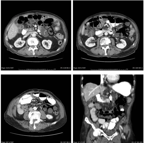
A 75-year- old man with stage 4 colon cancer was followed up during 2 years in our hospital. The patient was evaluated with abdominal computurize tomography for routine control. Oral and intravenous contrast material were given for examination. Diamaters of small bowel loops were increased and was approximately 38 milimeter. The air bubbles were seen in the wall of small bowels. In addition to, there were extensive air bubbles between the leaves of mesenteric. Contrast enhancement in the wall of small bowels was decreased in the left quadran. Superior mesenteric vein and artery were turn clock interwise (Figure 1-4) The patient had lung metastases, but had no
Karadeli et al. Cukurova Medical Journal
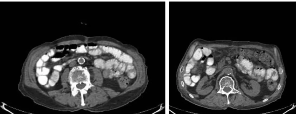
abdominal metastases. We thought that these findings were emergency problems. But, the patient were evaluated for emergency problems, his laboratory findings and physical examination were normal. The patient felt good. Abdominal CT without contrast material was performed after one week. Abnormal findings disappeared, abdominal
CT findings were normal (Figure 5,6). Later, we have learnt that the FOLFIRI was given to our patient for chemotheraphy. All thirtenn times, FOLFIRI chemotherapy was given, and last therapy was applied before two weeks. We thought that these findings might be due to FOLFIRI therapy.
Figure 1-4. Abdominal CT shows dilatation of small bowel loops and the air bubbles in the wall of small bowels, in addition to extensive air bubbles between the leaves of mesenteric . Coronal MIP shows extensive air bubbles in the mesenteric .
Cilt/Volume 41 Yıl/Year 2016 FOLFIRI
Figure 5-6. Second abdominal CT shows normal findings, emergency radiologic findings are disappeared.
DISCUSSION
The specific finding of gastrointestinal tract (GIT) perforation is extraluminal oral contrast, but its sensitivity is rather low (19–42%)3. The second
finding of perforation is free intraperitoneal air. CT is the most radiologic imaging modality for establishing even small amounts of free air. Additional CT signs that may also indicate the site of the perforation include discontinuity of the bowel wall on an enhanced scan and focal thickening of the bowel wall adjacent to extraluminal gas bubbles with localized mesenteric fatty infiltration 1-5.Interestingly, radiologic findings of our patient
were similar to findings of gastrointestinal tract (GIT) perforation. But physical examination of the patient was normal. C-reactive protein (CRP), sedimentation rate and amount of white blood cells were not abnormal. And one week later CT findings disappeared.
The FOLFIRI chemotherapy regimen was designed and is used almost strictly in patients with advanced, metastatic colorectal cancer. The combination chemotherapy regimen known as FOLFIRI consists of the following drugs: FOLolinic acid (leucovorin), Fluorouracil (5-FU), IRInotecan (irinotecan). The most common side effects are nausea and/or vomiting. Aggressive and proactive management of these symptoms with antiemetic medications is extremely important to mitigate these effects, Another very frequent problem with the FOLFIRI chemotherapy is diarrhea which, like the nausea, requires active management by one's oncology team
6,7. It has rarely been involved in hypersensitivity
reactions as flushing, pruritus, abdominal
pain,erythema, oedema developed over the face and thorax 8. FOLFIRI chemotherapy was given to our
patient for colorectal cancer. We thougth that these radiologic findings of our patient might be due to FOLFIRI therapy. He had no diarrhea, nause and vomiting, To our knowledge, this is the first case with radiologic findings mimicking acute abdomen due to FOLFIRI therapy in the literature
In conclusion, we have described side effects of FOLFIRI use that mimicking acute abdominal radiologic findings. If we see acute abdominal radiologic findings without complaint in oncology patients, we should think FOLFIRI use, especially,in patients with colorectal cancer.
REFERENCES
1. Maniatis V, Chryssikopoulos H, Roussakis A, Kalamara C, Kavadias S, Papadopoulos A et al. Perforation of the allimentary tract: evaluation with computed tomography. Abdom Imaging. 2000;25:373-9.
2. Yeung KW, Chang MS, Hsiao CP, Huang JF. CT evaluation of gastrointestinal tract perforation. Clin Imaging. 2004;28:329-3.
3. Hainaux B, Agneessens E, Bertinotti R, De Maertelaer V, Rubesova E, Capelluto E et al. Accuracy of MDCT in predicting site of gastrointestinal tract perforation. AJR Am J Roentgenol. 2006;187:1179-83.
4. Rubesin SE, Levine MS. Radiologic diagnosis of gastrointestinal perforation. Radiol Clin North Am 2003;41:1095-115.
5. Butela ST, Federle MP, Chang PJ, Thaete FL, Peterson MS, Dorvault CJ et al. Performance of CT in detection of bowel injury. AJR Am J Roentgenol. 2001;176:129-35.
Karadeli et al. Cukurova Medical Journal 6. Boyiadzis MM, Lebowitz PF, Frame JN, Fojo T.
Hematology-Oncology Therapy. New York, McGraw Hill, 2007.
7. Tournigand C, André T, Achille E, Lledo G, Flesh M, Mery-Mignard D et al. FOLFIRI followed by FOLFOX6 or the reverse sequence in advanced colorectal cancer: a randomized GERCOR study. J
Clin Oncol. 2004; 15: 229-37.
8. Florit-Sureda M, Conde-Estévez D, Vidal J, Montagut C. Hypersensitivity reaction caused by folinic acid administration: a case report and literature review. J Chemother. 2015; doi:10.1179/1973947815Y.0000000048.