Downloaded from https://journals.lww.com/prsgo by BhDMf5ePHKav1zEoum1tQfN4a+kJLhEZgbsIHo4XMi0hCywCX1AWnYQp/IlQrHD3tIQ5gQCIeyxoHNtHYz46smcqWIKq68Yk6GECN7AYm2hjLk9WTDkFGg== on 03/03/2020 Downloadedfrom https://journals.lww.com/prsgoby BhDMf5ePHKav1zEoum1tQfN4a+kJLhEZgbsIHo4XMi0hCywCX1AWnYQp/IlQrHD3tIQ5gQCIeyxoHNtHYz46smcqWIKq68Yk6GECN7AYm2hjLk9WTDkFGg==on 03/03/2020
www.PRSGlobalOpen.com
1
From the *Department of Plastic, Reconstructive and Aesthetic Surgery, Baskent University, Ankara, Turkey; and †Department of Plastic, Reconstructive and Aesthetic Surgery, Liv Hospital, Istanbul, Turkey.
Copyright © 2019 The Authors. Published by Wolters Kluwer Health, Inc. on behalf of The American Society of Plastic Surgeons. This is an open-access article distributed under the terms of the Creative Commons Attribution-Non Commercial-No Derivatives License 4.0
(CCBY-NC-ND), where it is permissible to download and share the
work provided it is properly cited. The work cannot be changed in any way or used commercially without permission from the journal. Plast Reconstr Surg Glob Open 2019;7:e2553; doi: 10.1097/
GOX.0000000000002553; Published online 31 December 2019.)
35-year Onset of a Squamous Cell Carcinoma Originating from Sacral
Pilonidal Sinus
Burak Ozkan, MD*; Harun Cologlu, MD†; Cagri A. Uysal, MD, PhD, FACS*; Nilgun M. Ertas, MD, FACS*
P
ilonidal sinus is a benign inflammatory disease char-acterized by fistulas with exudative discharge. This dis-ease is commonly seen in areas rich in hair follicles that penetrate the skin under direct pressure such as the sacro-coccygeal region in the predominantly male population.1Pilonidal sinuses can be transformed to malignancies in untreated, chronic conditions. Squamous cell carcinoma (SCC) is the most reported malignant alteration of piloni-dal sinus with 20 years of mean onset.2 We present a SCC
case with 35 years of unhealed pilonidal sinus history. A sixty-seven-year-old man presented to the outpatient clinic with an 8 × 7 cm2 ulcerative defect with fistulas and
exudative discharge in his sacrococcygeal region (Fig. 1). According to his history, he had multiple operations due to pilonidal sinus correction that started 35 years ago. Incisional biopsy was taken for excluding malignancy from the open wound. The biopsy result was compatible with squamous cell carcinoma. Systemic scans were per-formed; no loco regional lymph node enlargements and far metastatic findings were seen. The patient was taken to operation. All unhealed pilonidal sinus was excised by 2-cm clear margins including presacral fascia. The defect was reconstructed with gluteal transposition flap. The biopsy specimen result was confirmed as squamous cell carcinoma with clear margins. No postoperative complica-tions, relapses, and far metastasis were seen for 2 years of follow-up (Fig. 2).
The generally accepted SCC etiology is chronic inflam-mation and irritation of skin. Malignant transforinflam-mation should be suspected under recurrent, long-standing and rapidly growing cases, and cases with bleeding or excessive discharge.3 Fine needle biopsy or incisional biopsy should be
taken in different sites of the ulceration for diagnosis.4 Poor
outcome was reported in some cases with distant metasta-sis. Surgical excision with clear margins is the most curative treatment. Primary or secondary closure can be chosen. Ipsilateral or bilateral local skin flaps, fasciocutaneous or musculocutaneous flaps are preferred according to the
Fig. 1. Preoperative view shows 8 × 7 cm2 ulcerative pilonidal sinus.
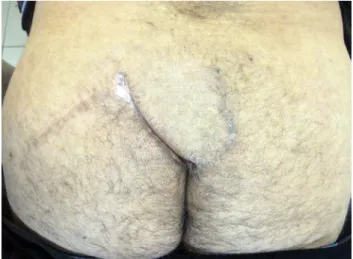
Fig. 2. Postoperative first year image shows well healed gluteal transposition flap.
PRS Global Open
•
2019
2
defect's size, depth, and surgeon's preference. Radiotherapy and chemotherapy are the reported treatment modalities in cases of recurrence or when surgery is not feasible.5
In summary, squamous cell carcinoma should be sus-pected in long-standing, unhealed pilonidal sinus cases.
Burak Ozkan, MD Department of Plastic, Reconstructive and Aesthetic Surgery, Baskent University Ankara 06900, Turkey E-mail: [email protected]
DISCLOSURE
The authors have no financial interest to declare in relation to the content of this article.
REFERENCES
1. Kulaylat MN, Gong M, Doerr RJ. Multimodality treatment of squamous cell carcinoma complicating pilonidal disease. Am
Surg. 1996;62(11):922–929.
2. Kim YA, Thomas I. Metastatic squamous cell carcinoma aris-ing in a pilonidal sinus. J Am Acad Dermatol. 1993;29(2 Pt 1): 272–274.
3. Pandey MK, Gupta P, Khanna AK. Squamous cell carcinoma aris-ing from pilonidal sinus. Int Wound J. 2014;11:354–356.
4. Williamson JD, Silverman JF, Tafra L. Fine-needle aspiration cytology of metastatic squamous-cell carcinoma arising in a pilonidal sinus, with literature review. Diagn Cytopathol. 1999;20: 367–370.
5. de Bree E, Zoetmulder FA, Christodoulakis M, et al. Treatment of malignancy arising in pilonidal disease. Ann Surg Oncol. 2001;8:60–64.