ORIGINAL ARTICLE
Comparative Evaluation of Conventional and OnyxCeph™
Dental Software Measurements on Cephalometric
Radiography
ABSTRACT
Objective: Cephalometry can be measured with traditionally conventional analysing methods (hand tracing), as well as using com-puters. Many dental softwares have been developed for this purpose. The reliability of these programs are often compared with the conventional method. The aim of the present study was to compare the conventional method of manual cephalometric analysis with a computerized one, OnyxCeph ™ (Image Instruments, Chemnitz, Germany) dental software.
Methods: Lateral cephalometric radiographs of 150 patients (75 males and 75 females) age range 12-34 were traced by two methods. Conventional method and computerized (OnyxCeph) cephalometric analysis method. 2 maxillar, 3 mandibular, 2 maxillo-mandibular, 3 vertical, 7 dental and 1 soft tissue parameters; 10 angular, 8 linear totally 18 cephalometric parameters were measured. Intra-class correlation coefficients were performed for both methods to assess the reliability of the measurements.
Results: The results 9 of 18 parameters were found statistically significant. They were Cd-A distance, Cd-Gn distance, Go-Me dis-tance, GoGnSN angle, ANS-Me disdis-tance, upper incisor-NA disdis-tance, lower incisor-NB disdis-tance, lower incisor-NB angle, overbite distance.
Conclusion: Despite some discrepancies in measured values between hand-tracing cephalometric analysis method and the OnyxCeph cephalometric analysis method, statistical differences were minimal and only Cd-A, Cd-Gn, Go-Me, ANS-Me, GoGnSN° were clinically important for cephalometric analysis OnyxCeph was evaluated as an efficient method to replace conventional method.
Keywords: Cephalometrics, manual tracing, onyxceph, computerized cephalometric program, reliability
INTRODUCTION
Cephalometric radiography is important diagnostic method that determines the morphology, development, and diagnosis in dental or skeletal abnormalities. It is used for treatment planning, evaluating the results of treat-ment, relationship between dental and cranial structures and identification of malocclusion (1-3). Three different methods are used to evaluate the cephalometric radiographs. The conventional cephalometric analysis is one of the methods, which is performed by tracing radiographic landmarks on acetate overlays and measuring linear and angular values. The second one is a computer-aided cephalometric analysis method, which uses scanners or digital cameras for exporting cephalometric images to measurement programs and anatomical structures marked with a mouse cursor on a computer monitor. The third method is a fully digital method, which transmits digital radiographs directly to a computer database, and a cephalometric program determines the anatomical structures and completes the cephalometric analysis by measuring distances and angles through automation (4-9).
Elif İzgi2 ,Filiz Namdar Pekiner1
1Department of Oral Diagnosis and Radiology, Medipol University School of Dentistry, İstanbul, Turkey 2Department of Oral Diagnosis and Radiology, Marmara University School of Dentistry, İstanbul, Turkey
Address for Correspondence: Filiz Namdar Pekiner, Department of Oral Diagnosis and Radiology, Marmara University School of Dentistry, İstanbul, Turkey
E-mail: [email protected]
©Copyright 2019 by Turkish Orthodontic Society - Available online at turkjorthod.org
Received: May 17, 2018 Accepted: November 23, 2018 Cite this article as: İzgi E, Pekiner FN. Comparative Evaluation of Conventional and OnyxCeph™ Dental Software Measurements on Cephalometric Radi-ography. Turk J Orthod 2019; 32(2): 87-95.
However, cephalometric analysis has some limitations. It produc-es errors during radiographic image acquisition or cephalomet-ric tracing. Cephalometcephalomet-ric errors can be categorized into projec-tion errors (acquisiprojec-tion), identificaprojec-tion errors, and measurement errors (2, 5, 10-13). Projection errors under magnification and distortion titles contain patient positioning errors, exposure position, differences in exposure parameters, collimation, bath conditions, and differences in film shooting methods. Errors during the digitization of the image are also considered (2, 5, 6, 11, 14-18). A study by Gaddam et al. (19) investigated the projec-tion errors in lateral cephalometric radiographs. Ten skulls and 8 cephalometric parameters were evaluated, and head rotations from 0° to −20° at 5° intervals in the vertical axis were performed. They concluded that according to the head rotation, angular measurements had fewer projection errors than linear measure-ments. Further evidence was from a study by Yoon et al. (20), which evaluated 17 skulls and 8 parameters to identify potential projection errors of lateral cephalometric radiographs according to the head rotation. Each skull was rotated from 0° to ±15° at 1° intervals in the vertical axis. The results were consistent with those of Gaddam et al. (19). Measurement errors were affected by the measuring device (ruler, protractor, etc.), technique (re-cording or archiving of measurements), or investigator (limita-tion in visual performance or fault in measuring) (2, 5, 6, 11, 18, 21). These errors mostly have been eliminated by the spread of digital analyzing methods (11). Landmark identification er-rors are the most common and important erer-rors in the analysis. These errors involve radiographic image quality (sharpness, blur, contrast, and noise), differences depending on the researchers (intra-observer: light, time constraints, psychological conditions; inter-observer: their experience or perspective differences), pre-cision of landmark identification, and reproducibility of the loca-tion. Errors less than 0.5 mm are considered acceptable anatomi-cal landmark errors (2, 5, 6, 8, 11, 15-18, 21, 22-25). The landmarks are located on the outline of the cranium, which are compara-tively easy to identify, whereas the internal structures were more difficult to identify because of the summation of superimposed anatomical details (14).
Computerized systems for cephalometric analysis were routinely performed, and many analysis programs have been developed to date (26). Various studies have been compared to a variety of computer-aided cephalometric analysis programs using the conventional cephalometric analysis method for reliability and reproducibility in literature (11, 12, 23, 27). However, no clear consensus has been achieved regarding the standard method. McClure et al. (28) evaluated 19 landmarks on 6 patients with an age range of 21–30 years to compare identification errors with the conventional analysis method and the Dolphin Imaging software. It was emphasized that even the statistically significant differences between the two methods of image acquisition were unlikely to attain clinical significance. Cavdar et al. (29) compared the conventional cephalometric analysis using Jiffy Orthodontic Evaluation (JOE®) and QuickCeph® computerized cephalometric programs and used 90 lateral cephalograms with 18 parameters. The authors justified that the computer-aided method may be preferable because of the benefits, such as time gain, archiving, and enhancement of radiographs. Akın et al. (30) compared intra
and interexaminer reliability of 19 parameters obtained from 60 lateral cephalometric radiographs using the conventional and QuickCeph computerized cephalometric analysis methods. It was determined that computerized cephalometric analysis did not increase the measurement error compared to the conven-tional method. Rusu et al. (31) assessed 39 lateral cephalograms with three different computerized programs: Planmeca Romexis, Orthalis, and AxCeph. They reported that Romexis and AxCeph give more reliable results than Orthalis.
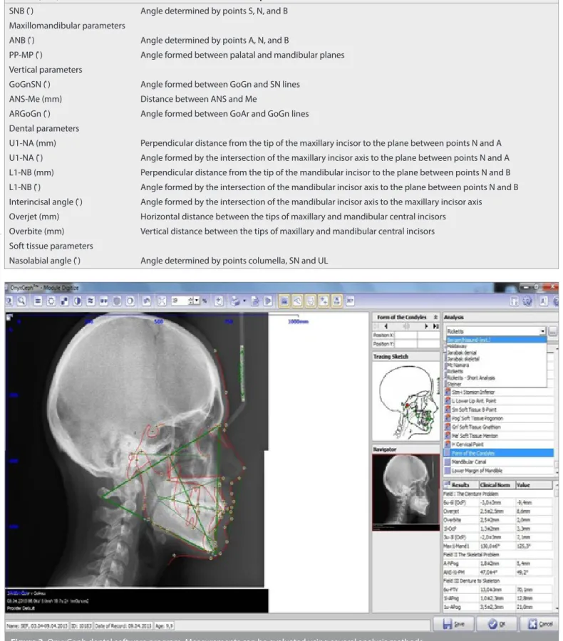
OnyxCeph™ dental Picture Archiving Communication Systems was developed for archiving, diagnostics, treatment planning, and patient education. This software program is based on two-dimensional (2D) and 3D data processing. Image import, image adjust (classify and crop image), cephalometric analysis and measurements, mirror image, model base (adjust models and attach base), segmentation (separation and completion) Ricketts Visual Treatment Objective, superimposition, image edit, data export, copy/save/send/show/print image, treatment simulation, slide show, online/offline reports are possible with this 2D and 3D image data (32).
Davoudian (32) examined and compared the reliability and re-producibility of digitization using the OnyxCeph imaging soft-ware with conventional techniques through 21 parameters in 30 lateral cephalograms. It was determined that all measurements showed good reliability in both methods except for the nasola-bial angle in the manual method.
Although researchers have studied several software programs based on computerized cephalometry, there are few studies based on the OnyxCeph software (32). The aim of the present study was to compare the conventional cephalometric analysis method and a computerized cephalometric analysis method with the OnyxCeph dental software.
METHODS
In this retrospective study, lateral cephalometric radiographic im-ages were gathered from a total of 150 participants (75 females and 75 males) with an age range of 12–34 years. The inclusion criteria were patients without any missing teeth based on the records for the period 2013–2016 in the archives of the Marma-ra University School of Dentistry, Department of Radiology. The exclusion criteria were participants with systemic disease, which may adversely affect their bone development; pathological con-ditions, such as cysts and tumors; and a history of a trauma or injury in the oral and maxillofacial regions. Ethical approval was obtained from the ethics committee of the Marmara University School of Medicine (Protocol 092015128 2015/9:128).
All lateral cephalometric radiographs were acquired from the same orthopantomogram (Promax, Planmeca Oy, 0080 Helsin-ki, Finland) using standard radiographic techniques (75 kV, 4.1 seconds, 10 mA).
Based on the cephalometric measurements, patients were grouped as those undergoing conventional techniques and
ital techniques (Onyx Ceph™). In the conventional method, the digital images were resized to a 1:1 scale using Adobe Photo-shop (Adobe Systems, San Jose, California, USA) and printed on an A4 paper using a laser printer (HP Laserjet P2035n). For stan-dardizing the analysis, there were no changes in the settings of resolution, contrast, and brightness before printing in the digital cephalometric radiographs.
For the conventional technique, 150 digital radiographs were manually traced on an A4 paper placed over the printed image. A 0.3 mm 2H lead pencil to trace all the required landmarks, a rul-er to draw lines, and a protractor to measure angles wrul-ere used. Bilateral structures were averaged to make a single landmark. For the digital technique, direct digital cephalometric radio-graphs were recorded in the Joint Photographic Experts Group format and transferred to the OnyxCeph3™ 3.1.54 (Image Instru-ments, Chemnitz, Germany) dental analysis software for a ceph-alometric analysis. Digital measurements were evaluated using a 23-inch Acer 1920×1080-pixel HP Reconstruction PC monitor. The identified anatomical points were marked in the program with an indicator on the mouse control. Before marking the an-atomical points, a ruler on the cephalostat was calibrated in the program, and thus standardization was provided in all cephalo-metric radiographs. Measurements were drawn automatically by the program after the marking the anatomical points.
All the tracings were performed by the same maxillofacial radiol-ogist. No more than 10 radiographs were traced per day to avoid examiner fatigue. To assess reliability, 30 randomly selected ra-diographs were retraced by the same investigator using the con-ventional cephalometric analyzing method. A time interval of more than 2 months elapsed between first and second analyses. The commonly used dental, skeletal, and soft tissue parameters in a cephalometric analysis were selected and the linear and an-gular measurements shown in Figure 1 were produced. A total of 18 anatomical landmarks with 5 planes and 8 linear and 10 angular measurements were evaluated. In these measurements, there were 2 maxillary parameters, 3 mandibular parameters, 2 maxillomandibular parameters, 3 vertical parameters, 7 dental parameters, and 1 soft tissue parameters (Table 1).
Cephalometric radiographs were divided into 3 groups (classes I, II, and III) according to the Angle classification, which is used for the classification of malocclusions. The ANB angle of 0° to 4° is class 1; ANB angle >4° is class 2; and ANB angle <4° is class 3. The conventional and digital methods were compared based on this classification.
The OnyxCeph dental software program was used to analyze lateral cephalometric radiographs after the anatomic landmarks were marked. The measurements can be evaluated using several
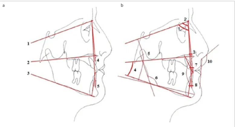
Figure 1. a, b. (a): 1. SN plane: Plane is passing through Sella and Nasion points. 2. PP (Palatal plane): Plane is passing through ANS and PNS points. 3. MP (Mandibular plane): Plane is passing through Gonion and Menton points. 4. NA plane: Plane is passing through Nasion and A points. 5. NB plane: Plane is passing through Nasion and B points. (b): 1.SNA°: Angle determined by points S, N, and A. 2. SNB°: Angle determined by points S, N, and B. 3. ANB°: Angle determined by points A, N, and B. 4. PP-MP°: Angle formed between palatal and mandibular planes. 5. GoGnSN°: Angle formed between GoGn and SN lines. 6. ArGoGn°: Angle formed between GoAr and GoGn lines. 7. U1NA°: Angle formed by the intersection of the maxillary incisor axis to the plane between points N and A. 8. L1NB°: Angle formed by the intersection of the mandibular incisor axis to the plane between points N and B. 9. İnterinsizal°: Angle formed by the intersection of the mandibular incisor axis to the maxillary incisor axis. 10. Nazolabial°: Angle determined by points columella, SN and UL
a b
analysis methods (Figure 2). The measured values were based on the drawing, deviation ratio according to the norm values. These rates are in different color tones. Green color indicates class 1 de-viation, red color class 2, and blue color class 3.
Pretreatment, intermediate stages, end of treatment analysis of the cases were evaluated using the OnyxCeph dental software program. Changes in the middle and final stages were deter-mined by the dynamic nature of the visual assessment of the
Figure 2. OnyxCeph dental software program. Measurements can be evaluated using several analysis methods Table 1. Measurements used for this study
Go-Me (mm) Distance between Go and Me points
SNB ( ̊) Angle determined by points S, N, and B
Maxillomandibular parameters
ANB ( ̊) Angle determined by points A, N, and B
PP-MP ( ̊) Angle formed between palatal and mandibular planes
Vertical parameters
GoGnSN ( ̊) Angle formed between GoGn and SN lines
ANS-Me (mm) Distance between ANS and Me
ARGoGn ( ̊) Angle formed between GoAr and GoGn lines
Dental parameters
U1-NA (mm) Perpendicular distance from the tip of the maxillary incisor to the plane between points N and A U1-NA ( ̊) Angle formed by the intersection of the maxillary incisor axis to the plane between points N and A L1-NB (mm) Perpendicular distance from the tip of the mandibular incisor to the plane between points N and B L1-NB ( ̊) Angle formed by the intersection of the mandibular incisor axis to the plane between points N and B Interincisal angle ( ̊) Angle formed by the intersection of the mandibular incisor axis to the maxillary incisor axis
Overjet (mm) Horizontal distance between the tips of maxillary and mandibular central incisors Overbite (mm) Vertical distance between the tips of maxillary and mandibular central incisors Soft tissue parameters
Nasolabial angle ( ̊) Angle determined by points columella, SN and UL
OnyxCeph dental software program. Thus, the alterations of the facial appearance can be easily interpreted during and after treatment.
Statistical Analysis
The statistical analyses were performed using the software Statis-tical Package for Social Sciences version 22.0 (IBM Corp.; Armonk, NY, USA). Shapiro Wilks test was used for evaluating the normal distribution of parameters. It was determined that the parame-ters were in accordance with the normal distribution. Descrip-tive statistical methods were used for each measurement (mean, standard deviation frequency). A student’s t-test was used for the evaluation of the digital and conventional measurements based on gender. A paired sample t-test was used to evaluate the statistical significance and compare differences between the digital and conventional measurement values. A p value of <0.05 was considered significant.
RESULTS
The total of 150 individuals’ (75 females and 75 males) cepha-lometric radiograph images were assessed in this study. The age ranged from 12.5 years to 33.7 years, the average age was 17:56±3:59 years.
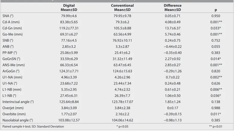
According to the statistical analysis, the average of the digital and conventional measurements and measuring differences are shown in Table 2. There was a statistically significant difference between averages of Cd-A distance, which is one of the maxillary parameters (p=0.001; p<0.01). The agreement between the two measurement methods was 48.9% (intra-class correlation coeffi-cient [ICC]: 0.489; 95% confidence interval [CI]:0.357–0.602). There was a statistically significant difference between the averag-es of Cd-Gn distance, which is one of the mandibular parameters
Table 2. Difference between digital and conventional measurement averages in all patients
Digital Conventional Difference
Mean±SD Mean±SD Mean±SD p
SNA (°) 79.99±4.6 79.95±9.78 0.05±0.71 0.950 Cd-A (mm) 83.38±5.65 79.3±6.2 4.08±0.49 0.001** Cd-Gn (mm) 119.2±77.31 105.5±8.88 13.7±6.37 0.033* Go-Me (mm) 69.31±6.27 63.56±4.99 5.74±0.46 0.001** SNB (°) 77.16±4.5 76.92±10.11 0.24±0.75 0.752 ANB (°) 2.85±3.2 3.3±2.87 −0.44±0.22 0.055 PP-MP (°) 25.06±5.99 25.41±6.2 −0.35±0.40 0.383 GoGnSN (°) 33.59±6.29 31.32±11.49 2.27±0.92 0.014* ANS-Me (mm) 66.33±6.54 63.47±6.45 2.85±0.27 0.001** ArGoGn (°) 124.31±7.71 124.6±15.63 −0.29±1.26 0.820 U1-NA (mm) 4.96±3.39 4.26±2.96 0.7±0.22 0.002** U1-NA (°) 23.68±7.22 23.44±7.34 0.24±0.48 0.626 L1-NB (mm) 5.35±2.95 4.74±2.52 0.61±0.21 0.006** L1-NB (°) 27.45±6.31 26.39±7.7 1.06±0.50 0.036* Interincisal angle (°) 125.64±8.84 123.78±17.07 1.85±1.24 0.138 Overjet (mm) 3.84±3.09 3.84±2.38 0±0.17 0.988 Overbite (mm) 1.77±2.07 2.16±2.2 −0.39±0.15 0.011* Nasolabial angle (°) 103.08±12.57 104.06±14.62 −0.98±1.13 0.385
Paired sample t-test; SD: Standard Deviation * p<0.05 ** p<0.01
Table 3. Distribution of cephalometric parameters according to the difference (md) * in conventional and digital measurements in all patients
md < 0.5 0.5 < md < 1.0 1.0 < md < 1.5 1.5 < md < 2.0 2.0 < md
SNA° U1NA (mm) L1NB° Interincisal angle° Cd-A (mm)
SNB° L1NB (mm) Cd-Gn (mm)
ANB° Nasolabial angle° Go-Me (mm)
PP - MP° GoGnSN°
ArGoGn° ANS - Me (mm)
U1NA° Overjet (mm) Overbite (mm)
*md (measurement difference): millimeter for linear measurements, degree for angular measurements
(p=0.033; p<0.05). The agreement between the two measurement methods was 0.6% (ICC: −0.006; 95% CI: −0.165–0.154). There was a statistically significant difference between the averages of Go-Me distance, which is one of the other mandibular parameters (p=0.001; p<0.01). The agreement between the two measurement methods was 50.7% (ICC: 0.570; 95% CI: 0.378–0.617).
There was a statistically significant difference between the aver-ages of the PP-MP angle, which is one of the maxillomandibular parameters (p=0.383; p>0.05). The agreement between the two measurement methods was 67.6% (ICC: 0.676; 95 % CI: 0.579– 0.754).
There was a statistically significant difference between the av-erages of other vertical parameters GoGnSN angle (p=0.014; p<0.05). The agreement between the two measurement meth-ods was 26.6% (ICC: 0.266; 95% CI: 0.111–0.408). There was a sta-tistically significant difference between the average of the oth-er voth-ertical parametoth-ers ANS-Me distance (p=0.001; p<0.01). The agreement between the two measurement methods was 87% (ICC: 0.870; 95% CI: 0.825–0.904).
There was a statistically significant difference between the av-erages of U1-NA distance, which is one of the dental parame-ters (p=0.002; p<0.01). The agreement between the two mea-surement methods was 63.9% (ICC: 0.639; 95% CI: 0.533–0.725). There was a statistically significant difference between the av-erage of the other dental parameters L1-NB distance (p=0.006; p<0.01). The agreement between the two measurement meth-ods was 53.6% (ICC: 0.536; 95% CI: 0.412–0.641). There was a sta-tistically significant difference between the average of the other dental parameters L1-NB angle (p=0.036; p<0.05). The agree-ment between the two measureagree-ment methods was 62.1% (ICC: 0.621; 95% CI: 0.512–0.710). There was a statistically significant difference between the average of the other dental parameters overbite distance (p=0.011; p<0.05). The agreement between the two measurement methods was 62.4% (ICC: 0.624; 95% CI: 0.516–0.713).
Considering the differences between the conventional and digi-tal measurements in Table 3, values less than 0.5 mm for dimen-sional parameters and the degree for angular parameters are follows: SNA angle, SNB angle ANB angle, PP-MP angle ArGoGn
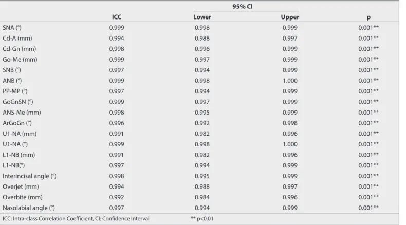
Table 4. Method error assessment for repeated conventional measurements in 30 patients
95% CI
ICC Lower Upper p
SNA (°) 0.999 0.998 0.999 0.001** Cd-A (mm) 0.994 0.988 0.997 0.001** Cd-Gn (mm) 0,998 0.996 0.999 0.001** Go-Me (mm) 0.999 0.997 0.999 0.001** SNB (°) 0.997 0.994 0.999 0.001** ANB (°) 0.999 0.998 1.000 0.001** PP-MP (°) 0.997 0.994 0.999 0.001** GoGnSN (°) 0.999 0.997 0.999 0.001** ANS-Me (mm) 0.998 0.995 0.999 0.001** ArGoGn (°) 0.996 0.992 0.998 0.001** U1-NA (mm) 0.991 0.982 0.996 0.001** U1-NA (°) 0.999 0.998 1.000 0.001** L1-NB (mm) 0.991 0.982 0.996 0.001** L1-NB(°) 0.997 0.994 0.999 0.001** Interincisal angle (°) 0.998 0.995 0.999 0.001** Overjet (mm) 0.994 0.988 0.997 0.001** Overbite (mm) 0.992 0.984 0.996 0.001** Nasolabial angle (°) 0.997 0.994 0.999 0.001**
ICC: Intra-class Correlation Coefficient, CI: Confidence Interval ** p<0.01
Table 5. Compatibility between classifications according to ANB in digital and conventional measurements
ANB Digital
ANB Conventional Class I Class II Class III Total
Class I 56 (37.3%) 17 (11.3%) 13 (8.7%) 86 (57.3%) Class II 12 (8%) 40 (26.7%) 3 (2%) 55 (36.7%) Class III 1 (0.7%) 0 (0%) 8 (5.3%) 9 (6%) Total 69 (46%) 57 (38%) 24 (16%) 150 (100%) McNemar p=0.003
92
angle, U1NA angle, overbite distance, and overjet distance. Pa-rameters with a difference value between 0.5 and 1.0 included U1NA distance, L1NB distance, and nasolabial angle. The only parameter with a difference value between 1.0 and 1.5 was the L1NB distance. The only parameter with a difference value be-tween 1.5 and 2.0 was the interincisal angle. Parameters with a difference value more than 2.0 were ANS-Me distance, Cd-A dis-tance, Cd-Gn disdis-tance, Go-Me disdis-tance, and GoGnSN angle. Randomly selected 30 cephalometric radiographs were repeated using the conventional techniques to control individual drawings and the level of measurement error for the assessment of the mea-surements used. Each parameter of recurrence coefficients (r²) was calculated. The results are shown in Table 4. For each measurement method, the error and the upper and lower limits of 95% CI were determined and provided in the table. The ICC for all samples was found to be close to 1.00. The results of the ICC analysis regarding the method showed an insignificant error and did not affect the results of conventional measurement.
In total, 56 patients were defined as class 1, 40 patients as class 2, and 8 patients as class 3 by the conventional and digital mea-surement as shown in table 5. Accordingly, there was no statis-tically significant compliance of digital and conventional mea-surements depending on the ANB angle between the classes (p=0.003; p<0.01). Overall, 69 patients (46%) were class 1, 57 patients (38%) were class 2, and 24 patients (16%) were class 3 in the digital measurement, while 86 patients (57.3%) were class 1, 55 patients (36.7%) were class 2, and 9 patients (6%) were class 3 in the conventional measurement. The kappa coefficient was 47.8% between the two measurement methods.
DISCUSSION
The cephalometric radiography analysis was divided into conven-tional or digital analysis. In the convenconven-tional cephalometric anal-ysis, numerous measurements could be waste of time. Currently, those time problems are eliminated by a software that allows a precise measurement of improved digital cephalometric analysis systems (7, 9, 12, 16, 25, 29, 33-37). Numerous studies have inves-tigated the differences between computer-aided cephalometric analysis programs and conventional cephalometric analysis in terms of reliability, accuracy, repeatability, and time (9, 12, 17, 25, 29, 30, 34, 36-38). In our study, the digital cephalometric analysis method (OnyxCeph) was compared to the conventional cepha-lometric analysis. We found that that digital method was faster and consistent with the other researches. In a study by Iseri et al. (39), 14 parameters were identified in 50 cephalometric radio-graphs, which was measured twice and compared for accuracy, repeatability, and time using the conventional analysis methods and computer-aided analysis method. There were no statistically significant differences between the average of primary and sec-ondary measurements in both methods. However, the comput-er-aided method provided significantly higher time gain, which was 7 times faster than conventional methods.
Uysal et al. (40) assessed inter- and intra-examiner reproducibil-ity in the conventional cephalometric and Dolphin
cephalomet-ric analyses methods. It was reported that although the com-puter-aided cephalometric analysis method was not effective to reduce inter- and intra-examiner error, it was preferable in terms of time gain for clinicians.
Inter-examiner errors were greater than intra-examiner errors; hence, to minimize errors, all the measurements in this study were measured by one examiner as in other studies (8, 16, 17, 25, 29, 32, 33, 38, 41, 42). In Naoumova and Lindman’s study (33), 30 patients (12 males and 18 females) with identified 25 landmarks were compared using the conventional cephalometric analysis methods with the digital cephalometric analysis method (FA-CAD, Ilexis AB, Linköping, Sweden). This study was conducted by a single researcher. The researchers’ reproducibility correlation coefficients of all variables were above 0.95 for the convention-al method and above 0.8 for FACAD. In our study, the ICC was examined for only the conventional method, and the ICC for all variables were above 0.9. The reproducibility of the individual re-searcher was high.
Several studies have found the main source of error in cephalo-metric analysis to be the identification of the landmarks (9, 14, 27). As the consensus of many researchers, the Frankfurt hori-zontal plane showed low coefficient of repeatability. Since it is difficult to detect as stated in many studies, any parameters including a reference from the Po or Or were not used in our study(6, 7, 9, 12, 34, 40).. Moreover, measurements including references points Cd , Cd-A and the Cd-Gn distance was used in our study and there was no statistically significant difference between the conventional cephalometric and OnyxCeph cepha-lometric measurement.
The nasolabial angle, which is a commonly used parameter in cephalometric analysis, was indicated as an angle which is dif-ficult to determinate and shows a low reproductibility as tated in many studies (6, 12, 34, 40, 43). Unlike other studies, the na-solabial angle did not show significant differences between the conventional cephalometric analysis and digital cephalometric analysis (OnyxCeph) method in our study. The reason for the high reproducibility of the nasolabial angle could be explained by the easily determined soft tissue and appropriate radiograph-ic contrast.
If complex parameters with multiple anatomical landmarks have low reproducibility, it is considered clinically insignificant (16). The GoGnSN angle contained in our study comprised 4 differ-ent cephalometric points and showed a statistically significant difference. However, in the light of previous information, these statistical differences are not considered reliable to achieve a clinical decision.
The ANB angle is used for the classification of malocclusion and for revealing the relationship between the upper and lower jaw in a sagittal direction (44). The ANB angle showed statistically sig-nificant differences between the conventional and digital tech-niques in our study. Although a complex parameter that con-tains multiple anatomical points that we can make the comment the reason for the high reproducibility in present study of the
ANB angle is the ease of detection landmarks of A, B and N. Akin et al. compared conventional and digital measurement methods with two different researchers. The ANB angle was found to be highly reliable for both researchers (30).
It has been suggested that the problem about repeatability is not related the measurement techniques, such as digital or con-ventional; it might be related to the parameters to be measured whether angular or linear (16). It was concluded that linear mea-surements have a higher error rate from angular meamea-surements due to the distortion on the image (13). Kumar et al. (36) com-pared the conventional and digital tracing methods using Bur-stone analysis. They stated that differences in the measurements of linear parameters were greater than those of the angular pa-rameters as the reason of that errors in calibration were not affect-ed angular values; it changaffect-ed the linear values. In a study by Tikku et al. (46), which compared the conventional and digital cephalo-metric measurement methods, a total of 26 parameters (13 linear and 13 angular) were assessed. Only the occlusal plane angle had a statistically significant difference between two methods. From this view point, it has been commented that linear measurements cause a higher statistically difference than angular measurements. It was reported that in a clinical situation, a reproducibility that is within 2° or 2 mm would probably not make a difference in the treat-ment and is insignificant for a clinical decision (13, 32). Our study is also consistent with similar studies, which showed a statistically significant difference in 9 parameters between the conventional and digital cephalometric analysis (OnyxCeph), where 7 of them are linear parameters (Cd-A, Cd-Gn, Go-Me, ANS-Me, U1NA, L1NB, and overbite) and 2 of them were angular parameters (GoGnSN°, L1NB°). CONCLUSION
Although the parameters that showed a statistically significant difference between conventional and digital method were available (Cd-A, Cd-Gn, Go-Me, GoGnSN°, ANS-Me, U1NA, L1NB, L1NB°, Overbite), the differences within 2° or 2 mm were insig-nificant for a clinical decision. The parameters that showed clin-ically significant differences were Cd-A, Cd-Gn, Go-Me, ANS-Me, GoGnSN°. It was concluded that considering many advantages of computer-aided cephalometric analysis, OnyxCeph software is preferable. Nevertheless, further studies are necessary to con-clude the reliability and reproducibility of digitization using On-yxCeph imaging software.
Ethics Committee Approval: Ethics committee approval was received for this study from the Ethics Committee of the Marmara University School of Medicine (Protocol 092015128 2015/9:128).
Informed Consent: Written informed consent was obtained from the patients who participated in this study.
Acknowledgement: This study was based on thesis of Dt. Elif İzgi. Peer-review: Externally peer-reviewed.
Author Contributions: Concept - E.İ., F.N.P.; Design - E.İ., F.N.P.; Data Collection and/or Processing - E.İ., F.N.P.; Analysis and/or Interpretation -
F.N.P.; Literature Search - E.İ., F.N.P.; Writing Manuscript - E.İ., F.N.P.; Critical Review - F.N.P.
Conflict of Interest: The authors have no conflict of interest to declare. Financial Disclosure: The authors declared that this study has received no financial support.
REFERENCES
1. Moyers RE, Bookstein FL. The inappropriateness of conventional cephalometrics. Am J Orthod 1979; 75: 599-617. [CrossRef]
2. Athanasiou AE, Droschl H, Bosch C. Data and patterns of transverse dentofacial structure of 6- to 15-year-old children: a posteroanteri-or cephalometric study. Am J Orthod Dentofacial Orthop 1992; 101: 465-71. [CrossRef]
3. Jung PK, Lee GC, Moon CH. Comparison of cone-beam computed tomography cephalometric measurements using a midsagittal projection and conventional two-dimensional cephalometric mea-surements. Korean J Orthod 2015; 45: 282-8. [CrossRef]
4. Houston WJB. The analysis of errors in orthodontic measurements. Am J Orthod 1983; 83: 382-90. [CrossRef]
5. Rudolph DJ, Sinclair PM, Coggins JM. Automatic computerized ra-diographic identification of cephalometric landmarks. Am J Orthod Dentofacial Orthop 1998; 113: 173-9. [CrossRef]
6. Sarı Z, Başçiftçi FA, Uysal T, Malkoç S. Comparison of Researchers and Techniques in Three Different Cephalometric Film Tracing Methods. Turk J Orthod 2002; 15: 99-107. [CrossRef]
7. Bruntz LQ, Palomo JM, Baden S, Hans MG. A comparison of scanned lateral cephalograms with corresponding original radiographs. Am J Orthod Dentofacial Orthop 2006; 130: 340-8. [CrossRef]
8. AlBarakati SF, Kula KS, Ghoneima AA. The reliability and reproducibility of cephalometric measurements: a comparison of conventional and digital methods. Dentomaxillofac Radiol 2012; 41: 11-7. [CrossRef]
9. Goracci C, Ferrari M. Reproducibility of measurements in tablet-as-sisted, PC-aided, and manual cephalometric analysis. Angle Orthod 2014; 84: 437-42. [CrossRef]
10. Ferreira JT, Telles Cde S. Evaluation of the reliability of computer-ized profile cephalometric analysis. Braz Dent J 2002; 13: 201-04.
[CrossRef]
11. Ongkosuwito EM, Katsaros C, van't Hof MA, Bodegom JC, Kui-jpers-Jagtman AM. The reproducibility of cephalometric measure-ments: a comparison of analogue and digital methods. Eur J Orthod 2002; 24: 655-65. [CrossRef]
12. Sayınsu K, Işık F, Trakyalı G, Arun T. An evaluation of the errors in ceph-alometric measurements on scanned cephceph-alometric images and conventional tracings. Eur J Orthod 2007; 29: 105-08. [CrossRef]
13. Navarro Rde L, Oltamari-Navarro PV, Fernandes TM, Oliveira GF, Conti AC, Almeida MR, Almeida RR. Comparison of manual, digital and lateral CBCT cephalometric analyses. J Appl Oral Sci 2013; 21: 167-76. [CrossRef]
14. Midtgård J, Björk G, Linder-Aronson S. Reproducibility of cephalo-metric landmarks and errors of measurements of cephalocephalo-metric cranial distances. Angle Orthod 1974; 44: 56-61.
15. McWilliam JS, Welander U. The effect of image quality on the identi-fication of cephalometric landmarks. Angle Orthod 1978; 48: 49-56. 16. Santoro M, Jarjoura K, Cangialosi TJ. Accuracy of digital and analogue
cephalometric measurements assessed with the sandwich tech-nique. Am J Orthod Dentofacial Orthop 2006; 129: 345-51. [CrossRef]
17. Roden-Johnson D, English J, Gallerano R. Comparison of hand traced and computerized cephalograms: landmark identification, measurement, and superimposition accuracy. Am J Orthod Dento-facial Orthop 2008; 133: 556-64. [CrossRef]
18. Vučinić P, Trpovski Z, Šćepan I. Automatic landmarking of cephalo-grams using active appearance models. Eur J Orthod 2010; 32: 233-41. [CrossRef]
19. Gaddam R, Shashikumar HC, Lokesh NK, Suma T, Arya S, Shwetha GS. Assessment of Image Distortion from Head Rotation in Lateral Cephalometry. J Int Oral Health 2015; 7: 35-40.
20. Yoon YJ, Kim KS, Hwang MS, Kim HJ, Choi EH, Kim KW. Effect of head rotation on lateral cephalometric radiographs. Angle Orthod 2001; 71: 396-403.
21. Shahidi Sh, Oshagh M, Gozin F, Salehi P, Danaei SM. Accuracy of computerized automatic identification of cephalometric land-marks by a designed software. Dentomaxillofac Radiol 2013; 42: 20110187. [CrossRef]
22. Houston WJB. The analysis of errors in orthodontic measurements. Am J Orthod 1983; 83: 382-90. [CrossRef]
23. Chen YJ, Chen SK, Chang HF, Chen KC. Comparison of landmark identification in traditional versus computer-aided digital cepha-lometry. Angle Orthod 2000; 70: 387-92.
24. Leonardi R, Giordano D, Maiorana F. An evaluation of cellular neu-ral networks for the automatic identification of cephalometric landmarks on digital images. J Biomed Biotechnol 2009; 717102.
[CrossRef]
25. Cutrera A, Barbato E, Maiorana F, Giordano D, Leonardi R. Reproduc-ibility and speed of landmarking process in cephalometric analy-sis using two input devices: mouse-driven cursor versus pen. Ann Stomatol 2015; 6: 47-52.
26. Iwanicki J, Mikulewicz M, Zietek M. Computer Technologies in Den-tofacial Orthopeadics - Historical Review. Dent Med Probl 2008; 45: 349-53.
27. Geelen W, Wenzel A, Gotfredsen E, Kruger M, Hansson LG. Repro-ducibility of cephalometric landmarks on conventional film, hard-copy, and monitor-displayed images obtained by the storage phos-phor technique. Eur J Orthod 1998; 20: 331-40. [CrossRef]
28. McClure SR, Sadowsky PL, Ferreira A, Jacobson A. Reliability of dig-ital versus conventional cephalometric radiology: A comparative evaluation of landmark identification error. Semin Orthod 2005; 11: 98-110. [CrossRef]
29. Cavdar K, Ciğer S, Öz AZ. A comparison of conventional and cepha-lometric methods. Clinical Dentistry and Research 2011; 35: 33-40. 30. Akın M, Tezcan M, İleri Z. Comparison of conventional and digital
cephalometric measurement methods. SDUSBED 2014; 5: 94-7. 31. Rusu O, Petcu AE, Dragan E, Haba D, Moscalu M, Zetu IN. Reliability
and accuracy of three different computerized cephalometric analy-sis software. Rev Med Chir Soc Med Nat Iasi 2015; 119: 248-56.
32. Davoudian M. The validity of computer assisted system for lateral cephalometric analysis in comparison with conventional method. II. Onyxceph Imaging Software. Iran J Orthod 2009; 4: 67-77. 33. Naoumova J, Lindman R. A comparison of manual traced images
and corresponding scanned radiographs digitally traced. Eur J Or-thod 2009; 31: 247-53. [CrossRef]
34. Celik E, Polat-Ozsoy O, Toygar Memikoglu TU. Comparison of ceph-alometric measurements with digital versus conventional cephalo-metric analysis Eur J Orthod 2009; 31: 241-6. [CrossRef]
35 Tsorovas G, Karsten AL. A comparison of hand-tracing and cephalomet-ric analysis computer programs with and without advanced features--ac-curacy and time demands. Eur J Orthod 2010; 32: 721-8. [CrossRef]
36. Kumar D, Singh R, Dua VS, Punia P, Solanki R, Khangwal M. Com-parison of Manual and Computerized Tracing Using Computer Soft-ware: Burstone Analysis. IJDHS 2014; 1: 131-140.
37. Segura FJE, Valverde AS, Ocampo AM, Angelares PRC. Comparative study between digital and manual cephalometry with digital radio-graphs. Rev Mex Ortodon 2014; 2: 93-6. [CrossRef]
38. Çelikoğlu M, Kılkış D. A Comparison of Conventional and Computerized Cephalometric Methods. Turk J Orthod 2012; 25: 105-12. [CrossRef]
39. Iseri H, Açıkbaş A, Yılmaz O. The Evaluation of Accuracy and Reliabil-ity of the measurement of Cephalometric Radiographs by Tracing and Direct Digitization. Turk J Orthod 1992; 5: 1-6. [CrossRef]
40. Uysal T, Baysal A, Yagci A. Evaluation of speed, repeatability and repro-ducibility of digital radiography with manual versus computer-assisted cephalometric analyses. Eur J Orthod 2009; 31: 523-8. [CrossRef]
41. Tan SS, Ahmad S, Moles DR, Cunningham SJ. Picture archiving and communications systems: a study of reliability of orthodontic ceph-alometric analysis. Eur J Orthod 2011; 33: 537-43. [CrossRef]
42. Erkan M, Gurel HG, Nur M, Demirel B. Reliability of four different computerized cephalometric analysis programs. Eur J Orthod 2012; 34: 318-21. [CrossRef]
43. Polat-Ozsoy O, Gokcelik A, Toygar Memikoglu TU. Differences in cephalometric measurements: a comparison of digital versus hand-tracing methods. Eur J Orthod 2009; 31: 254-9. [CrossRef]
44. Haydar S, Haydar B. Evaluation of the mandibular incisor positions in different cephalometric analyzes. Turk J Orthod 1999; 12: 162-9.
[CrossRef]
45. Steiner CC. Cephalometrics for you and me. Am J Orthod 1953; 39: 729-55. [CrossRef]
46. Tikku T, Khanna R, Maurya RP, Srivastava K, Bhushan R. Comparative evaluation of cephalometric measurements of monitor-displayed images by Nemoceph software and its hard copy by manual trac-ing. J Oral Biol Craniofac Res 2014; 4: 35-41. [CrossRef]