MATERIAL AND METHODS
Designed as a prospective study, this study enrolled a total of 42 patients who underwent knee arthroscopy. All participants were informed about the study and all of them gave informed consent prior to treatment. The patients were randomized on the basis of the rank of ad-mission, and the first group underwent a total of 4 kine-siotherapy sessions on postoperative second, eighth, sixteenth, and twenty-fourth days while the second group underwent sham taping therapy for mimicking kinesiotaping. All operations were carried out by two surgeons (M.E, E.K).
The study involved a total of 42 patients older than 18 years of age who underwent knee arthroscopy and agreed to participate in the study (Table 1). Patients with the following properties were excluded: Those with dermal hypersensitivity reaction against kinesiotherapy, extensive arthrosis, or extensive synovitis like pigmented villonodular synovitis; who were younger than 18 years of age; who INTRODUCTION
Knee arthroscopy is widely performed surgical technique for a variety of knee conditions including meniscus and cartilage surgery, assessment of synovial structures, and ligament reconstruction (6).
In addition to medical therapy, the main treatment methods used for swelling and pain, include elevation, ice applying, cryotherapy, elastic bandaging, and physio -therapy (2, 12).
Locally applied kinesiotaping reduces pain and swelling by regulating blood and lymphatic circulation at the region of interest. It is currently used as an ancillary treatment method for a wide array of disorders, from Achilles tendinitis to chronic muscular injuries (5, 7, 8, 11).
In this study we aimed to determine the efficacy of ki-nesiotaping for the control of pain and edema after knee arthroscopy, one of the most widely performed surgeries in orthopedic surgery.
Kinesiotaping Reduces Knee Diameter but Has No
Effect on Differences Pain and Edema
Following Knee Artroscopy
Kinesiotaping kolena následující po artroskopii zmenšuje obvod kloubu, ale nemá
efekt na rozdíly v bolesti a otoku
B. GÜLENÇ1, E. KUYUCU1, H. BİÇER2, S. G. GENÇ2, S. YALÇIN1, M. ERDİL1 1İstanbul Medipol University, Department of Orthopedics and Traumatology, İstanbul, Turkey 2İstanbul Medipol University, Department of Physiotherapy, İstanbul, Turkey
ABSTRACT
PURPOSE OF THE STUDY
Investigating the efficacy of kinesiotaping for the reduction of postoperative pain and swelling after invasive knee arthroscopy, which tend to develop in almost every patient undergoing this procedure.
MATERIALS AND METHODS
Kinesiotaping or sham taping was performed in a total of 42 patients who underwent invasive knee arthroscopy. Pain intensities; mid-thigh, knee, mid-calf, and ankle diameters; and VAS (Visual Analogue Scale) were recorded preoperatively and postoperatively.
RESULTS
However knee diameter was significantly greater in the control group compared to the group there is no significant differences were observed between the two groups' pain scores at the postoperative period. There were also no significant statistical differences between the two groups with respect to mid-thigh, calf, and ankle diameters.
CONCLUSIONS
Kinesiotherapy could be used as an effective treatment method to relieve knee effusion after knee arthroscopy. However, both groups did not have significant differences with respect to pain and edema levels.
underwent a knee ligament reconstruction such as anterior cruciate ligament reconstruction; who underwent post-operative epidural catheter insertion; and who had poor treatment compliance. Patients in both groups were moni -tored for one day postoperatively and discharged the reafter. Postoperative analgesia was achieved by weight-adjusted doses of 100 μg/day tramodol, 2 g/day tenoxicam, and intravenous 2 g/day paracetamol unless a contraindi-cation existed for any of these medicontraindi-cations. All patients were discharged on the first postoperative day on oral 1.5 g/day paracetamol for pain control and 100 mg acetylsalicylic acid for prophylaxis against deep vein thrombosis. Patients in both groups were invited for postoperative visits for an assessment of pain and measu -re ment of ext-remity diameters on 2nd, 8th, 16th, 24thdays and at 6th week. Leg diameter measurement was done from four reference regions, which included:
1. Mid-thigh (thigh diameter measured at a site 10 cm superior to the superior pole of the patella).
2. Knee joint (diameter measured at the middle of the patella).
3. Mid-calf (at a site 25 cm superior to the lower edge of the lateral malleolus).
4. Ankle (at a site 2 cm superior to the tip of the medial malleolus).
All diameter measurements were done from these points at each control visit.
Taping
Kinesiotaping was applied using a 5-cm wide Kinesio Tex®Tape Gold ™ (Kinesio USA, Albuquerque, NM, USA). Two Y-shaped tapes were prepared and applied to relieve the tension of the quadriceps muscle around the patella according to textbook (Fig. 1) (7). In order to further reduce tension, the hip was positioned into flexion and the knee into extension during the procedure. The tapes were applied with moderate (by extending the tapes length 1.25-times by stretching it from both sides) tension. Then, two kinesio tapes clipped in an un-dulating form were applied to the medial and lateral aspects of leg with mild tension with the knee in 20 degrees flexion to achieve lymphatic correction. The procedure was started from the second day on (Figs 1–2). Sham taping was applied and follow-up to control group with same manner. It was performed by two expert physiotherapists (H.B, G.G); it was repeated to both groups every three days to be terminated on 24th day. Pain level and leg diameters at the above-specified anatomic levels were measured at 2nd, 8th, 16th, 24th post-operative days and, finally, at 6thweek.
Statistical analysis
The descriptive statistics included mean, standard deviation, median, frequency, and percentage. Data dis-tribution was tested using the Kolmogorov-Smirnov test. Independent samples T-test and Mann-Whitney U test were used to compare independent quantitative variables. Dependent variables were analyzed using Wilcoxon test. SPSS 22.0 software package was used for all analyses.
RESULTS
This study included a total of 50 patients who underwent knee arthroscopy for knee disorders. Four patients were excluded for having degenerative arthritis, three patients for being lost to follow-up, and one patient for having skin reaction.
Mean ages of the study group and the control group 40.6 ± 10.5 and 42.3 ± 14.8 years, respectively. No sig-nificant differences existed between the study and control groups with respect to mean age, gender distribution, height, BMI (Body Mass Index) value, tourniquet amount, tourniquet duration, preoperative knee flexion value, and preoperative knee extension value (p< 0.05). (Table 1).
The kinesiotaping and control groups did not show sig-nificant differences with respect to preoperative, 2ndday, 8th day, 16th day, 24th day, and 6th week pain scores (p > 0.05). Both groups showed significant decreases of 2ndday, 8thday, 16thday, and 24thday pain scores compared to the preoperative pain score (p< 0.05) (Tables 2–8).
The two groups did not show any significant difference between the changes of pain scores at 2nd, 8th, 16th, and 24thdays. (p > 0.05) (Table 2).
Kinesiotaping and the control groups did not signifi-cantly differ in terms of preoperative, 2ndday, 8th day, 16th day, 24th day, and 6th week mid-thigh diameters. Both groups had statistically similar reductions in leg diameters (Table 3).
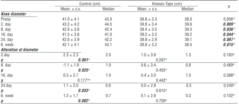
Kinesiotherapy and control groups did not show any significant difference between preoperative knee diameter (p > 0.05). On the other hand, whereas patients undergoing kinesiotherapy did not show a significant increase of knee diameter compared to the preoperative value, the control subjects were found to have a significant increase of knee diameter compared to the preoperative period (Tables 4, 5).
Fig. 1. Early kinesiotaping application over wound closure. Fig. 2. Late kinesiotaping application after removal of closure.
Control Kinesio-Tape
p Mean. s.s./n-% Median Mean. s.s./n-% Median
Age 42.25 ± 14.8 42.0 40.6 ± 10.5 41.0 0.665t Sex Women 7 33.3% 7 35.0% 0.910X2 Man 14 66.7% 13 65.0% Height 169.1 ± 8.0 168.5 170.3 ± 10.5 173.0 0.524m Weight 77.4 ± 12.8 74.0 82.2 ± 15.7 80.0 0.228m BMI 27.1 ± 4.7 26.5 28.4 ± 5.4 27.5 0.473m Tourniquet 274.7 ± 18.1 270.0 282.2 ± 15.9 280.0 0.149m Tourniquet Duration 35.7 ± 10.0 35.0 34.8 ± 12.7 35.0 0.629m
Preop Knee Flexion 136.7 ± 7.3 140.0 138.3 ± 4.9 140.0 0.450m
Preop Diz Extansion -2.4 ± 7.8 -5.0 -2.3 ± 3.0 0.0 0.317m ttest / mMann-Whitney U test / X2Chi-Square test
Table 1. General characteristics of the study and control groups
Control Kinesio-Tape
p Mean.± s.s. Median Mean.± s.s. Median
VAS Preop 4.3 ± 2.4 5.0 4.2 ± 2.1 4.0 0.701m 2. day 4.7 ± 2.0 5.0 4.9 ± 2.4 5.0 0.885m 8. day 2.9 ± 1.8 3.0 2.6 ± 1.4 2.5 0.563m 16. day 2.9 ± 2.0 3.0 1.9 ± 1.3 2.0 0.109m 24. day 1.4 ± 1.0 1.0 1.6 ± 1.3 2.0 0.646m 6. week 1.0 ± 0.9 1.0 1.2 ± 1.5 1.0 0.934m
Comparisons of VAS alteration
2. day 0.4 ± 2.3 0.0 0.9 ± 3.1 0.0 0.683m p 0.496w 0.430w 8. day -1.4 ± 2.8 -2.0 -1.4 ± 2.5 -1.5 0.712m p 0.040w 0.003w 16. day -1.5 ± 2.7 -2.0 -2.1 ± 2.8 -2.0 0.589m p 0.013w 0.001w 24. day -3.0 ± 2.4 -3.0 -2.4 ± 2.6 -2.0 0.399m p 0.000w 0.000w 6. week -3.4 ± 2.2 -4.0 -2.8 ± 2.7 -3.0 0.422m p 0.000w 0.000w
m Mann-Whitney U test / w Wilcoxon test
Table 2. Inter-group analysis of the changes in VAS score
Control (cm) Kinesio-Tape (cm)
p Mean.± s.s. Median Mean.± s.s. Median
Mid-thigh diameter Preop 47.8 ± 4.6 48.0 49.0 ± 6.0 50.7 0.465m 2. day 50.1 ± 4.8 50.2 49.2 ± 5.3 50.8 0.657m 8. day 49.6 ± 4.8 50.0 48.9 ± 5.0 49.5 0.764m 16. day 50.0 ± 4.5 50.2 48.0 ± 5.1 48.5 0.285m 24. day 50.5 ± 5.0 50.0 47.8 ± 4.8 48.5 0.127m 6. week 50.8 ± 5.3 50.1 48.6 ± 5.1 50.5 0.225m Alteration of diameter 2. day 2.3 ± 3.0 1.5 0.2 ± 4.3 -0.4 0.049m p 0.002w 0.794w 8. day -1.7 ± 2.8 1.0 -0.1 ± 4.2 0.1 0.120m p 0.003w 0.896w 16. day 2.1 ± 3.4 1.1 -1.1 ± 4.2 -0.8 0.003m p 0.003ww 0.150w 24. day 2.7 ± 3.9 1.0 -1.2 ± 5.0 -0.1 0.010m p 0.004w 0.276w 6. week 3.0 ± 4.1 1.0 -0.5 ± 4.0 0.2 0.006m p 0.000w 0.559w
m Mann-Whitney U test / w Wilcoxon test
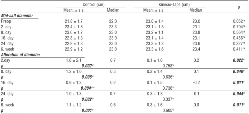
(p > 0.05). The kinesiotherapy group had a significantly reduced mid-calf diameter compared to the control group (p< 0.05) (Table 5). No sig-nificant differences were found be-tween the two groups with respect to the preoperative, 2ndday, 8th day, 16thday, 24thday, and 6thweek ankle diameters (p > 0.05) (Tables 5–8).
DISCUSSION
Patients who underwent kinesio-taping after knee arthroscopy showed no significant differences in pain scores compared to the control group. While the kinesiotherapy group had smaller knee diameters at the early and late preoperative periods, it had a smaller mid-calf diameter only at late postoperative
Control (cm) Kinesio-Tape (cm)
p Mean.± s.s. Median Mean.± s.s. Median
Knee diameter Preop 41.0 ± 4.1 43.0 38.8 ± 3.3 38.0 0.056m 2. day 43.3 ± 4.2 44.5 39.8 ± 3.4 39.6 0.009m 8. day 42.0 ± 3.8 42.4 39.4 ± 3.5 39.3 0.036m 16. day 41.5 ± 3.8 41.0 39.2 ± 3.2 39.2 0.044m 24. day 42.0 ± 3.9 42.2 38.8 ± 2.9 39.1 0.007m 6. week 42.1 ± 4.1 43.1 38.8 ± 3.2 38.5 0.015m Alteration of diameter 2.day 2.3 ± 2.3 2.0 1.0 ± 3.9 1.3 0.183m p 0.001w 0.287w 8. day -1.1 ± 1.9 1.0 0.6 ± 3.4 0.8 0.489m p 0.025w 0.455w 16. day 0.5 ± 2.7 1.0 0.4 ± 3.0 1.0 0.388m p 0.177ww 0.442w 24.day 1.1 ± 2.0 0.8 0.0 ± 2.8 0.3 0.240m p 0.033w 0.615w 6. week 1.2 ± 1.7 0.7 0.1 ± 2.8 0.3 0.102m p 0.002w 0.709w
m Mann-Whitney U test / w Wilcoxon test
Table 4. Inter-group analysis of the changes in mid-calf diameter
Table 5. Line graphics of VAS, mid-thigh and knee diameter
Table 6. Line graphics of mid-calf and ankle diameter
Kinesiotherapy and control groups did not show any significant difference with regard to preoperative, 2nd day, 8th day, 16th day, and 24th day mid-calf diameter
period. The two groups did not differ significantly with respect to thigh and ankle diameters.
Kinesiotaping has been used for years for knee condi-tions, particularly to achieve pain control, to relieve me-chanical problems like patellar instability, and to establish effective lymphatic circulation and mitigate swelling (4, 9, 10, 14).
The efficacy of kinesiotaping therapy after surgery has been assessed particularly for cruciate ligament tears and total knee prosthesis procedures (1, 3, 10).
In a prospective randomized controlled study, Balki et al studied 30 anterior cruciate ligament tears. They divided patients into two groups each containing 15 patients, one of which underwent lymphatic correction
and rectus femoris inhibition method. Then, one group was assigned to kinesiotaping and the other to sham taping, and both groups were compared for pain, knee swelling, and hamstring muscle strength as well as articular range of motion. At the end of two months, the kinesiotaping group fared significantly better than the control group. Our study exclusively dealt with the effect of diagnostic and invasive arthroscopy on pain and whole leg swelling. Bearing in mind the hypothesis stating that blood extravasated after arthroscopy causes diameter changes not only inside the knee joint, but also around the knee and calf region, we determined three more different reference points others than those around the knee. However, although we detected significant changes
Control (cm) Kinesio-Tape (cm)
p Mean.± s.s. Median Mean.± s.s. Median
Mid-calf diameter Preop 37.0 ± 3.3 38.0 35.7 ± 4.0 36.5 0.284m 2. day 39.2 ± 3.5 39.0 37.4 ± 4.4 37.8 0.151m 8. day 38.7 ± 3.3 38.6 36.7 ± 3.7 37.1 0.066m 16. day 38.6 ± 3.1 38.6 36.6 ± 4.2 36.3 0.148m 24. day 38.7 ± 3.2 38.5 36.2 ± 3.7 37.1 0.050m 6. week 39.0 ± 3.4 39.4 35.8 ± 4.0 36.0 0.011m Alteration of diameter 2.day 2.2 ± 2.5 1.7 1.7 ± 3.6 1.1 0.296m p 0.001w 0.032w 8. day 1.7 ± 2.3 1.0 0.9 ± 3.6 0.4 0.080m p 0.001w 0.421w 16. day 1.6 ± 2.4 1.0 0.8 ± 3.5 0.1 0.251m p 0.005ww 0.343w 24. day 1.7 ± 2.2 0.7 0.5 ± 3.0 0.2 0.017m p 0.002w 0.522w 6. week 2.0 ± 2.5 0.9 0.1 ± 2.8 0.3 0.004m p 0.001w 0.762w
m Mann-Whitney U test / w Wilcoxon test
Table 7. Inter-group analysis of the changes in thigh diameter
Control (cm) Kinesio-Tape (cm)
p Mean.± s.s. Median Mean.± s.s. Median
Mid-calf diameter Preop 21.8 ± 1.7 22.0 23.0 ± 1.4 23.0 0.052m 2. day 23.4 ± 1.8 23.3 23.1 ± 1.8 23.1 0.794m 8. day 23.0 ± 1.7 23.0 23.2 ± 1.1 23.8 0.564m 16. day 22.8 ± 1.3 23.0 23.1 ± 1.4 23.1 0.456m 24. day 22.9 ± 1.3 23.0 23.3 ± 1.3 23.6 0.327m 6. week 22.9 ± 1.2 23.0 23.3 ± 1.6 23.4 0.411m Alteration of diameter 2.day 1.6 ± 2.1 0.7 0.1 ± 1.6 0.2 0.022m p 0.002w 0.758w 8. day 1.2 ± 1.6 0.5 0.2 ± 1.4 0.1 0.040m p 0.006w 0.636w 16. day 0.9 ± 1.3 0.2 0.1 ± 1.5 -0.2 0.011m p 0.004ww 0.736w 24. day 1.0 ± 1.3 0.7 0.3 ± 1.3 0.1 0.044m p 0.002w 0.337w 6. week 1.1 ± 1.2 0.6 0.3 ± 1.6 0.0 0.011m p 0.001w 0.605w
m Mann-Whitney U test / w Wilcoxon test
in knee diameter and late-term calf diameter, we did not notice any significant differences in other diameters. Shai et al. compared kinesiotaping and classical bandaging among 41 patients with lymphedema after breast cancer surgery. They reported that kinesiotherapy was effective enough to replace bandaging for the treatment of edema. Unlike a study suggesting that it was an effective therapy for relieving upper extremity swelling and lymphedema, the diameter of whole leg except for knee diameter was not significantly altered (13).
Laborie et al. studied patients undergoing anterior cruciate ligament surgery for an assessment of early-term pain intensity and the amount of analgesics used at the postoperative period. They assigned 60 patients into two groups, one of which underwent kinesiotaping and the other was determined as the control group. They did not observe any significant differences with regard to early postoperative pain scores and the amount of anal-gesics although the kinesio group showed a better patient satisfaction than the control group. Our study likewise did not detect any significant difference between the two groups' pain scores, both at early postoperative period and 6th week. We therefore are of the opinion that kinesiotaping is not effective on pain control among patients undergoing knee arthroscopy (10).
The strength of the present study is that it is the first trial to assess the efficacy of kinesiotherapy after arthroscopy, one of the most commonly preferred operations in ortho-pedics clinics. Another strength is its prospective design, and inclusion a control group in a randomized fashion.
The main limitations of the study are the assessment of all patients undergoing arthroscopy, inability to form a ho-mogenous group, and the lack of an assessment of muscle strength and functional score besides pain and swelling
There are some patient related conditions that make tape application complicating like sweating, body hairs etc. and these conditions could be effect the results.
CONCLUSIONS
Compared to the control group, kinesiotherapy alone is not an effective method for pain relief after knee arthroscopy. On the other hand, it can be preferred par-ticularly for reducing knee effusion after surgery. Kine-siotaping has no effect on thigh, calf, and ankle diameters after arthroscopy.
Authors’ contributions:
BG planned the study and writing. HB and SGG coordinated the ap-plication of physiotherapy and kinesiotaping. EK and ME moderated and analysed the study. SY took the photos and coordinated statistics. Level of evidence 1, Prospective randomized controlled study. The medical ethics committee of the Istanbul Medipol Univercity (Istan-bul-Turkey) approved this study with the decision number 10840098-604.01.01-E.1698. Written informed consents were obtained from the patients for publication of this cases and accompanying images. No funding resources or acknowledgements.
Data sharing is not applicable to this article, as no data sets were ge-nerated or analysed during the study.
There is no conflict of interest in this study.
References
1. Balki S, Göktaş HE, Öztemur Z. Kinesio taping as a treatment method in the acute phase of ACL reconstruction: A double-blind, placebo-controlled study. Acta Orthop Traumatol Turc. 2016;50:628– 634.
2. Barlow T, Downham C, Barlow C. The effects of complementary therapies on post-operative pain control in ambulatory knee surgery: a systematic review. Complement Ther Med. 2013;2:529– 534.
3. Donec V, Kriščiūnas A. The effectiveness of Kinesio Taping®after
total knee replacement in early postoperative rehabilitation period. A randomized controlled trial. Eur J Phys Rehabil Med. 2014;50:363– 371.
4. Freedman SR, Brody LT, Rosenthal M, Wise JC. Short-term effects of patellar kinesio taping on pain and hop function in patients with patellofemoral pain syndrome. Sports Health. 2014;6:294–300.
5. Hong S, Shim J, Kim S, Namkoong S, Roh H. Effect of kinesio taping on the isokinetic muscle function in football athletes with a knee injury. J Phys Ther Sci. 2016;28:218–222.
6. Imbert R. Arthroscopy of the knee. Sem Hop Paris. 1961;37:854– 855.
7. Kase K, Wallis J, Kase T. Health & Personal care. Clinical therapeutic applications of the kinesio taping method. 3rdedition.
[Kinesio Taping Ass., Albuquerque, NM , 2013].
8. Kaya Mutlu E, Mustafaoglu R, Birinci T, Razak Ozdincler A. Does Kinesio Taping of the Knee Improve Pain and Functionality in Patients with Knee Osteoarthritis?: a randomized controlled clinical trial. Am J Phys Med Rehabil. 2017;96:25–33.
9. Kocyigit F, Turkmen MB, Acar M, Guldane N, Kose T, Kuyucu E, Erdil M. Kinesio taping or sham taping in knee osteoarthritis? A randomized, double-blind, sham-controlled trial. Complement Ther Clin Pract. 2015;21:262–267.
10. Laborie M, Klouche S, Herman S, Gerometta A, Lefevre N, Bohu Y. Inefficacy of Kinesio-Taping®on early postoperative pain after
ACL reconstruction: Prospective comparative study. Orthop Trau-matol Surg Res. 2015;101:963–967.
11. Lee JH, Yoo WG. Treatment of chronic Achilles tendon pain by Kinesio taping in an amateur badminton player. Phys Ther Sport. 2012;13:115–119.
12. Murgier J, Cassard X. Cryotherapy with dynamic intermittent compression foranalgesia after anterior cruciate ligament recon-struction. Preliminary study. Orthop Traumatol Surg Res. 2014;100:309–512.
13. Tsai HJ, Hung HC, Yang JL, Huang CS, Tsauo JY. Could kinesio tape replacethe bandage in decongestive lymphatic therapy for breast-cancer-related lymphedema? A pilot study. Support Care Cancer. 2009;17:1353–1360.
14. Wageck B, Nunes GS, Bohlen NB, Santos GM, de Noronha M. Kinesio Taping does not improve the symptoms or function of older people with knee osteoarthritis: a randomised trial. J Physiother. 2016;62:153–158.
Corresponding author:
Ersin Kuyucu Medipol University Tem Avrupa Göztepe çıkışı Bağcılar-Istanbul-Turkey