THE COMPARISON OF THE PRECISION OF DIFFERENT DENTAL
RADIOGRAPHIC METHODS IN MANDIBULAR PERI-IMPLANTARY
MEASUREMENTS: AN IN VITRO STUDY
Alt Çene İmplant Çevresi Ölçümlerinde Farklı Dental Radyografik Metodların
Hassasiyetlerinin Karşılaştırılması: in vitro çalışma
Hakan Bilhan
1, Onur Geckili
1, Selda Arat Bilhan
2, Fatih Ayçicek
1, Berkman Albayrak
3Pelin Bozbulut
3, Fatma Ünalan
1Received: 13/05/2014 Accepted:13/11/2014
ABSTRACT
Purpose: The objective of this in vitro study was to investigate and compare the precisions of several radiodiagnostic methods used in dentistry for the measurement of peri-implantary sites.
Materials and Methods: Six dental implants were placed in a human cadaver mandible. Periapical radiographs obtained with the parallel as well as the bisecting angle technique, digital and conventional panoramic radiographs were used for implant and periimplant bone measurements. The measurement results at each implant were statistically analyzed. Results: The ICC values for the inter-observer reliability were 0.79 for implant diameters and 0.96 for implant lengths. Statistical significance was not detected between the differences of the measurements of the 2 examiners from the original implant dimensions related to anatomic locations. For both of the examiner measurements, significantly less difference from the original implant dimensions was detected in the parallel technique compared to the other techniques (p<0.05).
Conclusions: The present study showed that the most precise peri-implant bone measurements can be obtained from periapical radiographies by using the parallel technique.
Keywords: Panoramic radiography, dental implantation, reproducibility of results, parallel technique, bisecting technique, measurement precision, periapical radiography
ÖZ
Amaç: Bu in vitro çalışmanın amacı implant çevresi alanların ölçümünde kullanılan çeşitli radyodiagnostik metodların hassasiyetlerini değerlendirmek ve karşılaştırmaktır.
Gereç ve Yöntem: Altı dental implant bir insan kadavrasına ait alt çeneye yerleştirildi. İmplant ve implant çevresi kemik ölçümleri için hem paralel hem de açıortay tekniği ile elde edilen periapikal radyografiler, dijital ve konvansiyonel panoramik radyografiler kullanıldı. Her implantın ölçüm sonuçları istatistiksel olarak analiz edildi.
Bulgular: Gözlemciler arası güvenilirlik için sınıf içi kolerasyon değerleri ; implant çapı için 0.79 , implant uzunluğu için 0.96 dır. 2 gözlemcinin ölçümleri ve anatomik konumlara ilişkin orjinal implant ebatları arasında istatistiksel olarak anlamlı bir fark bulunamamıştır. Her gözlemcinin ölçümünün, paralel teknik kullanılarak yapılan ölçümlerde diğer tekniklere göre orjinal implant ebatlarından anlamlı olarak daha az fark gösterdiği belirlenmiştir. Sonuç: Bu çalışma implant çevresi kemik ölçümünde paralel teknik ile elde edilen periapikal radyografinin en hassas ölçüm tekniği olduğunu göstermiştir.
Anahtar kelimeler: Panoramik radyografi, dental implantasyon, sonuçların tekrarlanabilirliği, paralel teknik, açıortay tekniği, ölçüm hassasiyeti, periapikal radyografi
1 Department of Prosthodontics Faculty of Dentistry Istanbul University 2 Department of Prosthodontics, Medipol Mega Hospital and University 3. Undergraduate Student, Faculty of Dentistry Istanbul University
Introduction
The measurement of the marginal alveolar bone level changes over time using radiographs has been reported to be an important parameter in the evaluation of dental implant success (1). Thus, radiography plays an important role in clinical routine practice and in research projects evaluating dental implants. It is difficult to determine small changes in bone height clinically, therefore radiography is an important help for detailed measurements. Marginal bone loss can be defined as the distance between the alveolar crest and the cement-enamel border or the border line of rough and polished surfaces in dental implants. Several intra- or extra-oral radiographic methods are used in dentistry. Intra- and extra-oral radiographs can be acquired conventionally as well as digitally. Periapical radiographies acquired by using bisecting angle or parallel techniques are often used in implant dentistry (2-4). Panoramic radiographs are widely used for evaluation of the condition of the bone around implants supporting mandibular overdentures (5-10). Panoramic radiograph includes both the jaws and the teeth and is a simple examination process (11), offering ease of operation and a shorter working time. It should be kept in mind that, especially in patients with severe alveolar resorption, the use of intraoral films may be difficult. On the other hand, there are several studies pointing out the disadvantages of panoramic radiographies in diagnostic imaging performed for detailed evaluation and quantification. Panoramic radiographs have been reported to be unsuitable because of the magnification, distortion, relative unsharpness and superimposition of the cervical spine (12, 13). Many authors only accept the results of measurements obtained from standardized intraoral radiographs.
The aim of this in vitro study was to investigate the precision of peri-implantary measurements on different 2-dimensional radiographic methods and help clinicians to decide which technique to use for radiographic measurements.
Materials and Methods
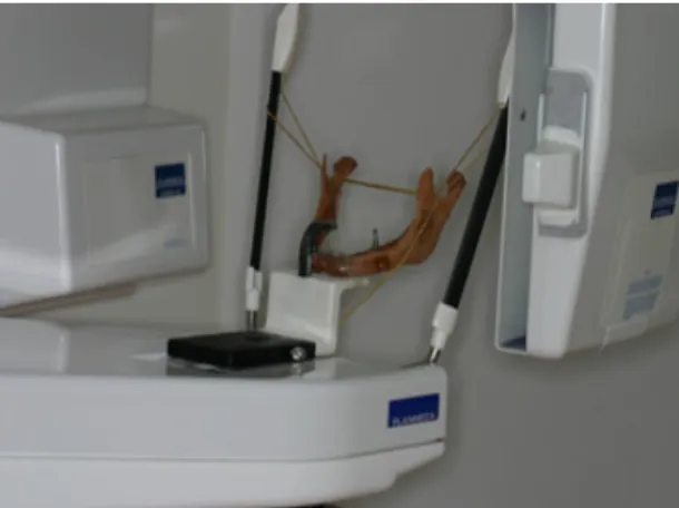
The mandible of a formalin fixed human cadaver (Figure 1) was used for the exposure with different radiographic methods. After removal of the mandible from the cadaver, six implant beds with a safe distance to each other (Figure 2) were prepared following the standard drilling protocol recommended by the
manufacturer (Straumann Dental Implants; Institut Straumann AG, Basel, Switzerland). Care was taken to spread the implants to all possible alveolar sites of the mandible. Six implants were inserted until the roughened surfaces were completely covered by the bone.
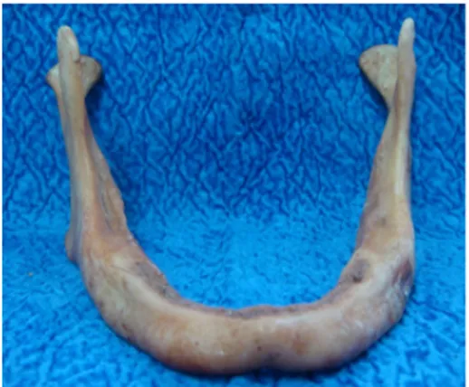
Figure 1. The mandible obtained from a formalin fixed human cadaver.
Figure 2. Six implants were placed with a safe
distance to each other.
Radiographic Evaluation and Bone Level Assessment After placement of the implants, following methods were used to obtain radiographies:
1. Conventional panoramic radiography (Planmeca, Proline XC, Helsinki, Finland) (Figure 3).
Figure 3. Conventional panoramic radiography of the mounted mandible.
2. Digital panoramic radiography (Morita Veraview IC5, J. Morita MFG. Corp., Kyoto, Japan).
3. Bisecting angle (Kodak Ultraspeed Dental Film, Eastman Kodak, Rochester, NY) with a Siemens Heliodent MD model X1744 (Sirona Dental Systems, GmbH D-64625, Bensheim, Germany).
4. Parallel technique (Kodak Ultraspeed Dental Film, Eastman Kodak, Rochester, NY) with a Siemens Heliodent MD model X1744 (Sirona Dental Systems, GmbH D-64625, Bensheim, Germany) X-ray machine set to 70 kV and 7 mA.
All the measurements were performed independently by two examiners blinded to the study protocol. Measurements were obtained from images of conventional radiographs which were scanned, digitized (Epson 1680 Pro, Seiko Epson Corporation, Nagano, Japan) and analyzed at 5x magnification using a software program (CorelDraw 11.0; Corel Corp and Coral Ltd, Ottawa, Canada). The implant diameters at the widest region and implant lengths, as specified by the manufacturer, were used as reference points, which were 4.8 mm and 10 mm respectively. To account for variability, implant diameters and lengths were measured and compared to the documented original dimensions, and ratios were calculated to adjust for distortion. A distortion coefficient was determined for each site and imaging technique. Inter-observer reliability was assessed via comparison of the measurements of two different examiners.
Statistical Analysis
Statistical Package for Social Sciences (SPSS) statistical software (ver. 15.0 for Windows;
SPSS, Inc., Chicago, IL, USA) was used in this study. Kolmogorov-Smirnov test was used to test normality of the distribution. Since the data did not meet the requirements for normal distribution, non-parametric Kruskal Wallis test was used for multiple comparisons. When signigicant differences are detected among study variables, Mann Whitney U test was employed for pairwise comparisons. Wilcoxon sign-test was performed to compare the examiners. Inter-observer reliability was calculated by the intra-class correlation coefficient (ICC). The results were assessed at 95% confidence interval, and p values less than 0.05 are considered significant.
Results
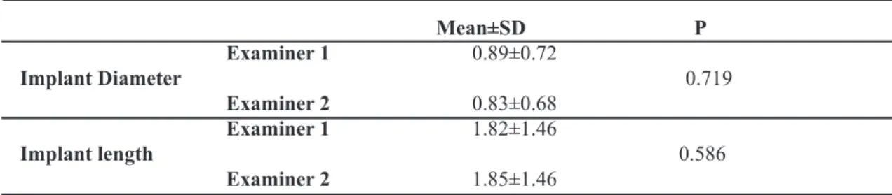
No statistically significant difference was detected between the differences of the overall measurements of the 2 examiners from the original implant lengths and implant diameters (p>0.05; Table 1). The ICC values for the inter-observer reliability were 0.79 for implant diameter and 0.96 for implant lengths These findings were qualified as excellent agreements according to the recommendations of Schuck (14). The measurements of 2 examiners for in all techniques are presented in Table 2.
Table 1. The differences of all of the measurements of the 2 examiners from the original implant dimensions. Mean±SD P Examiner 1 0.89±0.72 Implant Diameter 0.719 Examiner 2 0.83±0.68 Examiner 1 1.82±1.46 Implant length 0.586 Examiner 2 1.85±1.46
Wilcoxon sign test
Table 2. The differences of the measurements of the 2 examiners from the original implant dimensions using the bisecting, parallel and analog and digital panoramic techniques.
Technique Mean±SD p Examiner 1 0.58±0.46
0.173 Implant Diameter Bisecting Examiner 2 0.65±0.40 Technique Examiner 1 1.19±1.93 Implant length 0.249 Examiner 2 1.31±1.82 Examiner 1 0.05±0.04 Implant Diameter 0.600 Parallel Examiner 2 0.10±0.15 Technique Examiner 1 0.13±0.11 Implant length 0.753 Examiner 2 0.19±0.30 Examiner 1 1.67±0.29 Implant Diameter 0.917 Analog panoramic Examiner 2 1.68±0.33 Technique Examiner 1 3.40±0.60 Implant length 0.600 Examiner 2 3.44±0.68 Examiner 1 1.42±0.78 Implant Diameter 0.600 Digital panoramic Examiner 2 1.10±0.77 Technique Examiner 1 2.90±1.58 Implant length 0.917 Examiner 2 3.01±1.39
No statistical difference was detected between the different measurements of the 2 examiners from the original implant dimensions related to anatomic locations (p>0.05; Table 3).
For both of the examiner measurements, significantly less difference from the original implant dimensions was detected in parallel technique than the other techniques
(p<0.05; Table 4). The difference from the original implant dimensions measured by the bisecting angle technique were found significantly lower than the analog and digital panoramic techniques (p<0.05; Table 4). No significance was detected between the differences from the original implant dimensions found in analog and digital panoramic techniques (p>0.05; Table 4).
Table 3. The differences of the measurements of the 2 examiners from the original implant dimensions related to anatomic locations.
Examiner 1 Examiner 2
Anatomic Implant Diameter Implant length Implant Diameter Implant length
Location
Mean±SD Mean±SD Mean±SD Mean±SD
Tooth no: 31 0.81±0.63 (0.68) 1.65±1.29 (1.40) 0.77±0.72 (0.71) 1.57±1.48 (1.44) Tooth no: 33 1.07±0.87 (0.78) 2.22±1.73 (1.60) 1.12±0.94 (0.78) 2.38±2.04 (1.61) Tooth no: 36 0.41±0.54 (0.05) 0.85±1.10 (0.11) 0.51±0.50 (0.47) 1.04±1.02 (0.97) Tooth no: 41 1.08±0.74 (0.98) 2.20±1.51 (2.01) 1.06±0.69 (1.10) 2.17±1.42 (2.22) Tooth no: 43 1.20±0.83 (1.24) 2.46±1.69 (2.54) 0.76±0.71 (0.88) 2.38±1.54 (2.11) Tooth no: 46 0.76±0.72 (0.53) 1.56±1.47 (1.09) 0.78±0.65 (0.38) 1.60±1.33 (0.78) p 0.521 0.481 0.856 0.716
Kruskal Wallis Test
Table 4. Comparison of the techniques by subtracting the measured values from the original dimensions.
Examiner 1 Examiner 2
Technique Implant Diameter Implant length Implant Diameter Implant length
Mean±SD Mean±SD Mean±SD Mean±SD Bisecting Technique 0.81±0.63 (0.68) 1.65±1.29 (1.40) 0.77±0.72 (0.71) 1.57±1.48 (1.44) Parallel Technique 1.07±0.87 (0.78) 2.22±1.73 (1.60) 1.12±0.94 (0.78) 2.38±2.04 (1.61) Analog panoramic Technique 0.41±0.54 (0.05) 0.85±1.10 (0.11) 0.51±0.50 (0.47) 1.04±1.02 (0.97) Digital panoramic Technique 1.08±0.74 (0.98) 2.20±1.51 (2.01) 1.06±0.69 (1.10) 2.17±1.42 (2.22) TTechnique 1.20±0.83 (1.24) 2.46±1.69 (2.54) 0.76±0.71 (0.88) 2.38±1.54 (2.11) Technique 0.76±0.72 (0.53) 1.56±1.47 (1.09) 0.78±0.65 (0.38) 1.60±1.33 (0.78) p 0.521 0.481 0.856 0.716
Techniques Examiner 1 Examiner 2
Implant Diameter Implant length Implant Diameter Implant
length p p p p Bisecting/Parallel Techniques 0.037* 0.037* 0.019* 0.025* Bisecting/Analog panoramic Techniques 0.006** 0.006** 0.004** 0.004** Bisecting/Digital panoramic Techniques 0.050* 0.050* 0.262 0.037* Parallel /Analog panoramic Techniques 0.004** 0.004** 0.004** 0.004** Parallel /Digital panoramic Techniques 0.011* 0.016* 0.016* 0.004** Analog/Digital panoramic Techniques 0.631 0.631 0.150 0.631
Kruskal Wallis Test
Mann Whitney U Test * p<0.05 ** p<0.01
Discussion
The purpose of this study was to investigate the precision of various radiographic methods in evaluating bone around dental implants. Intraoral and panoramic radiographies were used to compare the precision of measurements in this study. It is noteworthy that the results are reproducible, the ICC values showing excellent inter-observer agreement for overall measurements of two examiners according to the recommendations of Schuck (14).
The peri-implant bone can be analyzed with the aid of vectoral software programs and various measurements on digital or digitized radiographies or by hand tracing of digital radiographic printouts. Two recent articles showed that in computerized tracing of direct digital radiographs compared with hand tracing of digital radiographic printouts, differences were minimal and clinically acceptable (15, 16). The digital method which was used in the present study is one of the most often used programs for this purpose. Although a 2000% magnification is recommended, our experience showed that in magnification levels
over 500% the image can lose its clarity and the precision of the measurements is lost. For this reason a 500% magnification was used in the present study during measurements. When using radiography techniques, it is essential to use minimal ionizing radiation doses to obtain images that have acceptable diagnostic quality.Standard methods such as panoramic radiographies or intraoral projections are mostly sufficient, although geometric distortions or magnification in the images may occur depending on the distance of the film to the object as well as position and factors related to the patient.
Although it had been stated that radiographs cannot always provide diagnostic information, even when a computer-aided analysis system is used (17), radiography plays an important role in clinical routine practice and in research projects evaluating dental implants (18, 19) The bone loss documented in two-dimensional radiodiagnostic methods is the reduction of the bone levels at the mesial and distal aspects of the implants, ignoring the so-called saucerization
of the crestal bone around the neck of the implants since only 2-dimensional imaging is used. To be able to obtain data, especially about the vestibular crestal bone changes, a 3-dimensional imaging technique such as the Cone Beam Computerized Tomography (CBCT) is reported to be necessary (20) and it is considered as the golden standard (2). During radiographic evaluation of a radiolucent zone bordering a metal implant, it should also be reminded that the influence of the Mach-band effect will most probably alter the outcome (21).
Panoramic radiographs are widely used for evaluation of the bone condition around implants (5-10). Although there are numerous studies pointing out the disadvantages of panoramic radiographies in diagnostic imaging (12, 13) and many authorities only accept the results of measurements obtained from standardized intraoral radiographs, it is known that a panoramic radiograph includes both of the jaws and the teeth and is a simple examination method (22). Given that the radiographs were of a high quality, Akesson (23) had concluded that for the assessment of the marginal bone level around teeth the radiographic examination of choice should be the panoramic radiograph, which is in accordance with a study by Persson et al. (24) A study by Kullman et al. (25) also showed that panoramic radiographs are as reliable as conventional intra-oral films when used to assess the point of bone attachment to implant threads. It was concluded in a recent study that vertical measurements had acceptable accuracy and reproducibility when a software-based calibrated measurement tool was used, confirming that digital panoramic radiography can be reliably utilized to determine the pre-operative implant length in premolar and molar mandibular segments (26). As stated in a recent study, there are only a few studies reporting on the measurement accuracy and on the distortion or magnification of digital panoramic radiography (27-30) and none that describes the impact of measurement accuracy on the pre-operative bone height evaluation prior to especially implant placement in the posterior mandible. Although most panoramic machines also might give varied and unreliable magnification, they are easier to obtain (4), since the panoramic exposure offers ease of operation and shorter working time. In spite of a higher resolution of the images, the most important disadvantage of intraoral radiographies might be that intraoral film-holders can be painful especially for edentulous patients with severe ridge resorption (31, 32). For highly atrophic mandibles
with unfavorable imaging conditions, rotational panoramic radiographs can be a useful alternative to intraoral small-format radiographs for evaluating peri-implant bone loss (32). On conventional panoramic radiographs, dental implants of known length are reported to be easily measured with a sliding caliper to determine the unit’s magnification factor (33). Variations in the degree of magnification in the horizontal and vertical planes may result in image distortion, showing a certain degree of vertical magnification due to projection geometry, are expected for conventional film-based panoramic radiography, as well as direct digital panoramic devices (12, 34-36). Due to varying magnification, reference objects with known dimensions are necessary to determine the exact magnification in a particular area (27, 33, 36). Nevertheless, the results of the present study point out the importance of variable distortions in the vertical dimension in measurements on panoramic radiographies. This could be important in implant planning, especially in determination of the implant length, since the planned length can be misleading and implants with undesired dimensions may be used which can lead to injury of anatomic structures such as the mandibular canal or the mental foramen.
According to the results of a recent study (37), the diagnostic performance of conventional and digital panoramic images was reported to be equal for the localization of mental and mandibular foramens. The vertical radiographic measurements were considered generally reliable and had correlated with direct measurements in mandible for conventional and digital panoramic radiography. In the present study the measurements in mandible for conventional and digital panoramic radiography were in correlation as well. Although there are numerous studies showing a sympathy for panoramic radiographies, the results of the present study statistically supported the superiority of the accuracy of measurements on intraoral radiographies and the most superior obtained with the parallel technique.
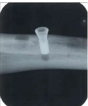
The comparison of both intraoral techniques has revealed that the parallel method gives much better images suitable for making precise measurements (Figure 4). The main problem with the bisecting angle method was that the placement of the film obviously was inexact in all exposures and the images failed to show the implant threads sharply (Figure 5). Additionally, the bone level at the implant shoulder was inexact, too, due to exposure geometry. In an experimental study, the errors (38) in radiographic
assessment of marginal bone height around osseointegrated implants had been analyzed and it was demonstrated that very small deviations of the X-ray beam from the perpendicular with respect to the long axis of the implant will compromise the accuracy of bone height measurements. It was pointed out that for taking periapical radiographs the use of the bisecting angle technique should be discouraged because of the inherent distortion of the resultant image. It was explained that the bisecting angle technique relies on a geometric trick to produce the image, but only a portion of the structures being imaged are dimensionally accurate. The long cone paralleling technique for taking periapical radiographs seems to be the technique of choice since it has advantages like reduced skin dose, reduced magnification, showing the true relationship between the bone height and adjacent teeth and the absence of superimposition of the zygoma over the upper molar region. It should be noted that to obtain optimum results from the long cone paralleling technique, the film-focal distance should be approximately 30 cm, as we have used in the present study.
Figure 4. The radiographs taken with the parallel technique give much better images which are suitable for making precise measurements.
Figure 5. Due to the difficulty of placement of the film in the cor-rect angle images will often fail to show the
implant threads sharply.
Digitizing radiographs were reported to be able to facilitate the quantification of bone changes (39). In a study that had been conducted for the detection of bone loss with different x-ray techniques, it had been reported that a pre-orientation with respect to the expected bone loss is possible using panoramic radiographs (32, 40). Additional intraoral films have reported to be useful where rapid changes of bone levels are expected (40). In the present study, we have used analogue radiographs.The daily routine forces us to use more practical methods such as the panoramic radiographies. As an argument it could be stated that the intra-oral radiographic technique can fail to depict the apical portion of the implant. However, this will not be a limitation because in cases where peri-implant bone level changes are evaluated, the region of interest is visualized. If accurate measurements on the radiographies are intended, the parallel technique seems to be the most reliable method to obtain high resolution images to be used for peri-implant bone measurements. Since panoramic and periapical radiographies do not provide three-dimensional information on bone quantity and anatomic structures, CBCT methods are recommended for the measurement of bone resorption around implants (20) and the current literature also inclined towards CBCT analysis (40). This may be regarded as the major limitation of this study and further studies investigating the precision of CBCT methods in
the same manner should be conducted.
Conclusion
Within the limitations of this in vitro study, it can be concluded that the most suitable radiographic method for precise mandibular periimplant bone measurements is the intraoral periapical radiography taken with the parallel technique. This technique is repeatable and gives a good interobserver reliability.
Disclosure
The authors declare that they do not have any affiliation with any product or company.
Source of funding
None declared
Conflict of interest
None declared
References
1. Chaytor DV, Zarb GA, Schmitt A, Lewis DW. The longitudinal effectiveness of osse-ointegrated dental implants. The Toronto study: bone level changes. Int J Periodontics Restorative Dent 1991;11(2):112–125. 2. Monsour PA, Dudhia R. Implant radiography and
radiology. Aust Dent J 2008;53 (Suppl 1):11-25. 3. Dula K, Mini R, van der Stelt PF, Buser D. The
radiographic assessment of implant patients: decision-making criteria. Int J Oral Maxillofac Implants 2001;16(1):80–89.
4. Mupparapu M, Singer SR. Implant imaging for the dentist. J Can Dent Assoc 2004; 70(1):32.
5. Donatsky O. Osseointegrated dental implants with ball attachments supporting over-dentures in patients with mandibular alveolar ridge atrophy. Int J Oral Maxillofac Im-plants 1993;8(2):162–166.
6. Boerrigter EM, van Oort RP, Raghoebar GM, Stegenga B, Schoen PJ, Boering G. A controlled clinical trial of implant-retained mandibular overdentures: clinical aspects. J Oral Rehabil 1997;24(3):182–190. 7. Versteegh PA, Van Beek GJ, Slagter AP, Ottervanger
JP. Clinical evaluation of man-dibular overdentures supported by multiple-bar fabrication: a follow-up study of two implant systems. Int J Oral Maxillofac Implants 1995;10(5):595–603.
8. Mericske-Stern R, Steinlin Schaffner T, Marti P, Geering AH. Peri-implant mucosal aspects of ITI implants supporting overdentures. A
five-year longitudinal study. Clin Oral Implants Res 1994;5(1):9–18.
9. Spiekermann H, Jansen VK, Richter EJ. A 10-year follow-up study of IMZ and TPS implants in the edentulous mandible using bar-retained overdentures. Int J Oral Maxil-lofac Implants 1995;10(2):231–243. 10. Leimola-Virtanen R, Peltola J, Oksala E, Helenius H,
Happonen RP. ITI titanium plas-ma-sprayed screw implants in the treatment of edentulous mandibles: a follow-up study of 39 patients. Int J Oral Maxillofac Implants 1995;10(3):373–378.
11. Akesson L, Hakansson J, Rohlin M, Zöger B. An evaluation of image quality for the assessment of the marginal bone level in panoramic radiography. A comparison of radi-ographs from different dental clinics. Swed Dent J 1993;17(1-2):9–21.
12. Batenburg RHK, Stellingsma K, Raghoebar GM, Vissink A. Bone height measure-ments on panoramic radiographs: the effect of shape and position of edentulous mandi-bles. Oral Surg Oral Med Oral Pathol Oral Radiol Endod 1997;84(4): 430-435. 13. Batenburg RH, Meijer HJ, Geraets WG, van der Stelt
PF. Radiographic assessment of changes in marginal bone around endosseous implants supporting mandibular overdentures. Dentomaxillofac Radiol 1998;27(4):221-224.
14. Schuck P. Assessing reproducibility for interval data in health related quality life ques-tionnaires: which coefficient should be used? Qual Life Res 2004;13(3):571–586.
15. Celik E, Polat-Ozsoy O, Toygar Memikoglu TU. Comparison of cephalometric meas-urements with digital versus conventional cephalometric analysis. Eur J Orthod 2009;31(3):241-246.
16. Naoumova J, Lindman R. A comparison of manual traced images and corresponding scanned radiographs digitally traced. Eur J Orthod 2009;31(3):247-253. 17. Adell R, Eriksson B, Lekholm U, Brånemark PI,
Jemt T. Long-term follow-up study of osseointegrated implants in the treatment of totally edentulous jaws. Int J Oral Maxillofac Implants 1990;5(4):347-359. 18. Albrektsson T, Sennerby L. State of the art in oral
implants. J Clin Periodontol 1991;18(6):474-481. 19. Brägger U, Burgin W, Fourmoussis I, Lang NP. Image
processing for the evaluation of dental implants. Dentomaxillofac Radiol 1992;21(4):208-212. 20. Duyck J, Rønold HJ, Van Oosterwyck H, Naert
I, Vander Sloten J, Ellingsen JE. The influence of static and dynamic loading on marginal bone reactions around osseointe-grated implants: an animal experimental study. Clin Oral Implants Res
2001;12(3):207-218.
21. Sewerin IP, Gotfredsen K, Stoltze K. Accuracy of radiographic diagnosis of peri-implant radiolucencies--an in vitro experiment. Clin Oral Implants Res 1997;8(4):299-304.
22. Misch CE, Perel ML, Wang HL, Sammartino G, Galindo-Moreno P, Trisi P, Steigmann M, Rebaudi A, Palti A, Pikos MA, Schwartz-Arad D, Choukroun J, Gutierrez-Perez JL, Marenzi G, Valavanis DK. Implant success, survival, and failure: the International Congress of Oral Implantologists (ICOI) Pisa Consensus Conference. Implant Dent 2008;17(1):5-15.
23. Akesson L. Panoramic radiography in the assessment of marginal bone level. Swed Dent J Suppl 1991;78:1–129.
24. Persson RE, Tzannetou S, Feloutzis AG, Brägger U, Persson GR, Lang NP. Compari-son between panoramic and intra-oral radiographs for the assessment of alveolar bone levels in a periodontal maintenance population. J Clin Periodontol 2003;30(9):833–839.
25. Kullman L, Al Asfour A, Zetterqvist L, Andersson L. Comparison of radiographic bone height assessments in panoramic and intraoral radiographs of implant patients. Int J Oral Maxillofac Implants 2007;22(1):96–100.
26. Vazquez L, Nizamaldin Y, Combescure C, Nedir R, Bischof M, Dohan Ehrenfest DM, Carrel JP, Belser UC. Accuracy of vertical height measurements on direct digital pano-ramic radiographs using posterior mandibular implants and metal balls as reference ob-jects. Dentomaxillofac Radiol 2013;42(2):20110429. 27. Schropp L, Stavropoulos A, Gotfredsen E, Wenzel
A. Calibration of radiographs by a reference metal ball affects preoperative selection of implant size. Clin Oral Investig 2009;13(4):375–381.
28. Schulze R, Krummenauer F, Schalldach F, d’Hoedt B. Precision and accuracy of meas-urements in digital panoramic radiography. Dentomaxillofac Radiol 2000;29(1): 52–56.
29. Park JB. The evaluation of digital panoramic radiographs taken for implant dentistry in daily practice. Med Oral Patol Oral Cir Bucal 2010;15(4):e663–666.
30. Kim YK, Park JY, Kim SG, Kim JS, Kim JD. Magnification rate of digital panoramic radiographs and its effectiveness for pre-operative assessment of dental implants. Den-tomaxillofac Radiol 2011;40(2):76–83.
31. Chiapasco M. Early and immediate restoration and
loading of implants in completely edentulous patients. Int J Oral Maxillofac Implants 2004;19 Suppl:76-91. 32. Zechner W, Watzak G, Gahleitner A, Busenlechner D, Tepper G, Watzek G. Rotational panoramic versus intraoral rectangular radiographs for evaluation of peri-implant bone loss in the anterior atrophic mandible. Int J Oral Maxillofac Implants 2003;18(6):873– 878.
33. Vazquez L, Nizam Al Din Y, Christoph Belser U, Combescure C, Bernard JP. Reliabil-ity of the vertical magnification factor on panoramic radiographs: clinical implications for posterior mandibular implants. Clin Oral Impl Res 2011;22(12):1420-1425. 34. Tronje G, Welander U, McDavid WD, Morris CR.
Image distortion in rotational pano-ramic radiography. I. General considerations. Acta Radiol Diagn (Stockh) 1981; 22(3A):295–299.
35. Tronje G, Eliasson S, Julin P, Welander U. Image distortion in rotational panoramic radiography. II. Vertical distances. Acta Radiol Diagn (Stockh) 1981;22(4):449–455.
36. McDavid WD, Dove SB, Welander U, Tronje G. Dimensional reproduction in direct digital rotational panoramic radiography. Oral Surg Oral Med Oral Pathol 1993;75(4): 523–527.
37. Peker I, Gungor K, Semiz M, Tekdemir I. Localization of mental and mandibular fora-mens on the conventional and digital panoramic images. Coll Antropol 2009;33(3):857-862.
38. Sewerin IP. Errors in radiographic assessment of marginal bone height around osseoin-tegrated implants. Scand J Dent Res 1990;98(5):428-433. 39. Van der Stelt PF. Computer assisted interpretation
in radiographic diagnosis. Dent Clin North Am 1993;37(4):683-696.
40. Kim TS, Obst C, Zehaczek S, Geenen C. Detection of bone loss with different X-ray techniques in periodontal patients. J Periodontol 2008;79(7):1141-1149.
Corresponding Author: Onur GECKILI
Department of Prosthodontics
Faculty of Dentistry Istanbul University 34093 - Capa, Istanbul, TURKEY Phone: +90-212-414 20 20 (ext.: 30316) e-mail: [email protected]
View publication stats View publication stats