Case Report / Olgu Sunumu
DO I:10.4274/tnd.75428 Turk J Neurol 2016;22:130-132
Lhermitte-Duclos Disease with Orthostatic Hypotension
Ortostatik Hipotansiyon ile Birlikte İzlenen Lhermitte-Duclos Hastalığı Olgusu
Nesrin Helvacı Yılmaz
1, Mehmet Şeker
2, Mehmet Onur Omaygenç
3, Umut Yaka
4, Nazan Eryiğit
5, Burak Yuluğ
11Medipol University Faculty of Medicine, Department of Neurology, İstanbul, Turkey 2Medipol University Faculty of Medicine, Department of Radiology, İstanbul, Turkey 3Medipol University Faculty of Medicine, Department of Cardiology, İstanbul, Turkey 4Medipol University Faculty of Medicine, Department of Neurosurgery, İstanbul, Turkey 5Çakmak Erdem Hospital, Clinic of Neurosurgery, İstanbul, Turkey
130
Lhermitte-Duclos disease is a rare cerebellar dysplastic gangliocytoma. The most common symptoms include headache, nausea, vomiting, blurred vision, and imbalance. The typical appearance on cranial magnetic resonance imaging is hyper-intensity on T2-weighted images and hypo-intensity on T1-weighted images. The disease generally presents with benign progress. Development of obstructive hydrocephalus is an indication for urgent surgical intervention and surgery outcomes are satisfactory. Orthostatic hypotension is a very rare clinical presentation of this syndrome and ours is the second case of orthostatic hypotension to be reported in the literature.
Keywords: Lhermitte-Duclos disease, orthostatic hypotension, cerebellum
Lhermitte-Duclos hastalığı serebellumun nadir görülen displastik ganliyositomuna verilen isimdir. En sık başvuru şikayetleri baş ağrısı, bulantı, kusma, dengesizlik ve görme bulanıklığıdır. Tanıda kranyal manyetik rezonans görüntülemede T2 ağırlıklı kesitlerde hiperintens, T1 ağırlıklı kesitlerde hipointens görünüm oldukça tipiktir. Sıklıkla iyi seyirlidir. Obstrüktif hidrosefali gelişimi acil cerrahi müdahale endikasyonudur ve sonuç yüz güldürücüdür. Ortostatik hipotansiyon bu sendromun çok nadir görülen klinik prezentasyonu olup bizim hastamız literatürde bildirilen ikinci olgudur.
Anahtar Kelimeler: Lhermitte-Duclos hastalığı, ortostatik hipotansiyon, serebellum
Sum mary
Öz
Introduction
Dysplastic gangliocytoma of the cerebellum, also known as
Lhermitte-Duclos disease (LDD), is a rare hamartomatous disorder
of the cerebellar cortex (1,2). It typically presents in young
adults but can also appear at all ages (3). It is known as a slowly
progressive and benign disease but rarely it can cause obstructive
hydrocephalus and careful follow-up is required. We report a
patient with LDD who was admitted to hospital with sudden and
short-term loss of consciousness.
Case Report
A woman aged 21 years was presented to our neurology clinic
reporting a sudden loss of consciousness lasting a few seconds,
which happened two weeks before admission. Her parents
reported no convulsions or urinary incontinence. She had reported
Ad dress for Cor res pon den ce/Ya z›fl ma Ad re si: Nesrin Helvacı Yılmaz MD, Medipol University Faculty of Medicine, Department of Neurology, İstanbul, Turkey Phone: +90 532 685 13 90 E-mail: [email protected]
Re cei ved/Ge lifl Ta ri hi: 27.02.2015 Ac cep ted/Ka bul Ta ri hi: 27.04.2015 ©Turkish Journal of Neurology, Published by Galenos Publishing House.
Turk J Neurol 2016;22:130-132 Helvacı Yılmaz et al.; Lhermitte-Duclos Disease
sudden blackouts of vision for a few years. In her medical history
she had had breath holding spells once per month when she was
between 11 months and 3 years of age. She had not taken any
drugs in that period. Her neurologic examination was normal.
Total blood count, liver, kidney and thyroid function tests, serum
levels of glucose, calcium, magnesium and vitamin B12, which
were performed for the differential diagnosis of syncope, were
normal. An awake electroencephalograph was performed for
thirty minutes and was normal. Her cardiologic examination,
electrocardiography, echocardiography and rhythm Holter tests
were normal. During the tilt table test, at the tenth minute
after provocation she had a spell of syncope and was diagnosed
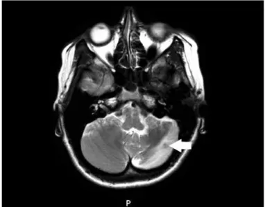
as having orthostatic hypotension. Cranial magnetic resonance
imaging (MRI) showed dysplastic gangliocytoma of the left
cerebellum (Figure 1, 2). Cranial MR angiography, and carotid
and vertebral artery Doppler ultrasonography were normal.
Physicians from the department of neurosurgery did not consider
a surgical operation.
Discussion
Gangliocytomas are benign intraparenchymal tumors
composed of neoplastic ganglion cells and stromal glial cells
(4). The pathologic changes in gangliocytoma of the cerebellum
that cause LDD are the loss of the middle Purkinje cell layer and
infiltration of the internal granular cell layer with large neurons
with vesicular nuclei and marked nucleoli, which enlarge cerebellar
folias (1,5). Findings of light and electron microscopes show that
these cells are hypertrophic granular cells (2).
The most common symptoms of LDD are headache, nausea,
vomiting, dizziness, imbalance, and blurred vision (6). Papilledema,
cranial nerve palsies, ataxia, and confusion can be found in the
neurologic examination (1,3). Neuroimaging is enough to make
the diagnosis (7). Cerebellar enlargement causes a typical striated
pattern that can be seen as hyper-intense in T2- and hypo-intense
in T1-weighted sequences of cranial MRI (8).
These tumors are slowly progressive and some patients can be
asymptomatic (9). It can take a couple of months to ten years to
manifest (10,11). However, the treatment options are controversial;
observation, performing biopsy, or resection (10,11). Sudden onset
headache, vomiting, and findings of hydrocephalus are indications
for surgery (12). The results of surgery are satisfactory but
recurrence can be seen (13).
Cowden sydrome (autosomal dominant inherited multiple
hamartoma syndrome) should be kept in mind in patients with
LDD (14). LDD is one of the major diagnostic criteria for Cowden
syndrome and these patients should be scanned for phosphatase
and tensin homolog gene mutations and cancers (15). Our patient
had no skin lesions or history of cancer.
Orthostatic hypotension is more common in elder patients but
it can also accompany neurologic disorders including multisystem
atrophy, Lewy body dementia, Parkinson’s disease, amyloidosis,
and diabetic autonomic neuropathy (16). Orthostatic hypotension
has also been reported with posterior fossa lesions, including
medullary tumors and infarctions (17,18). In our patient, who
was admitted with sudden and short-term loss of consciousness
and had normal neurologic examination, we considered that the
concurrence of syncope and tumor could have been a conincidence,
but when we searched the literature we found a patient with
orthostatic hypotension who had completely recovered after
resection of tumor (5). In our case, despite the lack of medullary
involvement, the most important clinical finding was orthostatic
hypotension. The increase in intracranial pressure is suggested to
indirectly cause orthostatic hypotension.
Conclusion
LDD is a very rare disorder and our patient is only the second
reported in the literature. To explain the relationship between
brain stem and spells of orthostatic hypotension, we suggest
fluorodeoxyglucose-positron emission tomograhy studies should
be performed.
131
Figure 1.
T1 hypointensity in the left posterior inferior cerebellar hemisphere in axial magnetic resonance imagingFigure 2.
T2 hyperintensity in the left posterior inferior cerebellar hemisphere in axial magnetic resonance imagingTurk J Neurol 2016;22:130-132 Helvacı Yılmaz et al.; Lhermitte-Duclos Disease
Ethics
Informed Consent: Consent form was filled out by all participants,
Peer-review: External and internal peer-reviewed.
Authorship Contributions
Surgical and Medical Practices: Nesrin Helvacı Yılmaz, Umut
Yaka, Nazan Eryiğit, Mehmet Onur Omaygenç, Concept: Nesrin Helvacı
Yılmaz, Burak Yuluğ, Nazan Eryiğit, Design: Nesrin Helvacı Yılmaz,
Mehmet Şeker, Burak Yuluğ, Data Collection or Processing: Nesrin
Helvacı Yılmaz, Mehmet Şeker, Analysis or Interpretation: Nesrin
Helvacı Yılmaz, Mehmet Şeker, Mehmet Onur Omaygenç, Literature
Search: Nesrin Helvacı Yılmaz, Mehmet Onur Omaygenç, Burak Yuluğ,
Writing: Nesrin Helvacı Yılmaz, Burak Yuluğ.
Conflict of Interest: No conflict of interest was declared by the authors.
Financial Disclosure: The authors declared that this study has received
no financial support.
References
1. Nowak DA, Trost HA, Porr A, Stölzle A, Lumenta CB. Lhermitte-Duclos disease (Dysplastic gangliocytoma of the cerebellum). Clin Neurol Neurosurg 2001;103:105-110.
2. Onder E, Arikök AT, Türkoğlu E, Alper M. Lhermitte-Duclos Disease: A Rare Lesion with Variable Presentations and Obscure Histopathology. Turk Patoloji Derg 2014.
3. Nowak DA, Trost HA. Lhermitte-Duclos disease (dysplastic cerebellar gangliocytoma): a malformation, hamartoma or neoplasm? Acta Neurol Scand 2002;105:137-145.
4. Türeyen K, Senol N, Sav A. Gangliocytoma associated with focal cortical dysplasia in a young-adult: a case report. Turk Neurosurg 2008;18:259-263. 5. Ruchoux MM, Gray F, Gherardi R, Schaeffer A, Comoy J, Poirier J.
Orthostatic hypotension from a cerebellar gangliocytoma (Lhermitte-Duclos disease). Case report J Neurosurg 1986;65:245-248.
6. Wei G, Zhang W, Li Q, Kang X, Zhao H, Liu X, Tang X, Wu Y, Han J, Yin H. Magnetic resonance characteristics of adult-onset Lhermitte-Duclos disease: An indicator for active cancer surveillance? Mol Clin Oncol 2014;2:415-420. 7. Shinagare AB, Patil NK, Sorte SZ. Case 144: Dysplastic cerebellar
gangliocytoma (Lhermitte-Duclos disease). Radiology 2009;251:298-303. 8. Meltzer CC, Smirniotopoulos JG, Jones RV. The striated cerebellum: an
MR imaging sign in Lhermitte-Duclos disease (dysplastic gangliocytoma). Radiology 1995;194:699-703.
9. Giorgianni A, Pellegrino C, De Benedictis A, Mercuri A, Baruzzi F, Minotto R, Tabano A, Balbi S. Lhermitte-Duclos disease. A case report Neuroradiol J 2013;26:655-660.
10. Milbouw G, Born JD, Martin D, Collignon J, Hans P, Reznik M, Bonnal J. Clinical and radiological aspects of dysplastic gangliocytoma (Lhermitte-Duclos disease): a report of two cases with review of the literature. Neurosurgery 1988;22:124-128.
11. Capone Mori A, Hoeltzenbein M, Poetsch M, Schneider JF, Brandner S, Boltshauser E. Lhermitte-Duclos disease in 3 children: A clinical long-term observation. Neuropediatrics 2003;34:30-35.
12. Bozbuga M, Gulec I, Suslu HT, Bayindir C. Bilateral Lhermitte-Duclos disease. Neurol India 2010;58:309-311.
13. Hashimoto H, Iida J, Masui K, Nishi N, Sakaki T. Recurrent Lhermitte-Duclos disease: Case report. Neurol Med Chir [Tokyo] 1997;37:692-696.
14. Riegert-Johnson DL, Gleeson FC, Roberts M, Tholen K, Youngborg L, Bullock M, Boardman LA. Cancer and Lhermitte-Duclos disease are common in Cowden syndrome patients. Hered Cancer Clin Pract 2010;8:6.
15. Pilarski R, Burt R, Kohlman W, Pho L, Shannon KM, Swisher E. Cowden syndrome and the PTEN hamartoma tumor syndrome: systematic review and revised diagnostic criteria. J Natl Cancer Inst 2013;105:1607-1616. 16. Maule S, Papotti G, Naso D, Magnino C, Testa E, Veglio F. Orthostatic
hypotension: evaluation and treatment. Cardiovasc Hematol Disord Drug Targets 2007;7:63-70.
17. O'Malley WE, O'Doherty DS, Auth TL. Orthostatic hypotension as a manifestation of posterior fossa tumor. Dis Nerv Syst 1970;31:846-850. 18. Korpelainen JT, Sotaniemi KA, Suominen K, Tolonen U, Myllylä VV.
Cardiovascular autonomic reflexes in brain infarction. Stroke 1994;25:787-792.