doi: 10.3389/fphar.2019.00340
Edited by: Flavio Rizzolio, Università Ca’ Foscari, Italy Reviewed by: Laura J. Blair, University of South Florida, United States Floriana Campanile, Università degli Studi di Catania, Italy *Correspondence: Can Murat Ünal [email protected]; [email protected]
Specialty section: This article was submitted to Experimental Pharmacology and Drug Discovery, a section of the journal Frontiers in Pharmacology Received: 04 December 2018 Accepted: 19 March 2019 Published: 05 April 2019 Citation: Ünal CM, Karagöz MS, Berges M, Priebe C, Borrero de Acuña JM, Wissing J, Jänsch L, Jahn D and Steinert M (2019) Pleiotropic Clostridioides difficile Cyclophilin PpiB Controls Cysteine- Tolerance, Toxin Production, the Central Metabolism and Multiple Stress Responses. Front. Pharmacol. 10:340. doi: 10.3389/fphar.2019.00340
Pleiotropic Clostridioides difficile
Cyclophilin PpiB Controls
Cysteine-Tolerance, Toxin Production, the
Central Metabolism and Multiple
Stress Responses
Can Murat Ünal1,2* , Mustafa Safa Karagöz2, Mareike Berges1,3, Christina Priebe1, José Manuel Borrero de Acuña1, Josef Wissing3,4, Lothar Jänsch3,4, Dieter Jahn1,3and Michael Steinert1,3,5
1Institut für Mikrobiologie, Technische Universität Braunschweig, Braunschweig, Germany,2Moleküler Biyoteknoloji Bölümü,
Türk-Alman Üniversitesi, Istanbul, Turkey,3Braunschweig Integrated Centre of Systems Biology, Braunschweig, Germany, 4Cellular Proteomics Research, Helmholtz Centre for Infection Research, Braunschweig, Germany,5Helmholtz Centre
for Infection Research, Braunschweig, Germany
The Gram-positive pathogen Clostridioides difficile is the main bacterial agent of nosocomial antibiotic associated diarrhea. Bacterial peptidyl-prolyl-cis/trans-isomerases (PPIases) are well established modulators of virulence that influence the outcome of human pathologies during infections. Here, we present the first interactomic network of the sole cyclophilin-type PPIase of C. difficile (CdPpiB) and show that it has diverse interaction partners including major enzymes of the amino acid-dependent energy (LdhA, EtfAB, Had, Acd) and the glucose-derived (Fba, GapA, Pfo, Pyk, Pyc) central metabolism. Proteins of the general (UspA), oxidative (Rbr1,2,3, Dsr), alkaline (YloU, YphY) and cold shock (CspB) response were found bound to CdPpiB. The transcriptional (Lrp), translational (InfC, RFF) and folding (GroS, DnaK) control proteins were also found attached. For a crucial enzyme of cysteine metabolism, O-acetylserine sulfhydrylase (CysK), the global transcription regulator Lrp and the flagellar subunit FliC, these interactions were independently confirmed using a bacterial two hybrid system. The active site residues F50, F109, and F110 of CdPpiB were shown to be important for the interaction with the residue P87 of Lrp. CysK activity after heat denaturation was restored by interaction with CdPpiB. In accordance, tolerance toward cell wall stress caused by the exposure to amoxicillin was reduced. In the absence of CdPpiB, C. difficile was more susceptible toward L-cysteine. At the same time, the cysteine-mediated suppression of toxin production ceased resulting in higher cytotoxicity. In summary, the cyclophilin-type PPIase of C. difficile (CdPpiB) coordinates major cellular processes via its interaction with major regulators of transcription, translation, protein folding, stress response and the central metabolism.
Keywords: Clostridium difficile, peptidyl-prolyl-cis/trans-isomerase (PPIase), cytotoxicity, interactomics, transcription
INTRODUCTION
The multi-resistant Gram-positive obligate anaerobe
Clostridioides (Clostridium) difficile is the main nosocomial bacterial agent of antibiotic treatment associated diarrhea (Hopkins and Wilson, 2018). The rise of C. difficile infection (CDI) coincides with the introduction of clindamycin, the first broad-band lincosamin against anaerobic Gram-negative pathogens (Bartlett et al., 1977; Lusk et al., 1978). Currently, first line of treatment is the use of antibiotics like vancomycin, metronidazole and fidaxomicin. However, high recurrence rates, especially after treatment with vancomycin or metronidazole, make this disease difficult to manage (Mullane, 2014;Hopkins and Wilson, 2018).
A major factor influencing the course and outcome of CDI is the composition of the host intestinal microbiome and, connected to it, the bile acid status of the infected individual. Patients undergoing a CDI typically have an antibiotic dependent change of their microbiome followed by a transition from antibacterial to less antibacterial bile acids in their gastrointestinal
tract (Theriot et al., 2016). Most commonly, CDI manifests
in form of pseudomembranous colitis, which is a strong inflammation of the large intestine. This is caused by massive tissue destruction and leukocytosis due to the production of two large glycosylating toxins, TcdA and TcdB. These exert their cytotoxic activity by inactivating small GTPases and disturbing the actin cytoskeleton dynamics leading to disruption of the gut epithelial barrier (Barbut et al., 2007; Rupnik et al., 2009;
Chandrasekaran and Lacy, 2017).
The production of TcdA and TcdB typically coincides with the entry of the bacteria into the stationary phase. Nevertheless, it is controlled by complex regulatory processes on multiple levels and by diverse environmental cues, including subinhibitory concentrations of antibiotics, temperature or the availability of carbon sources and amino acids (Dupuy and Sonenshein, 1998;
Bouillaut et al., 2015). One central metabolite in this respect is
L-cysteine that represses toxin production (Karlsson et al., 2000). Apart from that, L-cysteine also affects the expression of genes of amino acid biosynthesis, fermentation, energy metabolism, iron acquisition and stress response (Dubois et al., 2016; Gu et al., 2018). Furthermore, there is a strong interconnectedness between iron andL-cysteine dependent gene regulation circuits. Several of the genes of the latter contain Fur boxes in their promoters, which are binding sites for the global iron-responsive transcriptional ferric uptake regulator (Fur) (Dubois et al., 2016). If not metabolized effectively L-cysteine also has an inhibitory effect on growth onC. difficile as it was nicely shown in case of a mutant lacking the cysteine desulfidase CdsB (Gu et al., 2017).
Although there is strong correlation between CDI severity and the capability of the bacteria to produce TcdA and TcdB, this alone does not explain the virulence spectrum of C. difficile in its entirety. Accordingly, several other virulence factors such as extracellular proteases, surface layer proteins, a fibronectin bin-ding protein (Fbp68), a collagen binbin-ding protein (CbpA), type IV pili and flagella have been shown or are suspected to contribute to disease severity and host colonization (Geric et al., 2004;Janoir, 2016;Ünal and Steinert, 2016;Péchiné et al., 2018). Especially, the regulation of flagellation and its main structural component
flagellin (FliC) is crucial for modulating adherence to host cells, promoting colonization and eliciting immune responses via toll-like receptor 5 (Tasteyre et al., 2001;Dingle et al., 2011; Batah et al., 2016). Moreover, flagellation and toxin production are co-regulated by an intricate genetic switch adding up to the complexity of toxin-related pathogenicity ofC. difficile (Aubry et al., 2012;Anjuwon-Foster and Tamayo, 2017).
Currently, many single components and mechanisms of CDI that contribute to host colonization, disease outbreak and dissemination are known. However, their concerted action is still not fully understood. Bacterial PPIases (FKBPs, parvulins and cyclophilins) have in many instances been shown to modulate infectious processes (Ünal and Steinert, 2014). Among those are prominent representatives like Mip and Mip-like FKBPs, or parvulins, like SurA in Gram-negative as well as PrsA and PrsA2 in Gram-positive bacteria (Jacobs et al., 1993;Heikkinen et al., 2009;Behrens-Kneip, 2010;Alonzo et al., 2011;Obi et al., 2011;Ikolo et al., 2015;Ünal and Steinert, 2015). Additionally, recent studies have shown the participation of cyclophilins in the maturation of a secreted nuclease inStaphylococcus aureus as well as in virulence traits, like biofilm formation inEscherichia coli or in sliding motility and amoeba infection byLegionella pneumophila (Skagia et al., 2016; Wiemels et al., 2017; Rasch et al., 2018). The aim of our study was to identify the interaction partners of the sole cyclophilin of C. difficile (CdPpiB) and to characterize its contribution to bacterial physiology as well as virulence. Our results show an unprecedented relationship between bacterial cysteine metabolism and CdPpiB with far reaching implica-tions on virulence. Furthermore, a protein network controlling major cellular function including transcription, translation, protein folding, metabolism, and stress responses was uncovered.
MATERIALS AND METHODS
Bacterial Strains and Culture
Bacterial strains and plasmids used in this study are listed in
Table 1.C. difficile 6301erm (hereafter referred to as wild type or wt) and its derivatives were cultured in BHIS medium (brain-heart infusion broth (Carl Roth GmbH, Germany) supplemented
with 5 g/L yeast extract (BD BactoTM, United States) and
1 g/L L-cysteine (Sigma-Aldrich R, Germany) or TY-medium (30 g/L tryptone, 20 g/L yeast extract) under anaerobic conditions
(95% N2/5% H2). If necessary, 5 µg/mL erythromycin or
15 µg/mL thiamphenicol were added. Escherichia coli and
Bacillus megaterium were cultured in LB medium supplemented with 100µg/mL ampicillin, 10 µg/mL tetracycline or 30 µg/ml chloramphenicol at 37◦
C and 200 rpm. All antibiotics were purchased from Sigma-Aldrich R. Media were solidified by adding 1.5% (wt/vol) agar when needed.
In vivo Cross-Linking for the
Identification of PpiB
Interaction Partners
Possible interaction partners of PpiB were identified
by in vivo cross-linking as previously described
(Borrero-de Acuña et al., 2015). Briefly, C. difficile str. 6301erm1ppiB + PpiB-NStrep was grown for 8 h in 25 mL
TABLE 1 | Bacterial strains and plasmids used in this study.
Name Features References
C. difficile str. 6301erm Erythromycin-sensitive derivative of strain 630 DSMZ†(DSM 28645) (Hussain
et al., 2005)
C. difficile str. 6301erm1ppiB cd03310::ClosTron, ErmR This study
E. coli DH10β Strain for cloning and plasmid propagation, F−mcrA1(mrr-hsdRMS-mcrBC) 880dlacZ1M15
1lacX74 endA1 recA1 deoR 1(ara, leu)7697 araD139 galU galK nupG rpsL λ−
Grant et al., 1990
E. coli CA434 E. coli HB101 carrying the R702- conjugative plasmid (Tra+, Mob+) Purdy et al., 2002
E. coli BTH101 F−, cya-99, araD139, galE15, galK16, rpsL1 (StrR), hsdR2, mcrA1, mcrB1 Euromedex, France
B. megaterium MS941 Strain for recombinant production, deficient of the major secreted protease,1nrpM Wittchen and Meinhardt, 1995 pMTL82151 Shuttle vector for complementation of C. difficile mutants; pBP1-ori (Gram-pos.), ColE1 ori
(Gram-neg.), tra, MCS, CmR
Heap et al., 2009
pPpiB_NStrep pMTL82151 carrying wild type ppiB including its 250 bp upstream and 50 bp downstream regions and a N-terminal StrepII-tag
This study
pDSW1728 Ptet::mCherryOpt, CmR Ransom et al., 2015
pPpiB-tet mCherryOpt in pDSW1728 replaced by cd03310 by SacI and BamHI yielding Ptet::ppiB, CmR This study
pN-STREPXa1622 Vector for recombinant protein production in B. megaterium, PxylA-StrepII-Xa-MCS, TcR, AmpR Biedendieck et al., 2007
p119_PpiB pN-STREPXa1622 in which the ORF of ppiB (CD630_03310) was cloned using BglII and SphI This study p119_CysK pN-STREPXa1622 in which the ORF of cysK (CD630_15940) was cloned using BglII and SphI This study pKNT25 Low copy plasmid carrying the T25 subunit of adenylate cyclase with N-terminal MCS, KmR Euromedex, France
pKNTB pKNT25 carrying wt ppiB This study
pUT18 High copy plasmid carrying the T18 subunit of adenylate cyclase with N-terminal MCS, AmpR Euromedex, France
pUT18_CysK pUT18 carrying wt cysK of C. difficile str. 6301erm This study
pUT18_Lrp pUT18 carrying wt lrp of C. difficile str. 6301erm This study
pUT18_FliC pUT18 carrying wt fliC of C. difficile str. 6301erm This study
†Deutsche Sammlung von Mikroorganismen und Zellkulturen GmbH.
of BHIS and refreshed 1:1000 in 2 L BHIS. After 16 h 0.125 % (vol/vol) formaldehyde (AppliChem, Germany) was added for chemical cross-linking (30 min at 37◦
C). Excess formaldehyde
was quenched for 5 min at 37◦
C by the addition of glycine at a final concentration of 130 mM. Cells were harvested and
washed twice with PBS (4000 g for 20 min at 4◦
C). Finally, the cells were resuspended in 25 mL PBS supplemented with complete Protease Inhibitor Cocktail (Roche, Switzerland), and
homogenized using FRENCH R Press (Thermo, United States)
or glass beads and FastPrep R (MP Biomedicals, United States). Cell debris was removed by centrifugation (10,000 g, 10 min, 4◦
C), and filtering the supernatant through a 0.45 µm syringe filter. The supernatant was applied onto a 3 mL Strep-Tactin R column (iba Lifesciences, Germany) in order to purify PpiB and its cross-linked partners according to manual instructions. The elution fraction was concentrated using Amicon Ultra-0.5 mL centrifugal filters (Merck, Germany) with a molecular cut-off of 10 kDa. For analysis, the concentrated eluate was mixed with 4x SDS loading buffer and subjected to decrosslinking by heating at 95◦
C for 30 min. Proteins were separated on a 12% acrylamide gel, and stained using Coomassie stain [100 g/L (NH4)2SO4,
100 mL/L H3PO4, 20% vol/vol methanol]. Bands of interest were
analyzed by LC-MS as previously described (Borrero-de Acuña et al., 2016). Resulting protein identifications (hits) were filtered according to their overall coverage by the LC/MS approach and the overall number of unique peptides. Hits with at least 15% coverage and two or more unique peptides were taken into account as this indicated robust enrichment. Ribosomal proteins were discarded because of their intrinsically high abundance in
the cell and their known unspecific protein-protein interactions due to their overall strong basic character.
Bacterial Two Hybrid (BACTH) Screening
Protein-protein interactions were confirmed using the BACTH-system of Euromedex (France), which bases on an adenylate cyclase (CyaA) dependent reporter system. For this, ppiB, fliC
(CD630_02390), lrp (CD630_35440) and cysK (CD630_15940)
were cloned in frame with in two different vectors, each containing one subunit of the adenylate cyclase of Bordetella pertussis (Tables 1, 2). After confirmation of correct cloning, the CyaA-deficient strainE. coli BTH101 was transformed with
combinations of plasmids carrying ppiB and the respective
interaction partners, and transformants were selected on
LB-agar containing 50µg/mL kanamycin and 100 µg/mL ampicillin.
Single transformants were then streaked out on MacConkey-agar (BD DifcoTM, United States) supplemented with 50µg/mL
kanamycin and 100µg/mL ampicillin, incubated for 2 days at
30◦
C, and checked for pink coloring due to CyaA-activity.
Measuring the Interaction of PpiB and
Lrp and Their Amino Acid Substitution
Mutants by
β-Galactosidase Assay
Escherichia coli BTH101 carrying plasmid combinations that had been confirmed on MacConkey-agar were used in a β-galactosidase assay for quantitative comparison of interactions between amino acid mutants of PpiB and Lrp, respectively. For this, at least six clones of each transformation were picked and
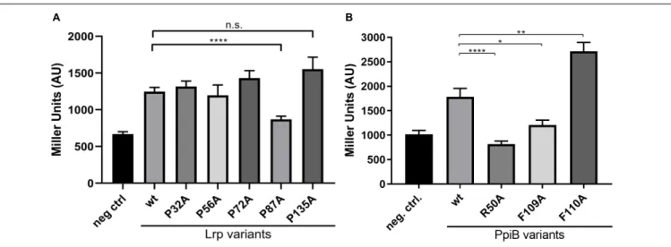
TABLE 2 | Primers used in this study.
Name Sequence† Features References
PpiB_KompF1 gatgagcttGAACCATTAATTGGGGATAATG HindIII, for cloning into pMTL82151 This study
PpiB_KompR1 atcatgcggccgcTTTATCCTCATTTTTAAGTA ATATATA NotI, for cloning into pMTL82151 This study
PpiB_NStrep_F cgggtggctccaTTCCATAATTTATTACCTTCCTTTTC For introducing N-terminal SrepII-Tag This study PpiB_NStrep_R cagtttgaaaaaAATAAAAATCCTATAGTAACTATAGAAATG For introducing N-terminal SrepII-Tag This study
PpiB_For4 TCTgagctcATGAAAAGGAAGGTAATAAATTATG SacI, for cloning into pDSW1728 This study
PpiB_Rev4 ATAggatccTTAGTTTTTTTCTACATCTGAGTAA BamHI, for cloning into pDSW1728 This study
CdPpiB_TH_For gactctagagGAAAATAAAAATCCTATAGTAAC XbaI, for cloning into BACTH vectors This study
CdPpiB_TH_Rev ctcggtaccgcGTTTTTTTCTACATCTGAGTAA KpnI, for cloning into BACTH vectors This study
CdLrp_TH_For gactctagagGATGTTACAGATTACAGAATC XbaI, for cloning into BACTH vectors This study
CdLrp_TH_Rev ctcggtaccgcCAATATTGATTTTGCTTGTATTG KpnI, for cloning into BACTH vectors This study
CdFliC_TH_For gactctagagAGAGTTAATACAAATGTAAGTG XbaI, for cloning into BACTH vectors This study
CdFliC_TH_Rev ctcggtaccgcTCCTAATAATTGTAAAACTCC KpnI, for cloning into BACTH vectors This study
CdCysK_TH_For gactctagagTTATATAATAACGCATTAGAGTT XbaI, for cloning into BACTH vectors This study
CdCysK_TH_Rev ctcggtaccgcAAATATTCCCATAGACATATAC KpnI, for cloning into BACTH vectors This study
Lrp_P32A_invF GCAGTTTCAGAAAGAGTCAAAAG This study
Lrp_P32A_invR tgcAGAAGTTAAACCAACTATTTTTCC This study
Lrp_P56A_invF GATTCATTAGGCAGAGTTATAAAG This study
Lrp_P56A_invR tgcGTTGACAATAGCTTTATATCCTTC This study
Lrp_P72A_invF gcaAGCAATGGATATACAGAATTTATTG This study
Lrp_P72A_invR AAGAGAAATATGAATAAATGCCTTTAT This study
Lrp_P87A_invF AGGATTGTAGAATGTCACCATAT This study
Lrp_P87A_invR tgcGTCCTTTGCAGCTGACTC This study
Lrp_P135A_invF gcaATACAAGCAAAATCAATATTGGC This study
Lrp_P135A_invR CGTTGATAGTATAACAGAGG This study
PpiB_R50A_invF GTAATACCAGGATTTATGATAC This study
PpiB_R50A_invR tgcATGAAATATTATTCCATTGTAATATC This study
PpiB_F109A_invF gcaTTCATAATGCATAAAAACTCACCAC This study
PpiB_F109A_invR TTGAGAACCAGCTGAATTAGGTG This study
PpiB_F110A_invF gcaATAATGCATAAAAACTCACCAC This study
PpiB_F110A_invR AAATTGAGAACCAGCTGAATTAG This study
PpiB_For3 atgagatcttaATGGAAAATAAAAATCCTATAGTAAC BglII, for cloning into pN-STREPXa1622 This study
PpiB_Rev3 tagcatgcTTAGTTTTTTTCTACATCTGAGTAAT SphI, for cloning into pN-STREPXa1622 This study
CdCysK_119_F1 gccagatctTGTTATATAATAACGCATTAGAG BglII, for cloning into pN-STREPXa1622 This study
CdCysK_119_R1 ccggcatgcTTAAAATATTCCCATAGACATATAC SphI, for cloning into pN-STREPXa1622 This study
EBS universal primer CGAAATTAGAAACTTGCGTTCAGTAAAC Heap et al., 2007
ErmRAM_F ACGCGTTATATTGATAAAAATAATAGTGGG Heap et al., 2007
ErmRAM_F ACGCGTGCGACTCATAGAATTATTTCCTCCCG Heap et al., 2007
†Restriction recognition sites are underlined; bases for the respective alanine exchange are highlighted.
grown o/n (30◦
C, 200 rpm) in 5 mL LB medium supplemented
with 50µg/mL kanamycin, 100 µg/mL ampicillin and 0.5 mM
IPTG (GERBU, Germany). Prior to the assay, bacterial growth was stalled by chilling the cultures for at least 30 min at 4◦
C and the OD600was determined. Bacteria of 200µL culture were
pelleted (3500 rpm, 5 min, 4◦
C), and resuspended in 900µL
of Z-Buffer (60 mM Na2HPO4, 40 mM NaH2PO4, 10 mM KCl,
1 mM MgSO4, and freshly added 50 mM β-mercaptoethanol,
pH 7.0). Bacteria were lysed by the addition of 50µL chloroform
and 50 µL 0.1% SDS (wt/vol) for 5 min at 30◦
C. After this,
200µl of o-nitrophenyl-β-D-galactoside (ONPG, Roth GmbH,
Germany) dissolved at 4 mg/mL in phosphate buffer (60 mM Na2HPO4, 40 mM NaH2PO4, pH 7.0) was added. The reactions
were kept for 12 min at 30◦
C and subsequently stopped by
the addition of 500 µL 1 M Na2CO3. After removal of cell
debris and chloroform by centrifugation (14000 rpm, 5 min, 4◦
C) absorption at 420 and 550 nm were recorded with a VarioSkanTM
(Thermo Fisher, United States) plate reader. β-galactosidase
activity, expressed in Miller Units, was calculated according to the following formula:
Miller Units =1000 × (OD420 − 1.75 × OD550)
t[min] × V[mL] × OD600
Construction of a ppiB Destruction
Mutant (
1ppiB) in C. difficile 6301erm
Using ClosTron
A synthetic vector containing the region of ppiB
help of the ClosTron website1 using the Perutka algorithm (Perutka et al., 2004). E. coli CA434 was transformed with customized vectors for mating with C. difficile 6301erm cells
as described previously (Heap et al., 2009, 2010). Mutants
were selected on BHIS containing 15 µg/mL erythromycin
and confirmed by PCR and sequencing using gene specific primers (Table 2).
Assessing Growth and Susceptibility
Toward Amoxicillin
Bacterial growth was monitored by refreshing cultures of C. difficile grown overnight (o/n) in 25 mL medium to a starting
optical density (OD600 nm) of 0.05 in 25 mL fresh medium.
Increase in OD600 nmwas measured starting with t0and every
60 min until the cultures reached stationary phase. For assessing amoxicillin susceptibility, o/n cultures of wild typeC. difficile and
its 1ppiB mutant were prepared in 20 mL BHIS medium. The
next morning the cultures were adjusted to an OD600 nmof 0.1
in BHIS medium. Amoxicillin stock solution was prepared fresh (2 mM in ddH2O) and a 1:1 serial dilution starting at 256µM
was prepared in 100 µl of BHIS distributed in the wells of a
96-well plate. Following this, 100µL of the bacterial suspension were added into each well resulting in a final OD of 0.05. Bacteria were grown for 6 h at 37◦
C under anaerobic conditions, and the
final OD was measured at 600 nm using a VarioSkanTM(Thermo
Fisher, United States) plate reader.
Complementation of C. difficile
1ppiB
With StrepII-Tagged PpiB and
Implementing a
Tetracycline-Inducible Promoter
In order to achieve a complementation of the mutant with wild type PpiB carrying a N-terminal StrepII-tag for affinity purification, ppiB was amplified including its 250 bp up- and 100 bp-downstream regions using the primer combination PpiB_KompF1/R1. This fragment was cloned into the
shuttle vector pMTL82151 with HindIII and BamHI yielding
pMTL_ppiB. This plasmid was used as a template for an inverse PCR by which an N-terminal StrepII-tag was added to PpiB with the primer combination PpiB_NStrep_F/R. The resulting PCR-product was gel purified, phosphorylated with T4 polynucleotide kinase (NEB) and re-ligated with T4 ligase (NEB) according to manufacturer’s instructions. Plasmids carrying the desired mutations were selected inE. coli DH10β and verified by
sequencing. For complementation of the 1ppiB mutant under
the control of a tetracycline-inducible promoter (Ptet), the ORF
of CdPpiB was cloned into pDSW1728 utilizing the restriction
sites SacI and BamHI. The 1ppiB mutant was transformed
by conjugation and selected using 15 µg/ml thiamphenicol.
For complementation studies the bacteria were cultured with 10 ng/mL anhydrotetracycline (AT), and a dose-dependent complementation was achieved by performing the assays with decreasing concentrations of TA starting at 400 ng/mL.
1http://www.clostron.com/
Recombinant Protein Production
For recombinant production,ppiB or cysK were amplified with
Q5-Polymerase (NEB GmbH, Germany) and cloned into the production vector pN-STREPXa1622 using the restriction sites BglII and SphI yielding N-terminally StrepII-tagged proteins (Biedendieck et al., 2007). For recombinant protein production,
an o/n culture of transformed B. megaterium MS941 was
prepared, and the next morning refreshed 1:100 in 300 ml LB medium containing 10µg/mL tetracycline (Biedendieck et al.,
2011). The cells were induced at an OD600 nm of ∼0.4 by
the addition of D-xylose (Carl Roth GmbH, Germany) at a
final concentration of 0.5 % (w/v). After 6 h, the cells were harvested (3000g, 15 min, 4◦
C), and the pellet was once washed with 1x PBS (5000 g, 15 min, 4◦
C). Cells were resuspended in
25 mL ddH2O, and disrupted by FRENCH
R
Press (Thermo, United States). Immediately after the disruption, 10x wash buffer was added to a final concentration of 1x. Cell debris was removed by centrifugation (10000 g, 20 min, 4◦
C) and the supernatant was filtered through a 0.45 µm syringe filter. StrepII-tagged PpiB or CysK were purified from the supernatant using Strep-Tactin R
sepharose (iba Lifesciences, Germany) according to manufacturer’s instructions. Concentration and buffer change of purified proteins were achieved by centrifugation (5000 g, RT) using Vivaspin R 20 ultrafiltration units (Sartorius, Germany) with 30 or 10 kDa molecular weight cut offs. Proteins were
stored in 100 mM HEPES (pH 7.0) at −20◦
C. Protein yield and purity were analyzed by SDS-page, and protein concentration was determined by absorption at 280 nm.
CysK Activity Assay
Enzymatic activity of recombinantly produced CysK ofC. difficile was measured photometrically as previously described with slight modifications (Tai et al., 1993; Saavedra et al., 2004). Briefly, the assays were performed in a 96-well format in 100 µL final volume. As substrates O-Acetyl-L-serine hydrochloride (OAS; Sigma-Aldrich R, A6262) and 5-Thio-2-nitrobenzoic acid (TNB) were used. 20 mM OAS stock solution was freshly prepared in 100 mM HEPES (pH 7.0). Fresh TNB-stock solution was
prepared by dissolving 10 mM 5,50
-Dithiobis(2-nitrobenzoic acid) (DTNB; Sigma-Aldrich R, D8130) and 15 mM dithiothreitol in 100 mM HEPES (pH 7.0). Each reaction contained 2 mM
OAS and 50 µM TNB. Following the addition of CysK at a
final concentration of 7µM, the reactions were monitored in a VarioSkanTM(Thermo Fisher, United States) plate reader at 30◦
C
by measuring the OD412 nm every 4 min. If needed CysK was
denatured by incubating aliquots for 30 min at 56◦
C.
Determination of Toxin Concentration in
Supernatants
For determining the toxin production, exponentially growing
cultures were adjusted to an OD600 nm of 0.05 in 10 mL of
fresh BHIS and grown at 37◦
C under anaerobic conditions. After 48 h, the culture supernatants of 2 mL of each culture were harvested (5000 g, 5 min, RT) and sterile filtered using 0.2µm syringe filters. Toxin concentrations in culture supernatants
ELISA Kit (tgc-E002-1) of tgcBIOMICS (Germany) following the manual instructions.
Cytotoxicity Assay
In order to visualize the cytotoxic activity of C. difficile culture supernatants, NIH-3T3 mouse fibroblast cells (ATCC R
CRL-1658TM, United States) were cultured in DMEM (Gibco,
United States) supplemented with 10% FBS at 37◦
C and 5% CO2. For the assay, 105 cells/mL were seeded on glass cover
slips in 24-well plates and cultured for 2 days. In parallel, C. difficile were grown in anaerobic chamber for 24 h, and the cell free supernatant containing secreted toxins was harvested by centrifugation (8000g, 5 min, RT). This supernatant was diluted to 0.1 % (vol/vol) in cell culture medium. Adherent NIH-3T3 cells were washed once with 1 mL PBS, and 1 mL medium with bacterial culture supernatant was added. After incubation for 2 h, medium was removed and the cells were fixed for 15 min at RT with 2% (wt/vol) PFA in 1x PBS, and subsequently permeabilized with 0.1 % (vol/vol) Triton X-100 in 1x PBS with 1% (wt/vol) BSA for 5 min. Cells were stained with PBS containing 1µg/mL DAPI (Sigma-Aldrich R) and 1:1000 diluted Phalloidin-Alexa488 (Abcam, United Kingdom). The mounted samples were analyzed with a confocal microscope (Leica R SP8, Germany) using an 63x oil immersion objective.
RESULTS
Identification of the Protein Interaction
Partners of CdPpiB
A protein blast search using the PpiB sequence ofBacillus subtilis str. 168 (Uniprot ID: P35137) against the most recently annotated C. difficile str. 6301erm genome identified a single potential cyclophilin encoded by the gene locus CDIF630erm_00459 (CD630_00330 inC. difficile 630). This gene is flanked upstream by a gene potentially encoding for a patatin-like phospholipase, and downstream by a gene encoding the putative exosporium glycoprotein BclA1 (Dannheim et al., 2017a). An alignment of the potential C. difficile cyclophilin with bacterial cyclophilins
of E. coli str. K12 (EcPpiB; P23869), B. subtilis (BsPpiB;
P35137) and Staphylococcus aureus str. NCTC8325 (SaPpiB;
Q2FZU9) as well as eukaryotic representative members of yeast
FIGURE 2 | In vivo cross-linking reveals putative interaction partners of CdPpiB. Shown is a representative analytical SDS-PAGE with the elution fraction after the in vivo cross-linking and the subsequent Strep-tag affinity purification of PpiB-StrepII and its putative interaction partners. Bands denoted with an asterisk were cut out analyzed by mass spectrometry. The bracket indicates bands that also occurred in control experiments without cross-linking and were excluded from the analysis.
(ScCyp1; P14832) and human (HsCypA; P62937) revealed high amino acid sequence similarities between the different proteins. An exceptional high degree of conservation was observed for amino acid residues involved in PPIase activity (Figure 1). Accordingly, this gene was selected for destruction by ClosTron,
which resulted in the generation of a 1ppiB mutant in the
C. difficile str. 6301erm background.
Next, we identified in vivo protein interaction partners of CdPpiB using an interactomics approach by using N-terminally StrepII-tagged PpiB as bait and formaldehyde as a cross-linker. A preparative SDS-PAGE of the purified protein complexes revealed in total, seven distinct protein bands that were cut out and prepared for LC-MS analysis (Figure 2).
This yielded 39 unique hits, and as expected, PpiB was found to be with 83% the best covered protein among those.
FIGURE 1 | CdPpiB is a classical cyclophilin-type PPIase. The alignment of amino acid sequences of cyclophilin type PPIases of the Gram-positive species S. aureus (SaPpiB; Q2FZU9), C. difficile (CdPpiB; Q18D70) and B. subtilis (BsPpiB; P35137) as well as the Gram-negative model organism E. coli (EcPpiB; P23869), and the yeast (ScCyp1; P14832) and human CypA (HsCypA; P62937) reveals highly conserved amino acids along the PPIase active surface (marked by a black line). The alignment was performed using the T-Coffee webserver and visualized by Jalview 2.10.3b1. Amino acids that are conserved to 100% are highlighted by color coding according to Taylor (Taylor, 1986;Notredame et al., 2000;Waterhouse et al., 2009). Amino acids that have been mutated in CdPpiB are marked by an asterisk.
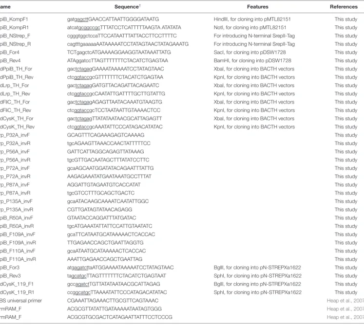
TABLE 3 | Putative interaction partners of CdPpiB identified by in vivo cross-linking.
Gene Coverage # unique
Class Name Accession-Nr.† locus† (%) peptides Function
Metabolism CysK ARE62524.1 01767 40 13 Cysteine synthase activity
GapA ARE64122.1 03466 36 14 Glyceraldehyde-3-phosphate dehydrogenase
Fba ARE61306.1 00531 34 11 Fructose-1,6-bisphosphate aldolase
Pyc ARE60920.1 00083 32 40 Pyruvate carboxylase
EtfA1 ARE61304.1 00529 28 10 Isocaproyl-CoA dehydrogenase, electron transfer
flavoprotein alpha subunit
AcdB ARE61302.1 00527 24 8 Isocaproyl-CoA dehydrogenase, catalytic subunit
PduL ARE63606.1 02937 24 5 Phosphate propanoyltransferase
Putative NUDIX-family hydrolase
ARE61693.1 00909 24 3 Hydrolase
Pfo ARE63605.1 02936 23 25 Pyruvate synthase
NifU-like protein ARE62199.1 01433 23 3 [Fe–S]-cluster assembly protein
AdhE2 ARE63909.1 03250 20 17 Aldehyde-alcohol dehydrogenase
LdhA ARE61297.1 00522 20 6 D-lactate dehydrogenase
PfkA ARE64384.1 03700 20 4 ATP-dependent 6-phosphofructokinase
EtfA3 ARE61965.1 01196 19 6 Electron transfer flavoprotein subunit alpha
Putative 5-nitroimidazole reductase
ARE62385.1 01623 19 3 Putative 5-nitroimidazole antibiotic resistance protein
HadA ARE61298.1 00523 18 6 Isocaprenoyl-CoA:2-hydroxyisocaproate
CoA-transferase
IorB ARE63301.1 02620 18 3 Indolepyruvate oxidoreductase, subunit beta
EtfB1 ARE61303.1 00528 17 4 isocaproyl-CoA dehydrogenase, electron transfer
flavoprotein beta subunit
Pyk ARE64383.1 03699 16 7 Pyuvate kinase
Stress Response Rbr1 ARE61727.1 00944 58 11 Iron ion binding, oxidoreductase activity, rubrerythrin
Dsr ARE61729.1 00946 36 4 In silico predicted desulfoferrodoxin
Rbr2 ARE62400.1 01638 35 6 Iron ion binding, oxidoreductase activity, rubrerythrin
Rbr3 ARE62452.1 01692 33 6 In silico predicted reverse rubrerythrin
UspA ARE61714.1 00930 22 3 Putative universal stress protein A
YloU ARE63487.1 02813 20 2 Putative alkaline-shock protein
Alkaline shock protein (YqhY)
ARE62115.1 01349 17 2 Alkaline shock protein
CspB ARE62276.1 01511 15 2 Cold shock protein
Transcription/ Translation
Lrp ARE64537.1 03859 30 4 AsnC-family transcriptional regulator
InfC ARE61586.1 00798 17 2 Translation initiation factor IF-3
RFF ARE63058.1 02368 16 2 Ribosome-recycling factor
Protein folding Putative phage protein ARE61884.1 01109 29 5 Protein-export protein, SecB-like
GroS ARE61098.1 00314 23 2 10 kDa chaperonin
DnaK ARE63387.1 02707 22 13 Chaperone protein, unfolded protein response
Transport ABC transporter substrate- binding protein
ARE61772.1 00993 34 8 ABC-type transport system, sugar-family extracellular solute-binding protein Biotin carboxyl carrier
protein
ARE62848.1 02143 23 5 In silico predicted biotin carboxyl carrier protein
UPF0145 protein ARE62649.1 01899 23 2 Putative heavy metal binding
Motility/Surface FliC ARE61143.1 00361 54 19 Flagellin
SlpA ARE63719.1 03056 19 11 S-layer precursor protein
†as annotated in str. 6301erm byDannheim et al. (2017a)(GenBank: CP016318.1).
The majority of the remaining 38 hits that resembled proteins interacting either directly or indirectly in larger complexes with CdPpiB belonged to the functional groups metabolism (19 hits, 50%) and stress response (8 hits, 21%). The rest of the hits were distributed among transcription/translation,
protein folding and transport (3 hits and 8% each) as well as motility/surface (2 hits 5%) (Table 3). Among the retrieved potential interaction partners were LdhA, AcdB, HadA, EtfA1, EtfA3, and EtfB1. These enzymes represent the complete
et al., 2006). Similarly, the also detected pyruvate carboxylase Pyc, pyruvate-ferredoxin oxidoreductase Pfo, aldehyde-alcohol dehydrogenase AdhE2 and pyruvate kinase Pyk as well as the glyceraldehyde-3-phosphate GapA and fructose-1,6-bisphosphate aldolase Fba are central enzymes of carbon metabolism (Dannheim et al., 2017b). Finally, theO-acetylserine sulfhydrylase/cysteine synthase (CysK), was among the metabolic proteins the one with the highest sequence coverage. It represents the last enzyme of cysteine biosynthesis (Dubois et al., 2016).
A whole battery of stress response proteins was also found attached to CdPpiB, including the two rubyerythrins Rbr1 and Rbr2 and the desulfoferrodoxin Dsr, which are involved in oxidative stress response (Kawasaki et al., 2009;Riebe et al., 2009). Furthermore, the cold shock protein CspB and the two alkaline shock proteins YloU and YqhY as well as the universal stress
protein UspA were among the interacting proteins (Nyström
and Neidhardt, 1993; Söderholm et al., 2011; Derman et al., 2015; Tödter et al., 2017). The chaperones GroS and DnaK are part of multiple stress responses (LaRossa and Van Dyk, 1991). Key players of translational quality control, the ribosome-recycling factor RRF, translation initiation factor IF-3 InfC and most interestingly, the pleiotropic transcriptional regulator Lrp were further proteins associated with CdPpiB (Brinkman et al., 2003;Ohashi et al., 2003;Laursen et al., 2005). The screen also yielded flagellin (FliC, 54% coverage) and surface layer protein (SlpA, 19% coverage), two major virulence associated proteins (Tasteyre et al., 2001; Merrigan et al., 2013). Finally, a sugar-family solute binding protein, a putative heavy metal binding protein (UPF0145) and a predicted biotin carrier protein were identified as transporter or transporter associated proteins cross-linked to CdPpiB (Table 3).
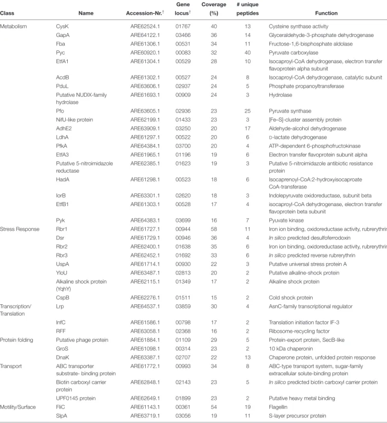
CdPpiB Interacts With Lrp, FliC and
CysK in a Bacterial Two Hybrid System
In order to verify direct interactions between candidate proteins identified by our interactomic studies, we applied bacterial two hybrid (BACTH) based protein-protein interaction tests. For this, we chose Lrp as it is a global transcriptional regulator, and as there are no reports on functional interactions between bacterial PPIases and transcriptional regulators. Another candidate was the metabolic enzyme with the highest coverage, CysK, that participates in cysteine metabolism which is central to virulence
of C. difficile (Dubois et al., 2016). FliC was chosen as
it is a well-established virulence factor in many pathogenic bacteria including C. difficile (Tasteyre et al., 2001). For all the tested proteins a clear-cut interaction with CdPpiB was observed, indicated by pink coloring of the resulting clones on MacConkey-agar verifying our findings from the interactomics study (Figure 3).
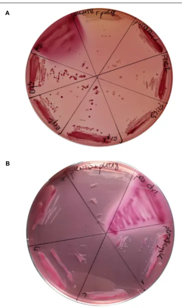
An Intact PPIase Domain of CdPpiB Is
Required for the Interaction With
P87 Substrate of Lrp
Following the confirmation of the interaction via the BACTH screen, we wanted to analyze whether these interactions may be dependent on the PPIase activity of CdPpiB. For this purpose, we
FIGURE 3 | Confirmation CdPpiB interaction partners with BACTH on MycKonkey-Agar. (A) E. coli BTH101 cells transformed with wt ppiB carrying pKNTB in combination with pUT18_FliC carrying wt fliC or pUT18_Lrp carrying wt lrp were streaked out on MacKonkey-agar and grown for 2 days at 30◦C. (B) E. coli BTH101 cells transformed with wt ppiB carrying pKNTB in
combination with pUT18_CysK carrying wt cysK were streaked out on MacKonkey-agar and grown for 2 days at 30◦
C. Purple coloring was indicative of protein interaction due to adenylate cyclase activity. On each plate E. coli BTH101 carrying empty pUT18 plasmid in combination with pKNTB served as negative control, whereas colonies carrying the pUT18-zip and pKT25-zip plasmids delivered by the manufacturer served as positive control. At least three colonies of each transformation were tested. Representative plates of three separate transformations are shown.
chose Lrp for a detailed study as it is with 15 kDa comparably small and has only five proline residues. As PPIases act on the peptidyl-prolyl bonds in proteins, we exchanged all five proline residues in Lrp by alanine generating the single amino acid substitution mutants P32A, P56A, P72A, P87A and P135A. The interactions between CdPpiB and the generated Lrp-variants were quantified in β-galactosidase assays using the outlined
FIGURE 4 | CdPpiB targets P87 in CdLrp and this interaction depends on conserved amino acids. (A)β-galactosidase activities of E. coli BTH101 clones carrying combinations of wt CdPpiB and single proline exchange mutants of CdLrp. (B)β-galactosidase activities of E. coli BTH101 clones carrying combinations of wt CdLrp and single amino acid exchange mutants of CdPpiB. Clones carrying wt CdPpiB and the empty companion vector pKNT25 served as negative control in both experiments. Shown are mean and SEM of three independent experiments with at least five clones each. Statistical significance was calculated by unpaired t-Test with Welch’s correction (∗
p ≤ 0.05,∗∗
p ≤ 0.01,∗∗∗∗
p ≤ 0.0001, n.s., not significant).
BACTH system. Among the five Lrp-variants only variant P87A showed a significantly reduced β-galactosidase activity. It was with 868.8 ± 43.55 MU 30% less than the interaction
with wild type Lrp which had an average β-galactosidase
activity of 1246 ± 59.06 MU indicating a specific interaction between the two proteins (Figure 4A). In order to prove that the PPIase domain is involved in the overall process, we went on by exchanging highly conserved single amino acid residues within the active site of CdPpiB that are known from homologous cyclophilins of Gram-positive and Gram-negative bacteria (Göthel et al., 1996; Skagia et al., 2017a; Wiemels et al., 2017). Accordingly, we generated the CdPpiB variants R50A, F109A and F110A (s.a. Figure 1). For all three mutants, significant differences were observed regarding the interaction with Lrp as assessed byβ-galactosidase activity (Figure 4B).
Destruction of ppiB Leads to Sensitivity
to Envelope Stress and
L-Cysteine in
C. difficile
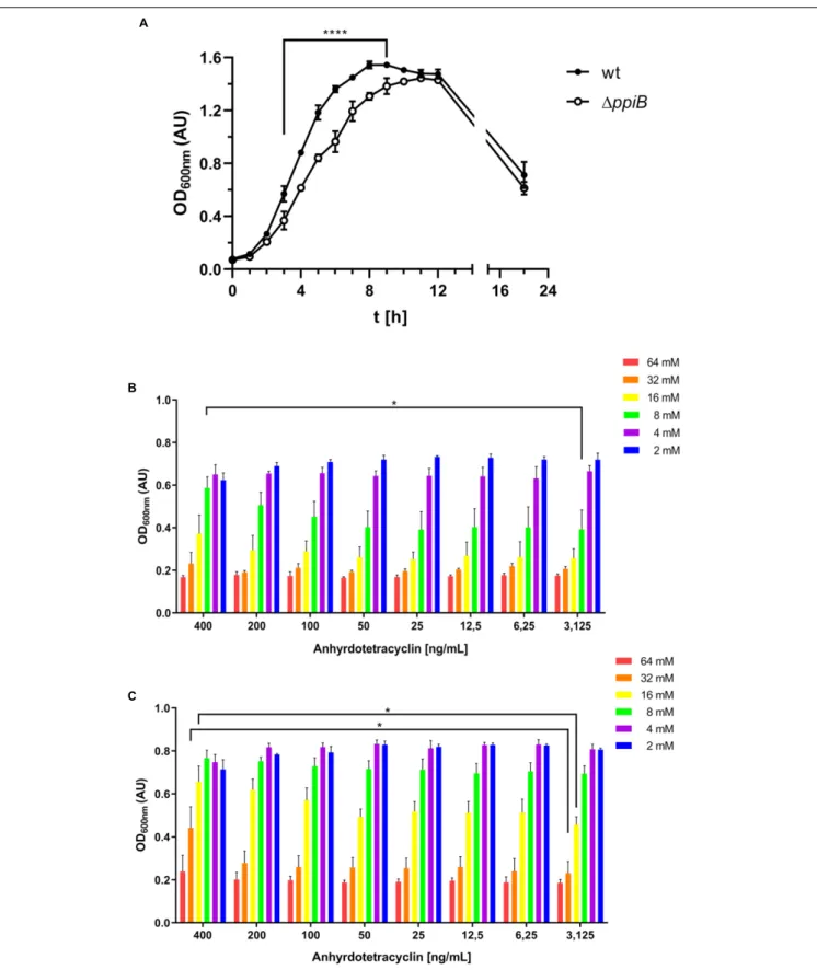
As our interactomic data revealed that CdPpiB interacts with several stress response proteins, we performed a literature survey of available transcriptomic data. By this, we found thatppiB was the only PPIase ofC. difficile that was significantly upregulated under cell wall stress caused by the beta-lactam amoxicillin (Emerson et al., 2008). Accordingly, we tested the susceptibility of the 1ppiB mutant in a serial dilution assay, and registered that the mutant was significantly more susceptible to amoxicillin concentrations ≥ 32µM supporting our findings regarding the involvement of CdPpiB in stress tolerance (Figure 5).
We further went on characterizing the generated 1ppiB
mutant by performing growth assays using the standard rich medium BHIS, which is supplemented with 0.5% (wt/vol) yeast extract and 0.1% (wt/vol) L-cysteine. Here, the 1ppiB
mutant displayed a significantly delayed growth in BHIS medium containing L-cysteine. Slower growth in the exponential phase resulting in a later entry into stationary phase (9 h in wt vs. 12 h in 1ppiB) was observed. The final OD after 22 h, were comparable for both strains (Figure 6A). When the assays were repeated with plain BHI or with BHIS lackingL-cysteine, no difference between
wild type and the 1ppiB mutant were observed. These tests
identified L-cysteine as the cause for the observed phenotype.
In order to verify the ppiB mutation as sole cause for the
observed differences, we complemented the mutant with an intactppiB gene in trans and brought ppiB under the control of the tetracycline promoter (Ptet) allowing fine-tuned expression
under the control of anhydrotetracycline (aTc). The wild type
FIGURE 5 | CdPpiB-deficiency increases susceptibility toward amoxicillin. Wild type and its isogenic1ppiB-mutant were grown in the presence of decreasing concentrations of amoxicillin (Amx) and bacterial growth was measured at 600 nm. The graph depicts mean ± SEM of three separate experiments performed in duplicates. Statistical significance was calculated by unpaired t-Test (∗
p ≤ 0.05,∗ ∗
FIGURE 6 | CdPpiB confers cysteine tolerance to C. difficile. (A) Destruction of ppiB causes growth defect in BHIS as assessed by the change in the OD600 nmover
time. (B) Complementation of the1ppiB mutant through induction with 400 ng/mL anhydrotetracycline allows the bacteria to cope with 8 mML-cysteine when grown in BHI supplemented with different concentrations of cysteine. (C) Stepwise over-expression of ppiB with increasing amounts of anhydrotetracycline raises the tolerance of the wild type toward 16 and 32 mML-cysteine. Shown are means ± SEM of three independent experiments performed in duplicate. Statistical significance was calculated by unpaired t-Test (∗p ≤ 0.05,∗ ∗ ∗ ∗p ≤ 0.0001).
strain and its isogenic 1ppiB mutant were transformed, and grown in the presence of different concentrations of L-cysteine
(2–62 mM) in combination of increasing concentrations of aTc (3,625-400 ng/mL) (Figures 6B,C). When fully induced
with 400 ng/mL, the 1ppiB mutant was able to cope with
8 mML-cysteine, which equals to the concentration in BHIS. Its growth under full induction was significantly better compared to reduced induction with 200 ng/mL or less aTc (Figure 6B). OverexpressingppiB in the wild type resulted in an even higher tolerance of up to 32 mM L-cysteine and a significantly better growth compared to lower induction levels. In case of 16 mM
L-cysteine the wild type overexpressing ppiB reached optical densities comparable to the wild type gown at 8 mML-cysteine
without ppiB overexpression (Figure 6C). These observations
corroborated the interactomic data where CysK was the major metabolic protein interacting with CdPpiB.
CdPpiB Restores CysK Activity
After Heat Inactivation
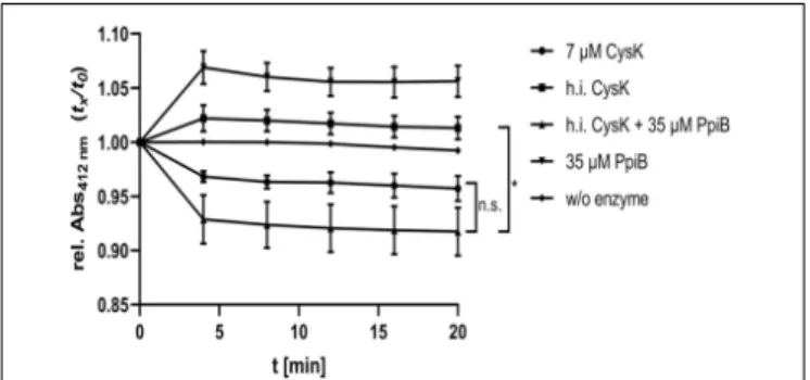
One important function of PPIases is the restoration of enzyme activity after protein inactivation by unfavorable chemical or physical conditions. In our interactomic study, we identified and verified CysK, the terminal enzyme in L-cysteine biosynthesis, as an interaction partner of CdPpiB. Accordingly, we tested the influence CdPpiB on heat inactivated CysK, the last enzyme of cysteine biosynthesis. Activity of recombinantly produced CysK was measured in a photometric assay at 412 nm, and was
shown to be active at 7 µM concentration. Next, CysK was
mildly heat denatured at 56◦
C for 30 min and its enzymatic activity was measured with or without five-fold excess of CdPpiB. As expected, heat denatured CysK showed no activity. But,
the addition of 35 µM CdPpiB to the reaction efficiently
restored its enzymatic activity (Figure 7). This indicates that CdPpiB functionally interacts with CysK and promotes its structural stability.
FIGURE 7 | CdPpiB restores CysK-activity after heat denaturation. O-acetyl-sulfhydrylase activity of recombinant CysK was measured by the decrease of OD412at 30◦C. Activity of 7µM recombinant CysK diminished
after inactivation at 56◦C for 30 min, and could be restored by the addition of
recombinant PpiB in 5-times molar excess. Shown are mean and SEM of three independent measurements proteins of two different productions. Statistical significance was calculated by unpaired t-Test with Welch’s correction (∗
p ≤ 0.05, n.s., not significant).
Deletion of ppiB Increases Toxin
Production and Cytotoxic Activity
Considering that CysK is a central enzyme in L-cysteine
metabolism and that this amino acid is also a potent suppressor of toxin production, we tested the influence of CdPpiB on the
cellular amounts of TcdA and TcdB under in vitro conditions
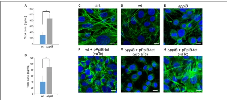
(Karlsson et al., 2000;Dubois et al., 2016). We quantified both toxins in stationary cultures of wild type and theppiB mutant using ELISA. Both toxins were found in significantly higher concentrations in the culture supernatants of theppiB mutant. While the wild type strain revealed in average 309 ng/mL TcdA in its culture supernatant, the ppiB mutant produced 2.8-fold more TcdA (868 ng/mL) (Figure 8A). Similarly, TcdB was 2.3-fold higher concentrated in the growth supernatant of the mutant (89 ng/mL) compared to the wild type (40 ng/mL) (Figure 8B). In accordance with the amounts of toxins found in the culture supernatant, the broth from mutant growth was considerably more active at 0.1% (vol/vol) dilution, causing stronger actin depolymerization and cell rounding of NIH-3T3 cells (Figures 8C–E). Similar results were obtained for the mutant complemented with theppiB gene in trans, however, without gene induction by aTc (Figure 8G). Wild type levels of toxins and corresponding cytotoxic effect similar to wild type were observed when the complemented mutant was induced with 400 ng/mL aTc (Figure 8H). Cells treated with supernatants from wild type bacteria overexpressingppiB exhibited no visual abnormalities of the cellular morphology (Figure 8F). Hence, CdPpiB not only
interacts with components of the L-cysteine metabolism and
supports the growth in the presence of L-cysteine, but it also affects toxin production inC. difficile.
DISCUSSION
The aim of this study was to gain insights into the physiological and virulence related functions of the cyclophilin CdPpiB of the nosocomial pathogenC. difficile. Our in vivo interactomics approach in combination with bacterial two hybrid testing allowed us the identification and verification of a specific set of interaction partners that are major control proteins of cell physiology and virulence. The majority of the interaction partners belonged to the category of energy and central metabolism (Table 3). Here, several functionally related proteins were identified, including LdhA, EtfA1, AcdB, EtfA3, HadA, and EtfB1. These enzymes are part of the same Stickland reaction involving L-leucine catabolism. By this, C. difficile harnesses metabolic energy from the oxidation of one equivalent ofL-leucine to isovalerate, CO2 and NH4+. In return, two equivalents of L-leucine are reduced to isocapronate and NH4+(Stickland, 1934; Britz and Wilkinson, 1982; Kim et al., 2006). This pathway is coupled via NAD+
and ferredoxin to the RNF complex responsi-ble for proton/sodium gradient formation required for ATP generation inC. difficile (Kim et al., 2004;Aboulnaga et al., 2013). Another group of proteins of putative interaction partners belonged to the primary carbon metabolism. These included pyruvate carboxylase Pyc, pyruvate-ferredoxin oxidoreductase Pfo, aldehyde-alcohol dehydrogenase AdhE2 and pyruvate
FIGURE 8 | Deletion of ppiB leads to higher toxin titres and cytotoxicity. (A) TcdA and (B) TcdB concentrations were measured by ELISA in 48 h old culture supernatants. Both toxins accumulated to significantly higher titres in culture supernatants in the isogenic1ppiB mutant. Shown are means ± SD of three independent experiments performed in duplicate. Statistical significance was calculated by unpaired t-Test (∗
p ≤ 0.05).(C–H) PpiB influences the cytotoxic activity in the culture supernatant. (C) In untreated control NIH-3T3 cells the actin cytoskeleton is organized in stressfibers as it is typical for epithelial cells. (D) Cell-free supernatants from 24 h old wild type cultures that were diluted to 0.1% (vol/vol) in cell culture medium have visually no detrimental effect on the actin cytoskeleton. (E) Supernatants of the1ppiB mutant cause disintegration and rounding up of the cells at the same dilution. (F) Overexpression of ppiB by the addition of 400 ng/mL anhydrotetracycline (aTc) in a tetracycline-inducible system has no detrimental effect on the cells in the wild type background. (G) The complemented strain exerts comparable cytotoxic activity as the1ppiB mutant in the absence of aTc. (H) Overexpression of ppiB by the addition of aTc reduces the cytotoxic effect in the1ppiB background to wild type levels. Shown are representative views of two separate experiments performed in triplicates. Actin cytoskeleton was stained green with Alexa488-coupled phalloidin, while nuclear DNA was stained blue with DAPI. Scale bars correspond to 10µm.
kinase, which control the flux around the pyruvate knot of the central metabolism. On the other hand, the glyceraldehyde-3-phosphate GapA and fructose-1,6-bisphosphate aldolase represent key control points of glycolysis (Dannheim et al., 2017b). Interestingly, gapA and adhE2 were also up-regulated under cysteine rich conditions (Dubois et al., 2016; Gu et al., 2018). Thus, CdPpiB potentially controls the major fluxes of the central metabolism ofC. difficile.
The second largest group of interaction partners belonged to the group of stress response, including proteins protecting against temperature, oxidative and alkaline shock. In line with
this, the 1ppiB mutant was more susceptible to amoxicillin,
which causes cell wall stress. This finding was also in accordance with a previous microarray study, whereppiB was significantly upregulated in amoxicillin treated C. difficile (Emerson et al.,
2008). Currently, we do not know how CdPpiB may influence
this phenotype. However, the well-known chaperones DnaK and GroS as well as YloU and YqhY were also upregulated in case of amoxicillin stress (Emerson et al., 2008). InS. aureus DnaK is part of the cell wall stress stimulon, anddnaK mutants have a higher susceptibility toward the beta-lactam oxacillin (Pechous et al., 2004;Singh et al., 2007). Recently, DnaK was identified as a binding partner of EcPpiB in functional complementation and co-precipitation studies inE. coli, where it positively influenced the enzymatic activity and correct localization of DnaK (Skagia et al., 2016). Hence, it is conceivable that, also in C. difficile, CdPpiB is part of the DnaK stress response network. This
feature might further be supported by the interactions with YloU and YqhY, which are close homologs of the alkaline shock protein 23 (Asp23) of S. aureus that is also associated with cell envelope stress (Müller et al., 2014;Tödter et al., 2017). EcPpiB is connected to another cell envelope related phenomenon, namely, cell division by directly interacting with FtsZ and probably further client proteins (Skagia et al., 2017b). Our interactomic study did not identify FtsZ as interaction partner in C. difficile. This might indicate that cyclophilins perform species-specific duties despite their high degree of conservation.
Several oxidative stress response proteins of the group of (Rbr1, Rbr2, and Rbr3) and a desulfoferrodoxin (Dsr) were found to bind to CdPpiB. Rubrerythrins are small proteins that have NADH-dependent peroxidase activity and act in concert with rubredoxins and desulfoferrodoxins (Mishra and Imlay, 2012). The homologs of Rbr1 and Dsr are important for conferring
O2-tolerance to C. acetbutylicum under the control of the
transcriptional regulator PerR, which located between these two genes inC. difficile (Hillmann et al., 2008;Kawasaki et al., 2009;
Dannheim et al., 2017a). As these proteins act in close vicinity for efficient electron transfer, it is likely that CdPpiB is involved in complex formation or stability supporting their physiological function. Moreover, oxidative stress genes are upregulated in the presence of L-cysteine in C. difficile (Dubois et al., 2016;
Gu et al., 2018).
Two well-established virulence partners, namely the flagellar subunit FliC and the surface layer protein SlpA, were among
the CdPpiB interaction partners. In case of FliC this interaction was confirmed by BACTH indicating a functional relationship between these proteins. Two oligomeric states of FliC seem to co-exist: the monomer which has been found in diverse cellular compartments and the polymeric organelle which is employed for motility, attachment and penetration (Campodónico et al., 2010). In fact, the implications of FliC monomers in invasiveness and virulence to the host are not restrained to C. difficile but
apply to many other bacteria. For instance, in Pseudomonas
aeruginosa or Salmonella typhimurium FliC appears to be the most prominent virulence factor in terms of inflammasome and
immune response activation (Duncan and Canna, 2018;Garcia
et al., 2018). Interestingly, FliC was shown to interact with DnaK in the periplasm of P. aeruginosa, and in C. difficile a1dnaK mutant had four-fold diminished fliC and four-fold increased groS expression (Borrero-de Acuña et al., 2016;Jain et al., 2017). This supports our idea of CdPpiB being a crucial part of the chaperone network ofC. difficile.
Next to some transport related proteins, very crucial factors of translational fidelity, IF-3 and RRF, were detected by interactomics. IF-3 is involved in the stabilization of the 30S ribosomal subunit, enabling mRNA binding to the 30S subunit and warranting the accuracy of the first aminoacyl-tRNA binding (Hua and Raleigh, 1998). The RRF, on the other hand, facilitates the dissociation of ribosomes from mRNA after termination of translation. RRF was together with elongation factor P one of the two highly upregulated translation factors inB. subtilis (Ohashi et al., 2003). As a foldase CdPpiB might, indeed, be in close vicinity of ribosomes and assist folding of newly synthesized proteins. However, there are currently no reports supporting this. Also, there is with trigger factor a known ribosome associated protein with a FKBP-domain that was shown to accompany proteins during their maturation following translation (Göthel et al., 1998;Kawagoe et al., 2018). This ubiquitous protein is also
present in C. difficile (CDIF630erm_03607/CD630_33060) and
is accordingly expected to be the major PPIase that facilitates protein folding. Nevertheless, it could be that CdPpiB does not associate with newly synthesized proteins but is involved in the recycling of translation factors for efficient ribosomal fidelity.
One of the most interesting binding partners was the transcriptional regulator Lrp. Hence, we analyzed the putative interaction between CdPpiB and Lrp in more detail and showed that this interaction depends on the PPIase active site residues. Mutating the highly conserved R50 and F109 weakened, while a F110A substitution significantly strengthened the association between both proteins. In return, out of the five proline residues of CdLrp only mutating P87 reduced the interaction of both proteins indicating a specific PPIase client protein relationship. Lrp-type proteins are global transcriptional regulators that consist of a classical N-terminal helix-turn-helix DNA-binding domain and a C-terminal substrate binding/ activation domain and typically act as specific regulators of amino acid metabolism-related genes (Brinkman et al., 2003).
However, in B. subtilis LrpC, a close homolog of CdLrp, was
also shown to be important for DNA-repair (López-Torrejón
et al., 2006). More interestingly, deleting decR (STM0459), a very close homolog of CdLrp in Salmonella typhimurium,
rendered the bacteria more susceptible to L-cysteine (Oguri et al., 2012). Hence, the cysteine-dependent phenotypes of the 1ppiB mutant might be due to the participation of CdPpiB on multiple levels ranging from gene regulation to enzymatic activity. The proline residue at position 87 is in the vicinity of the substrate binding/activation domain. Considering that peptidyl-prolyl-cis/trans-isomerization results in conformational changes in proteins, it is possible that CdPpiB assists CdLrp in changing its conformation upon binding its substrate or that it regulates the conformational equilibrium of CdLrp for a fine-tuned gene expression. Currently, only for eukaryotic PPIases examples of gene regulatory actions in the form of transcription
complex stabilization or mRNA maturation are known (Hanes,
2015;Thapar, 2015). To the best of our knowledge, this is the first time a direct interaction between a PPIase and a transcriptional regulator with direct implications on gene regulation has been shown in bacteria.
Interestingly and also in accordance with the interactomic
findings, the 1ppiB mutant showed two L-cysteine dependent
phenotypes: a higher susceptibility toward L-cysteine and
accumulation of more toxin in the supernatant. Typically added
to the medium to promote growth, L-cysteine has also some
detrimental effects because of the generated H2S during its
degradation. As a result, deleting cysteine catabolizing genes
results in decreased cysteine tolerance in bacteria (Oguri
et al., 2012; Gu et al., 2017). Cysteine is also among several metabolic stimuli that inhibit toxin production in C. difficile on transcriptional level, and it does so via its degradation byproducts pyruvate and H2S (Karlsson et al., 2000;Dubois et al., 2016). That cysteine tolerance and regulation of toxin expression converges via the cysteine metabolism was recently shown in a mutant lacking the cysteine desulfhydrase CdsB. This mutant was impaired in its growth in the presence of 5 mML-cysteine and its toxin production was unaffected byL-cysteine (Gu et al.,
2017). These observations overlap with the 1ppiB mutant in
our study, and are further supported by the identification of CysK as an interaction partner of CdPpiB. As aO-acetylserine sulfhydrylase CysK catalyzes the synthesis of L-cysteine from O-acetyl-L-serine and sulfide (Kredich et al., 1979). But it was also speculated that at higher L-cysteine concentrations CysK
catalyzes the opposite reaction and contributes to L-cysteine degradation by desulfhydration (Auger et al., 2005;Awano et al., 2005; Dubois et al., 2016). This was further supported by the observation that in C. difficile CysK was together with CysE one of the two transcriptionally most up-regulated genes of the cysteine metabolism in L-cysteine treated C. difficile (Dubois et al., 2016; Gu et al., 2018). In our study, we showed that CdPpiB interacts with CysK and it restores the enzymatic activity of denatured CysK. Considering this and the similarities between the phenotypes, we propose that CdPpiB contributes toL-cysteine tolerance ofC. difficile by stabilizing the metabolic network on the enzymatic level. Here again, a cooperative action
with other chaperones cannot be excluded as dnaK and groS
were also among the highly up-regulated genes in the presence of
L-cysteine (Dubois et al., 2016). Furthermore, a direct or indirect influence on the transcription of toxin genes by CdPpiB cannot be excluded and would need further analysis.
Taken together, CdPpiB is a key regulator that acts at the protein folding and modification level and controls central cellular processes. We also suggest that targeting CdPpiB as a therapeutic strategy aiming at virulence reduction may prove intricate, since in the absence of PpiB the bacteria become more toxigenic. Our findings confirm the contribution of the PPIase domain to its regulatory activity. But recent findings also suggest that cyclophilin function in bacteria does not solely rely on PPIase activity (Skagia et al., 2017b; Keogh et al., 2018). Accordingly, whether the PPIase-activity itself or some domains other than the PPIase domain are crucial for a certain interaction or even biological function still need to be evaluated in the future.
AUTHOR CONTRIBUTIONS
CÜ, JBdA, LJ, DJ, and MS conceived the experiments. CÜ, MK, MB, and CP conducted the experiments. JW performed the MS/LC analysis. CÜ, MK, MB, LJ, DJ, and MS analyzed the data. CÜ, DJ, and MS drafted and finalized
the manuscript. All authors reviewed and approved the final manuscript.
FUNDING
This work was funded by the Federal State of Lower Saxony, Niedersächsisches Vorab CDiff and CDInfect projects (VWZN2889/3215/3266) as well as by the German Research Foundation and the Open Access Publication Funds of the Technische Universität Braunschweig.
ACKNOWLEDGMENTS
We would like to thank Drs. Rebekka Biedendieck, Sarah Wienecke, and Tobias Knuuti for pN-STREPXa1622 and their technical support regarding recombinant production
in B. megaterium, and B.Sc. Lisa Maria Held for her
technical support.
REFERENCES
Aboulnaga, E.-H., Pinkenburg, O., Schiffels, J., El-Refai, A., Buckel, W., and Selmer, T. (2013). Effect of an oxygen-tolerant bifurcating butyryl coenzyme A dehydrogenase/electron-transferring flavoprotein complex fromClostridium difficile on butyrate production in Escherichia coli. J. Bacteriol. 195, 3704–3713. doi: 10.1128/JB.00321-13
Alonzo, F., Xayarath, B., Whisstock, J. C., and Freitag, N. E. (2011). Functional analysis of the Listeria monocytogenes secretion chaperone PrsA2 and its multiple contributions to bacterial virulence.Mol. Microbiol. 80, 1530–1548. doi: 10.1111/j.1365-2958.2011.07665.x
Anjuwon-Foster, B. R., and Tamayo, R. (2017). A genetic switch controls the production of flagella and toxins in Clostridium difficile. PLoS Genet. 13:e1006701. doi: 10.1371/journal.pgen.1006701
Aubry, A., Hussack, G., Chen, W., KuoLee, R., Twine, S. M., Fulton, K. M., et al. (2012). Modulation of toxin production by the flagellar regulon inClostridium difficile. Infect. Immun. 80, 3521–3532. doi: 10.1128/IAI.00224-12
Auger, S., Gomez, M. P., Danchin, A., and Martin-Verstraete, I. (2005). The PatB protein ofBacillus subtilis is a C-S-lyase. Biochimie 87, 231–238. doi: 10.1016/j. biochi.2004.09.007
Awano, N., Wada, M., Mori, H., Nakamori, S., and Takagi, H. (2005). Identi-fication and functional analysis of Escherichia coli cysteine desulfhydrases. Appl. Environ. Microbiol. 71, 4149–4152. doi: 10.1128/AEM.71.7.4149-4152. 2005
Barbut, F., Gariazzo, B., Bonné, L., Lalande, V., Burghoffer, B., Luiuz, R., et al. (2007). Clinical features ofClostridium difficile-associated infections and molecular characterization of strains: results of a retrospective study, 2000-2004.Infect. Control Hosp. Epidemiol. 28, 131–139. doi: 10.1086/511794 Bartlett, J. G., Onderdonk, A. B., Cisneros, R. L., and Kasper, D. L. (1977).
Clindamycin-associated colitis due to a toxin-producing species ofClostridium in hamsters.J. Infect. Dis. 136, 701–705. doi: 10.1093/infdis/136.5.701 Batah, J., Denève-Larrazet, C., Jolivot, P.-A., Kuehne, S., Collignon, A., Marvaud,
J.-C., et al. (2016).Clostridium difficile flagella predominantly activate TLR5-linked NF-κB pathway in epithelial cells. Anaerobe 38, 116–124. doi: 10.1016/j. anaerobe.2016.01.002
Behrens-Kneip, S. (2010). The role of SurA factor in outer membrane protein transport and virulence.Int. J. Med. Microbiol. IJMM 300, 421–428. doi: 10. 1016/j.ijmm.2010.04.012
Biedendieck, R., Borgmeier, C., Bunk, B., Stammen, S., Scherling, C., Meinhardt, F., et al. (2011). Systems biology of recombinant protein production usingBacillus megaterium. Methods Enzymol. 500, 165–195. doi: 10.1016/B978-0-12-385118-5.00010-4
Biedendieck, R., Yang, Y., Deckwer, W.-D., Malten, M., and Jahn, D. (2007). Plasmid system for the intracellular production and purification of affinity-tagged proteins inBacillus megaterium. Biotechnol. Bioeng. 96, 525–537. doi: 10.1002/bit.21145
Borrero-de Acuña, J. M., Jänsch, L., Rohde, M., Timmis, K. N., Jahn, D., and Jahn, M. (2015). “Interatomic characterization of protein–protein interactions in membrane-associated mega-complexes,” inHydrocarbon and Lipid Microbiology Protocols Springer Protocols Handbooks, eds T. McGenity, K. Timmis, and B. Nogales (Berlin: Springer).
Borrero-de Acuña, J. M., Rohde, M., Wissing, J., Jänsch, L., Schobert, M., Molinari, G., et al. (2016). Protein network of thePseudomonas aeruginosa denitrification apparatus.J. Bacteriol. 198, 1401–1413. doi: 10.1128/JB.00055-16 Bouillaut, L., Dubois, T., Sonenshein, A. L., and Dupuy, B. (2015). Integration of metabolism and virulence inClostridium difficile. Res. Microbiol. 166, 375–383. doi: 10.1016/j.resmic.2014.10.002
Brinkman, A. B., Ettema, T. J. G., de Vos, W. M., and van der Oost, J. (2003). The Lrp family of transcriptional regulators.Mol. Microbiol. 48, 287–294. doi: 10.1046/j.1365-2958.2003.03442.x
Britz, M. L., and Wilkinson, R. G. (1982). Leucine dissimilation to isovaleric and isocaproic acids by cell suspensions of amino acid fermenting anaerobes: the Stickland reaction revisited.Can. J. Microbiol. 28, 291–300. doi: 10.1139/m82-043
Campodónico, V. L., Llosa, N. J., Grout, M., Döring, G., Maira-Litrán, T., and Pier, G. B. (2010). Evaluation of flagella and flagellin ofPseudomonas aeruginosa as vaccines.Infect. Immun. 78, 746–755. doi: 10.1128/IAI.00806-09
Chandrasekaran, R., and Lacy, D. B. (2017). The role of toxins inClostridium difficile infection. FEMS Microbiol. Rev. 41, 723–750. doi: 10.1093/femsre/ fux048
Dannheim, H., Riedel, T., Neumann-Schaal, M., Bunk, B., Schober, I., Spröer, C., et al. (2017a). Manual curation and reannotation of the genomes ofClostridium difficile 6301erm and C. difficile 630. J. Med. Microbiol. 66, 286–293. doi: 10. 1099/jmm.0.000427
Dannheim, H., Will, S. E., Schomburg, D., and Neumann-Schaal, M. (2017b). Clostridioides difficile 6301erm in silico and in vivo - quantitative growth and extensive polysaccharide secretion.FEBS Open Bio 7, 602–615. doi: 10.1002/ 2211-5463.12208
Derman, Y., Söderholm, H., Lindström, M., and Korkeala, H. (2015). Role of csp genes in NaCl, pH, and ethanol stress response and motility in Clostridium botulinum ATCC 3502. Food Microbiol. 46, 463–470. doi: 10.1016/j.fm.2014.09.004
Dingle, T. C., Mulvey, G. L., and Armstrong, G. D. (2011). Mutagenic analysis of theClostridium difficile flagellar proteins, FliC and FliD, and their contribution