Published online 2019 July 31. Research Article
Neutrophil-Lymphocyte Ratio and the Platelet Parameters as
Biomarkers of Atopic Dermatitis Severity in Children
Ozlem Bostan Gayret
1, *, Hikmet Tekin Nacaro ˘glu
2, Meltem Erol
1and Ahmet ¸
Sener
11Department of Pediatrics, Bagcilar Training and Research Hospital, Health Sciences University, Istanbul, Turkey 2Department of Pediatric Allergy, Istanbul Medipol University, Istanbul, Turkey
*Corresponding author: Health Sciences University, Bagcilar Training and Research Hospital, Department of Pediatrics, Istanbul, Turkey. Tel:+90-5327633326,
+90-2124404000-1344, Fax: +90-2124404242, Email: [email protected]
Received2019 March 14; Revised 2019 June 18; Accepted 2019 June 27. Abstract
Background:Atopic dermatitis (AD) is a chronic inflammatory skin disease with a specific immune and inflammatory mechanism. This study investigates inflammatory biomarkers and their correlation with disease severity.
Objectives: The aim of this study is to determine the relationship between platelet parameters [(mean platelet volume (MPV), platelet distribution width (PDW)], the neutrophil-lymphocyte ratio (NLR), the platelet-lymphocyte ratio (PLR), and AD severity in children.
Methods:In this retrospective study, we reviewed patients diagnosed with AD and a healthy control group at the department of pediatrics at Health Sciences in a university-affiliated hospital in Istanbul, Turkey, between January 2015 and December 2016. The study included 79 children with AD and 75 healthy controls. AD severity was graded using the Scoring Atopic Dermatitis (SCORAD) index. Complete blood count was measured, and NLR and PLR were calculated. NLR, PLR, MPV, PDW were compared between AD patients and healthy controls, and the correlations between these indexes and clinical characteristics were analyzed.
Results:No significant difference was observed between patients and controls for MPV, NLR, and PLR (P = 0.708, P = 0.340, P = 0.179, respectively). In the AD group, PDW was lower than controls (17.39±1.45, 18.04±1.65, P = 0.006). In the severe AD group, MPV was higher (7.62±1.81, 6.64±1.16, P = 0.035) and PDW was lower than in the mild AD group (16.52±1.49, 17.93±1.44, P = 0.0001). Conclusions:Mean MPV and PDW levels are correlated with atopic dermatitis severity in children.
Keywords:Atopic, Biomarkers, Child, Dermatitis, Eczema, Lymphocytes, Neutrophils, Platelet, Turkey
1. Background
Atopic dermatitis (AD) is a skin disease with eczema-tous and pruritic lesions (1). AD affects 10% - 20% of children in developed countries (2).
The pathophysiology of AD is not well understood. Like other allergic diseases, AD is caused by interactions be-tween genetic and environmental factors (3). Correlations have been reported between AD severity in children and biomarkers such as serum thymus, activation-regulated chemokine, and serum interleukin 10, 17, and 23 (4). How-ever, these markers cannot be routinely examined.
When a platelet is activated, its volume increases. Platelets with increased volume contain denser granules and have a greater capacity to induce inflammation. Mean platelet volume (MPV) is a measure of thrombocyte vol-ume, and increases in MPV are correlated with increases in platelet function and activity. Hence it can be used as an inflammatory indicator (5-7). Platelet distribution width
(PDW) reflects an alteration in platelet size, similar to other platelet indexes (8). Recently, MPV and PDW have been highlighted as useful markers of several inflammatory dis-eases (9).
Neutrophil-lymphocyte ratio (NLR) and platelet-lymphocyte ratio (PLR) are inexpensive indicators to evaluate, which can quickly and easily detect inflamma-tory reactions (10). It has been reported that there is a correlation between PLR and the severity of rheumatoid arthritis and systemic lupus erythematosus (11). NLR has been associated with chronic inflammation in such conditions as psoriasis and rheumatoid arthritis (12,13). In children, some studies have reported that NLR, PLR, and MPV were higher in patients with atopic dermatitis, allergic rhinitis, and asthma. These studies emphasized that this might indicate the presence of inflammation in allergic diseases and be associated with disease severity (14-16).
A few studies conducted on children have shown a
relation between AD and platelet parameters, NLR, and PLR (16-18). In our study, we investigated the correlation be-tween platelet parameters, NLR, PLR, and AD severity in children, as assessed with the Scoring Atopic Dermatitis (SCORAD) index.
2. Objectives
This study aimed to identify useful and easily calcu-lated indicators of systemic inflammation in AD; evaluated potential correlations between inflammatory markers and disease severity.
3. Methods 3.1. Patients
We retrospectively reviewed all patients diagnosed with AD and a healthy control group at the department of pediatrics at Health Sciences University, Bagcilar Training and Research Hospital, which is a general tertiary referral governmental hospital, in Istanbul, Turkey, between Jan-uary 2015 and December 2016. We collected demographic data, AD signs and symptoms, and initial laboratory data from patient medical records. The inclusion criteria were: an AD diagnosis and an age in the range of 1 - 60 months. The study excluded children who were diagnosed with sep-sis, obesity, hyperlipidemia, diabetes mellitus, hyperten-sion, chronic renal disease, nephrotic syndrome, inflam-matory bowel disease, or chronic inflaminflam-matory disease as well as those who had received systemic steroid treat-ment before blood count analysis. Patients with immuno-logic disorders were also excluded. The control group was made up of healthy volunteers from the same age group. Healthy subjects were children who went to the hospital for routine check-ups. Children with any sign of infection, systemic illness, or inflammation were excluded from the control group. After applying these criteria to 250 poten-tial participants, the study included 154 children: 79 chil-dren with AD and 75 healthy chilchil-dren. AD was diagnosed following Hanifin and Rajka (19) and severity was graded using SCORAD: < 25 was classified as mild, 25 - 50 was classi-fied as moderate, and > 50 was classiclassi-fied as severe (20). The local ethics committee of the same institute approved our study (2016/494). The study was conducted according to the Declaration of Helsinki. We obtained written informed consent from the parents.
3.2. Laboratory Analysis
Complete blood count was measured within approx-imately 60 minutes after blood sampling with a Coulter LH 780 Analyzer and a Coulter Hmx Hematology Analyzer
(Beckman Coulter, Inc., CA, USA) using the original method and reagents. Based on data from the complete count anal-ysis, NLR was calculated by dividing the percentage of neu-trophils by the percentage of lymphocytes. PLR was cal-culated by dividing the percentage of platelets by the per-centage of lymphocytes.
3.3. Statistical Analyses
Data were analyzed with IBM SPSS Statistics for Win-dows, version 24.0 (IBM, Armonk, NY, USA). Descriptive statistics are presented as means, standard deviations, median, and IQR values for continues data and frequen-cies and percentages for categorical data. Kolmogorov-Smirnov tests showed the data were not normally dis-tributed, so Mann-Whitney U tests were used to compare the variables of two independent groups. The chi-square tests and, where applicable, Fisher’s exact test were con-ducted to assess differences between categorical variables. Since the variables were not normally distributed, Kruskal-Wallis tests were used to compare more than two groups. Mann-Whitney U tests were used to test the significance of pairwise differences with the Bonferroni correction, to adjust for multiple comparisons. For multivariate anal-ysis, possible factors identified with univariate analysis were further used in logistic regressions to determine in-dependent predictors of AD outcome. Hosmer-Lemeshow goodness-of-fit statistics were used to assess model fit. The difference was regarded as significant if P < 0.05.
4. Results
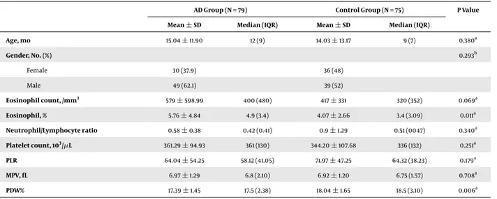
The patient group comprised 49 (62.1%) male and 30 (37.9%) female children with a mean age of 15.04±11.90 months. The control group comprised 39 (52%) male and 36 (48%) female children with a mean age of 14.03±13.17 months. There was no difference between the age or gen-der groups (P > 0.05). The patient group had significantly higher eosinophil counts, percentages of eosinophils, and lower PDW counts than the control group. There were no significant differences between the MPV, NLR, or PLR of the patients and the controls (Table 1). To determine the factors most affected by AD, logistic regression analysis was per-formed using percentages of eosinophil and PDW levels as variables. This showed that percentages of eosinophil (P = 0.069) and PDW (P = 0.006) were the factors most affected by AD (Table 2).
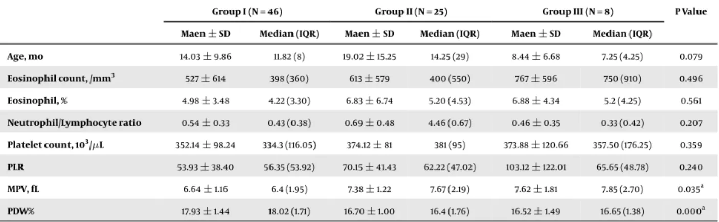
Based on the SCORAD index, AD patients were divided as mild (Group I: SCORAD < 25), moderate (Group II: SCO-RAD 25 - 50), and severe (Group III: SCOSCO-RAD > 50). No sig-nificant differences were found between groups in terms of age, eosinophil counts, or eosinophils percentage. How-ever, the mean MPV and PDW differed significantly among
Table 1.Comparison of Socio-Demographic Data and Laboratory Results for Patients and Control Groupsa , b
AD Group (N = 79) Control Group (N = 75) P Value Mean±SD Median (IQR) Mean±SD Median (IQR)
Age, mo 15.04±11.90 12 (9) 14.03±13.17 9 (7) 0.380a Gender, No. (%) 0.293b Female 30 (37.9) 36 (48) Male 49 (62.1) 39 (52) Eosinophil count, /mm3 579±598.99 400 (480) 417±331 320 (352) 0.069a Eosinophil, % 5.76±4.84 4.9 (3.4) 4.07±2.66 3.4 (3.09) 0.011a Neutrophil/Lymphocyte ratio 0.58±0.38 0.42 (0.41) 0.9±1.29 0.51 (0047) 0.340a Platelet count, 103/µL 361.29±94.93 361 (130) 344.20±107.68 336 (132) 0.251a PLR 64.04±54.25 58.12 (41.05) 71.97±47.25 64.32 (38.23) 0.179a MPV, fL 6.97±1.29 6.8 (2.10) 6.92±1.20 6.75 (1.57) 0.708a PDW% 17.39±1.45 17.5 (2.38) 18.04±1.65 18.5 (3.10) 0.006a
Abbreviations: AD, atopic dermatitis, PLR, platelet-lymphocyte ratio, MPV, mean platelet volume, PDW, platelet distribution width.
aResult from the Mann-Whitney U test. bResult from the chi-square test.
Table 2.Logistic Regression Analysis of Factors Associated with ADa
Parameters B Standard Error Wald P Value Odds Ratio 95% CI for exp(B)
Lower Upper
Percentages of eosinophils 0.137 0.056 5.909 0.015 1.147 1.027 1.281
PDW -0.283 0.110 6.571 0.010 0.754 0.607 0.936
Constant 4.407 1.955 5.082 0.024 82.061
Abbreviations: AD, atopic dermatitis, platelet distribution width.
aUsed together, the constant, percentages of eosinophil, and PDW can predict AD with statistically significance (P < 0.05). Lower PDW reduces the risk of AD with a ratio
of 0.755 at a 95% CI (0.607 - 0.936; P = 0.010). A higher percentage of eosinophil increases the risk of AD with a ratio of 1.147 at a 95% CI (1.027 - 1.281; P = 0.015).
groups. Mean MPV was higher (P = 0.035), and PDW was lower (P = 0.0001) in the severe AD group (Table 3). To de-termine the factors that most affected by SCORAD scores, logistic regression analysis was performed with MPV and PDW. This showed that the PDW level was the factor that most affected SCORAD in AD patients (Table 4).
5. Discussion
In our study, MPV levels were significantly higher, and PDW levels were significantly lower in children with AD and tracked AD severity. Although AD is an inflamma-tory disease and NLR and PLR reflect inflammation values, these variables showed no significant differences between groups.
MPV and PDW are frequently used as measures of platelet sizes and as inflammatory markers in various dis-eases (21). Cytokines released during inflammation cause increases in megakaryocyte ploidy and volume as well as the production of large quantities of platelets (22). These
platelets are large with variable sizes (23, 24). MPV is an easy test that reflects platelet function or activity (25). The correlation between the MPV and disease activity has been reported in chronic spontaneous urticaria, juvenile idiopathic arthritis, systemic lupus erythematosus, and rheumatoid arthritis (26-29). The relationship between AD and MPV has been investigated in a few recent studies (16,
17). While MPV was found to be higher among AD chil-dren (17) in one study, no relationship was found between AD severity and MPV. The same conclusion was reached by Jiang and Ma (16). However, our data do show a relation be-tween atopic dermatitis severity and MPV.
PDW, an index of platelet size heterogeneity, increases during platelet activation (30). Isik et al. (31) found that PDW was an adverse acute-phase reaction in patients with active rheumatoid arthritis. An inverse relationship be-tween ulcerative colitis activity and PDW was identified by Ozturk et.al (32). Topal et al. (17) reported that PDW was lower in AD, but it was not associated with disease sever-ity. Our study also found that PDW was low in AD children,
Table 3.Comparison of Socio-Demographic Data and Laboratory Findings of Patients with Mild (Group I), Moderate (Group II), and Severe Atopic Dermatitis (Group III) Group I (N = 46) Group II (N = 25) Group III (N = 8) P Value Maen±SD Median (IQR) Maen±SD Median (IQR) Maen±SD Median (IQR)
Age, mo 14.03±9.86 11.82 (8) 19.02±15.25 14.25 (29) 8.44±6.68 7.25 (4.25) 0.079 Eosinophil count, /mm3 527±614 398 (360) 613±579 400 (550) 767±596 750 (910) 0.496 Eosinophil, % 4.98±3.48 4.22 (3.30) 6.83±6.74 5.20 (4.53) 6.88±4.34 5.2 (4.25) 0.561 Neutrophil/Lymphocyte ratio 0.54±0.33 0.43 (0.38) 0.69±0.48 4.46 (0.67) 0.46±0.35 0.33 (0.42) 0.207 Platelet count, 103/µL 352.14±98.24 334.3 (116.05) 374.12±81 381 (95) 373.88±120.66 357.50 (176.25) 0.359 PLR 53.93±38.40 56.35 (53.92) 70.15±41.43 62.22 (47.02) 103.12±122.01 65.65 (48.78) 0.240 MPV, fL 6.64±1.16 6.4 (1.95) 7.38±1.22 7.67 (2.19) 7.62±1.81 7.85 (2.70) 0.035a PDW% 17.93±1.44 18.02 (1.71) 16.70±1.00 16.4 (1.76) 16.52±1.49 16.65 (1.38) 0.000a
Abbreviations: PLR, platelet-lymphocyte ratio, MPV, mean platelet volume, PDW, platelet distribution width.
aStatistical significance was defined at P < 0.05.
Table 4.Logistic Regression Analysis of Factors Associated with the SCORAD Index in AD Patientsa
SCORAD Score B Standard Error Wald P Value Odds ratio 95% Confidence Interval for exp(B)
Lower Upper Group II Intercept 10.274 5.701 3.247 0.072 MPV 0.118 0.244 0.233 0.629 1.125 0.697 1.816 PDW -0.677 0.268 6.396 0.011 0.508 0.301 0.859 Group III Intercept 9.065 8.214 1.218 0.270 MPV 0.248 0.371 0.449 0.503 1.282 0.620 2.651 PDW -0.732 0.388 3.555 0.059 0.481 0.225 1.029
Abbreviations: AD, atopic dermatitis, MPV, mean platelet volume, PDW, platelet distribution width; SCORAD, severity was graded using the scoring atopic dermatitis.
aWhen the reference category is mild, only PDW is a statistically significant factor for predicting SCORAD group (P < 0.05). Compared to the mild group, the moderate
group has lower PDW values, higher SCORAD indices, and an odds ratio of 0.755 with a CI of 95% (0.608 - 0.937; P = 0.011).
and additionally, it correlates with severity.
NLR has been used as an indicator in many inflamma-tory diseases (12,13). Neutrophilic inflammation has also been shown to occur with eosinophilic inflammation in AD patients. A positive correlation between potent neu-trophil chemoattractant gene set scores and neuneu-trophils of AD patients has been demonstrated (33). Neutrophilic inflammation was detected in lesioned areas but not in perilesional areas. Furthermore, a correlation was found between the number of neutrophils and crust presence. In addition to assessing the extent of lesions in AD, the SCORAD scale is used to assess lesion severity. The crust is also a parameter of density. These results show that trophilia is more common in severe AD. When high neu-trophil counts are present, NLR is higher. We found no re-lationship between AD severity and NLR. Comprehensive trails are required to determine their relationship clearly.
PLR has been associated with the severity of many
dis-eases (13,14,34-37). The few studies on PLR in AD patients have reported that PLR is correlated with disease severity (16,18), but our study did not identify this correlation.
One limitation of this study is the small number of par-ticipants. The low number of patients was due to the study being conducted at a single center and the study’s retro-spective design. We did not make groups based on intrin-sic and extrinintrin-sic atopic dermatitis. Another limiting factor is that there was no threshold value for MPV and PDW, such as CRP. These are the weak points of this study. However, we believe that our study is important because few other studies have evaluated MPV, PDW, NLR, and PLR in AD chil-dren and compared them to disease severity. Therefore, our study is important for filling this gap in the literature. As mentioned above, we think that MPV and PDW will be useful for investigating AD severity, as they are for many other inflammatory diseases.
5.1. Conclusions
Our study found no differences in mean MPV, NLR, and PLR values between AD patients and the control group. However, MPV and PDW differed significantly between se-vere and mild AD patients. MPV and PDW were correlated with AD severity and can be used to track severity. However, more studies are required to examine the relationship be-tween NLR, PLR, and disease severity.
Footnotes
Authors’ Contribution: Study concept and design:
Ozlem Bostan Gayret and Hikmet Tekin Nacaro ˘glu; acquisition of data: Hikmet Tekin Nacaro ˘glu and Ah-met Sener; analysis and interpretation of data: Meltem Erol, Ahmet Sener and Ozlem Bostan Gayret; drafting of the manuscript: Ozlem Bostan Gayret; critical revision: Meltem Erol; technical and material support: Ahmet Sener, Meltem Erol, and Hikmet Tekin Nacaro ˘glu; supervision: Hikmet Tekin Nacaro ˘glu.
Conflict of Interests: The authors declare no conflict of interest.
Ethical Considerations:2016/494.
Funding/Support: None.
References
1. Weidinger S, Novak N. Atopic dermatitis. Lancet. 2016;387(10023):1109– 22. doi:10.1016/S0140-6736(15)00149-X. [PubMed:26377142]. 2. Totri CR, Diaz L, Eichenfield LF. 2014 update on atopic
der-matitis in children. Curr Opin Pediatr. 2014;26(4):466–71. doi:
10.1097/MOP.0000000000000109. [PubMed:24926800].
3. Peng W, Novak N. Pathogenesis of atopic dermatitis. Clin Exp Allergy. 2015;45(3):566–74. doi:10.1111/cea.12495. [PubMed:25610977]. 4. Esaki H, Takeuchi S, Furusyo N, Yamamura K, Hayashida S, Tsuji
G, et al. Levels of immunoglobulin E specific to the major food allergen and chemokine (C-C motif) ligand (CCL)17/thymus and activation regulated chemokine and CCL22/macrophage-derived chemokine in infantile atopic dermatitis on Ishigaki Island. J
Dermatol. 2016;43(11):1278–82. doi: 10.1111/1346-8138.13360. [PubMed:
27028543].
5. Herter JM, Rossaint J, Zarbock A. Platelets in inflammation and im-munity. J Thromb Haemost. 2014;12(11):1764–75. doi: 10.1111/jth.12730. [PubMed:25224706].
6. Sert A, Aypar E, Odabas D. Mean platelet volume in acute rheumatic fever. Platelets. 2013;24(5):378–82. doi:10.3109/09537104.2012.701029. [PubMed:22757773].
7. Makay B, Turkyilmaz Z, Unsal E. Mean platelet volume in children with familial Mediterranean fever. Clin Rheumatol. 2009;28(8):975–8. doi:
10.1007/s10067-009-1148-5. [PubMed:19283330].
8. Kaito K, Otsubo H, Usui N, Yoshida M, Tanno J, Kurihara E, et al. Platelet size deviation width, platelet large cell ratio, and mean platelet vol-ume have sufficient sensitivity and specificity in the diagnosis of immune thrombocytopenia. Br J Haematol. 2005;128(5):698–702. doi:
10.1111/j.1365-2141.2004.05357.x. [PubMed:15725092].
9. Gasparyan AY, Ayvazyan L, Mikhailidis DP, Kitas GD. Mean platelet vol-ume: A link between thrombosis and inflammation? Curr Pharm Des. 2011;17(1):47–58. doi:10.2174/138161211795049804. [PubMed:21247392].
10. Wang D, Yang JX, Cao DY, Wan XR, Feng FZ, Huang HF, et al. Preop-erative neutrophil-lymphocyte and platelet-lymphocyte ratios as in-dependent predictors of cervical stromal involvement in surgically treated endometrioid adenocarcinoma. Onco Targets Ther. 2013;6:211– 6. doi: 10.2147/OTT.S41711. [PubMed: 23525143]. [PubMed Central:
PMC3604973].
11. Uslu AU, Kucuk A, Sahin A, Ugan Y, Yilmaz R, Gungor T, et al. Two new inflammatory markers associated with Disease Activity Score-28 in patients with rheumatoid arthritis: neutrophil-lymphocyte ratio and platelet-lymphocyte ratio. Int J Rheum Dis. 2015;18(7):731–5. doi:
10.1111/1756-185X.12582. [PubMed:25900081].
12. Wu Y, Chen Y, Yang X, Chen L, Yang Y. Neutrophil-to-lymphocyte ra-tio (NLR) and platelet-to-lymphocyte rara-tio (PLR) were associated with disease activity in patients with systemic lupus erythematosus. Int
Immunopharmacol. 2016;36:94–9. doi: 10.1016/j.intimp.2016.04.006.
[PubMed:27111516].
13. Fu H, Qin B, Hu Z, Ma N, Yang M, Wei T, et al. Neutrophil-and platelet-to-lymphocyte ratios are correlated with disease ac-tivity in rheumatoid arthritis. Clin Lab. 2015;61(3-4):269–73. doi:
10.7754/Clin.Lab.2014.140927. [PubMed:25974992].
14. Dogru M, Evcimik MF, Cirik AA. Is neutrophil-lymphocyte ratio as-sociated with the severity of allergic rhinitis in children? Eur Arch
Otorhinolaryngol. 2016;273(10):3175–8. doi:10.1007/s00405-015-3819-y.
[PubMed:26525883].
15. Dogru M, Yesiltepe Mutlu RG. The evaluation of neutrophil-lymphocyte ratio in children with asthma. Allergol Immunopathol
(Madr). 2016;44(4):292–6. doi: 10.1016/j.aller.2015.09.005. [PubMed:
26777420].
16. Jiang Y, Ma W. Assessment of neutrophil-to-lymphocyte ratio and platelet-to-lymphocyte ratio in atopic dermatitis patients. Med
Sci Monit. 2017;23:1340–6. doi: 10.12659/msm.900212. [PubMed:
28306706]. [PubMed Central:PMC5367851].
17. Topal E, Celiksoy MH, Catal F, Karakoc HT, Karadag A, Sancak R. The platelet parameters as inflammatory markers in preschool children with atopic eczema. Clin Lab. 2015;61(5-6):493–6. doi:
10.7754/Clin.Lab.2014.140930. [PubMed:26118181].
18. Batmaz SB. Simple markers for systemic inflammation in pedi-atric atopic dermatitis patients. Indian J Dermatol. 2018;63(4):305–10. doi: 10.4103/ijd.IJD_427_17. [PubMed: 30078874]. [PubMed Central:
PMC6052751].
19. Hanifin JM, Rajka G. Diagnostic features of atopic dermatitis. Acta
Derm Venereol (Stockh). 1980;92:44–7.
20. [No authors listed]. Severity scoring of atopic dermatitis: the SCO-RAD index. Consensus Report of the European Task Force on Atopic Dermatitis. Dermatology. 1993;186(1):23–31. doi: 10.1159/000247298. [PubMed:8435513].
21. Leader A, Pereg D, Lishner M. Are platelet volume indices of clini-cal use? A multidisciplinary review. Ann Med. 2012;44(8):805–16. doi:
10.3109/07853890.2011.653391. [PubMed:22413913].
22. Mattia G, Vulcano F, Milazzo L, Barca A, Macioce G, Giampaolo A, et al. Different ploidy levels of megakaryocytes generated from peripheral or cord blood CD34+ cells are correlated with dif-ferent levels of platelet release. Blood. 2002;99(3):888–97. doi:
10.1182/blood.v99.3.888. [PubMed:11806991].
23. Oncel MY, Ozdemir R, Yurttutan S, Canpolat FE, Erdeve O, Oguz SS, et al. Mean platelet volume in neonatal sepsis. J Clin Lab Anal. 2012;26(6):493–6. doi:10.1002/jcla.21552. [PubMed:23143634]. 24. Artunc Ulkumen B, Pala HG, Calik E, Oruc Koltan S. Platelet
distribu-tion width (PDW): A putative marker for threatened preterm labour.
Pak J Med Sci. 2014;30(4):745–8. doi:10.12669/pjms.304.4991. [PubMed:
25097509]. [PubMed Central:PMC4121690].
25. Park Y, Schoene N, Harris W. Mean platelet volume as an indicator of platelet activation: Methodological issues. Platelets. 2002;13(5-6):301– 6. doi:10.1080/095371002220148332. [PubMed:12189016].
26. Milovanovic M, Nilsson E, Jaremo P. Relationships between platelets and inflammatory markers in rheumatoid arthritis. Clin Chim
Acta. 2004;343(1-2):237–40. doi: 10.1016/j.cccn.2003.12.030. [PubMed:
15115702].
27. Yavuz S, Ece A. Mean platelet volume as an indicator of disease activity in juvenile SLE. Clin Rheumatol. 2014;33(5):637–41. doi: 10.1007/s10067-014-2540-3. [PubMed:24567240].
28. Gunes A, Ece A, Sen V, Uluca U, Aktar F, Tan I, et al. Correlation of mean platelet volume, neutrophil-to-lymphocyte ratio, and dis-ease activity in children with juvenile idiopathic arthritis. Int J Clin
Exp Med. 2015;8(7):11337–41. [PubMed: 26379946]. [PubMed Central:
PMC4565329].
29. Magen E, Mishal J, Zeldin Y, Feldman V, Kidon M, Schlesinger M, et al. Increased mean platelet volume and C-reactive pro-tein levels in patients with chronic urticaria with a positive autologous serum skin test. Am J Med Sci. 2010;339(6):504–8. doi:
10.1097/MAJ.0b013e3181db6ed5. [PubMed:20400886].
30. Vagdatli E, Gounari E, Lazaridou E, Katsibourlia E, Tsikopoulou F, Labrianou I. Platelet distribution width: A simple, practical and spe-cific marker of activation of coagulation. Hippokratia. 2010;14(1):28– 32. [PubMed:20411056]. [PubMed Central:PMC2843567].
31. Isik M, Sahin H, Huseyin E. New platelet indices as inflammatory parameters for patients with rheumatoid arthritis. Eur J
Rheuma-tol. 2014;1(4):144–6. doi:10.5152/eurjrheumatol.2014.140023. [PubMed:
27708900]. [PubMed Central:PMC5042243].
32. Ozturk ZA, Dag MS, Kuyumcu ME, Cam H, Yesil Y, Yilmaz N, et al. Could platelet indices be new biomarkers for inflammatory bowel diseases?
Eur Rev Med Pharmacol Sci. 2013;17(3):334–41. [PubMed:23426536].
33. Choy DF, Hsu DK, Seshasayee D, Fung MA, Modrusan Z, Martin F, et al. Comparative transcriptomic analyses of atopic dermati-tis and psoriasis reveal shared neutrophilic inflammation. J Allergy
Clin Immunol. 2012;130(6):1335–43 e5. doi: 10.1016/j.jaci.2012.06.044.
[PubMed:22920495]. [PubMed Central:PMC3511596].
34. Peng W, Li C, Zhu WJ, Wen TF, Yan LN, Li B, et al. Prognostic value of the platelet to lymphocyte ratio change in liver cancer. J Surg Res. 2015;194(2):464–70. doi:10.1016/j.jss.2014.12.021. [PubMed:25577142]. 35. Ayca B, Akin F, Okuyan E. Platelet to lymphocyte ratio as a
prog-nostic marker in primary percutaneous coronary intervention.
Platelets. 2015;26(8):816. doi:10.3109/09537104.2015.1015410. [PubMed:
25807323].
36. Kim DS, Shin D, Lee MS, Kim HJ, Kim DY, Kim SM, et al. Assessments of neutrophil to lymphocyte ratio and platelet to lymphocyte ra-tio in Korean patients with psoriasis vulgaris and psoriatic arthri-tis. J Dermatol. 2016;43(3):305–10. doi:10.1111/1346-8138.13061. [PubMed:
26381893].
37. Pan L, Du J, Li T, Liao H. Platelet-to-lymphocyte ratio and neutrophil-to-lymphocyte ratio associated with disease activity in patients with Takayasu’s arteritis: A case-control study. BMJ Open. 2017;7(4). e014451. doi:10.1136/bmjopen-2016-014451. [PubMed:28473512]. [PubMed Cen-tral:PMC5623399].