122
J PMR Sci 2018;21(3):122-5
ranscutaneous electrical nerve stimulation (TENS) is a widely used electro-therapeutic procedure for pain treatment that functions by applying electrical stimulation to the skin. TENS was originally in-troduced as a pain treatment in the early 1970s.1In clinical practice, it is
Effect of the Conventional Transcutaneous
Electrical Nerve Stimulation on Heart Rhythm
AABBSS TTRRAACCTT OObbjjeeccttiivvee:: This study aims to investigate the effect of transcutaneous electrical nerve stimulation (TENS) therapy with application to the left shoulder (which is the closest area to the heart in daily practice) and to measure the electrical activity of the heart. MMaatteerriiaall aanndd MMeetthhooddss:: Forty-four patients who had left shoulder pain (29 females, 15 males; mean age 53 years) were en-rolled in the study. Fifteen sessions of physical therapy were applied daily from monday to friday over a period of 3 weeks. The electrocardiography (ECG) was measured three times: At the begin-ning of the treatment (first), just towards the end of the first TENS session (second), and at the end of the fifteenth session (third). The QTc dispersion was calculated using the difference between the maximum and minimum QTc. RReessuullttss:: The study was completed with 41 patients. No statisti-cally significant difference was observed between the QTc interval measurements (first&second: P=0.597, second&third: P=0.601, first&third: P=0.762). No significant differences were noted in heart rate between the first (mean: 78.3±7.9), second (mean: 78.6±6.8), and third (mean: 79.2±7.4) measurements. CCoonncclluussiioonn:: Although treatment of pain with TENS is relatively safe, the literature is not clear on the cardiac effects. This study demonstrated that conventional TENS application to the left shoulder has no deleterious effects on the cardiac rate and rhythm.
KKeeyywwoorrddss:: Transcutaneous electrical nerve stimulation; physical therapy modalities; electrocardiography, heart
Ö
ÖZZEETT AAmmaaçç:: Bu çalışmamızda, sol omuza uygulanan transkutanöz elektrik stimülasyonunun (TENS) (günlük pratik kullanımda kalbe en yakın alan) kalp ritmi üzerine etkisini incelemeyi amaç-ladık. GGeerreeçç vvee YYöönntteemmlleerr:: Sol omuz ağrısı şikayeti olan 44 hasta (29 kadın, 15 erkek; ortalama yaş 53 yıl) çalışmaya dahil edildi. Hastalara üç hafta süreyle, haftada 5 gün olmak üzere, on beş seans fizik tedavi programı uygulandı. Tedavi başlangıcında, ilk TENS uygulamasının hemen sonrasında ve 15. seans sonunda olmak üzere 3 kez elektrokardiyografi (EKG) ölçümü yapıldı. QTc dispersi-yonu maksimum ile minimum QTc arasındaki farklılık kullanılarak hesaplandı. BBuullgguullaarr:: Çalışma 41 hasta ile tamamlandı. QTc aralıkları ölçümleri arasında istatistiksel olarak anlamlı farklılık göz-lenmedi (birinci/ikinci uygulama: P=0.597, ikinci&üçüncü: P=0.601, birinci&üçüncü: P=0.762). Kalp hızları arasında da ölçümler bakımından istatistiksel olarak anlamlı farklılık saptanmadı: bi-rinci (ortalama: 78.3±7.9), ikinci (ortalama: 78.6±6.8) ve üçüncü (ortalama: 79.2±7.4) ölçümler. SSoonnuuçç:: Ağrı tedavisinde TENS uygulaması her ne kadar güvenli bir yöntem olsa da, literatürde kalp üzerine etkisi tam olarak net değildir. Bu çalışmada, sol omuza uygulanan konvansiyonel TENS te-davisinin kardiyak ritm ve kalp hızı üzerine olumsuz bir etkisinin olmadığı görülmüştür. AAnnaahh ttaarr KKee llii mmee lleerr:: Transkutanöz elektrik sinir stimülasyonu; fizik tedavi yöntemleri;
elektrokardiyografi, kalp
Mehmet AĞIRMAN,a,b
Halil İbrahim AYDINc
aDepartment of Physical Medicine and Rehabilitation,
İstanbul Medipol University Faculty of Medicine, İstanbul
Clinics of
bPhysical Medicine and Rehabilitation, cCardiology,
Oltu State Hospital, Erzurum
Ge liş Ta ri hi/Re ce i ved: 20.09.2017 Ka bul Ta ri hi/Ac cep ted: 16.05.2018 Ya zış ma Ad re si/Cor res pon den ce: Mehmet AĞIRMAN
İstanbul Medipol University Faculty of Medicine,
Department of Physical Medicine and Rehabilitation, İstanbul,
TURKEY/TÜRKİYE [email protected]
Cop yright © 2018 by Türkiye Fiziksel Tıp ve Rehabilitasyon Uzman Hekimleri Derneği
DOI: 10.31609/jpmrs.2017-58107
123
Mehmet AĞIRMAN et al. J PMR Sci 2018;21(3):122-5
used for relief of acute or chronic musculoskeletal, inflammatory, and neuropathic pain. Although several theories have been put forward to explain the mechanism of the TENS effect, the main theory is the gate control theory and release of endoge-nous opioids.2,3 Treatment of pain with TENS is
relatively safe, inexpensive, and easy to use. Con-traindications and side effects reported in the lit-erature for TENS are mostly skin irritation, cardiac risks, pregnancy, local infection, etc.4,5
TENS application over the anterior chest wall is not recommended in patients with cardiac dis-ease, arrhythmias, or a cardiac pacemakers.5
How-ever, in clinical practice, clinicians are hesitant to apply this treatment to the neck or shoulder areas in patients with arrhythmias or cardiac pacemak-ers, even though these areas are away from the chest. The “relative safety” of administering TENS, according to the literature, is determined mostly by TENS therapy that has no adverse outcomes.5,6Our
aim in this study was to investigate the adverse ef-fect of TENS therapy and to measure the electrical activity of the heart in patients with left shoulder pain.
MATERIAL AND METHODS
Forty-four patients who had left shoulder pain (29 females, 15 males; mean age 53 years, range 32 to 75 years) were enrolled in the study between May 2015 and October 2015. All patients signed written informed consent forms to participate in the study, which was approved by the local ethics committee. The inclusion criteria were patients aged between 18 and 80 years who had undergone left shoulder physical therapy including a hot pack (30 min), TENS (30 min), and ultrasound (5 min). The exclu-sion criteria were patients with previous cardiac rhythm disorders, pacemakers, pregnancy, and neuroendocrine diseases such as diabetes and hypo/hyper thyroid. Fifteen sessions of physical therapy were applied daily from Monday to Friday over a period of 3 weeks. Conventional TENS was applied with four standard electrodes, which were placed on the anterior and superior areas of the left shoulder. The stimulation was a frequency of 80 Hz with a pulse width of 0.2 ms. The intensity of the
stimulation was increased until a perceptible tin-gling sensation was experienced by the patient. ECGs were recorded with a standard 12-lead ECG at a paper speed of 25 mm/s and an amplification of 10 mm/mV. A single observer, who was unaware clinical findings, analyzed the ECGs. The QT in-terval was measured to the nadir of the curve be-tween the T and U waves. In addition, the QTc interval was calculated using Bazett’s formula: QTc=QT/ interval.7The QTc dispersion was
calcu-lated using the difference between the maximum and minimum QTc.
The ECG was measured three times: At the be-ginning of the treatment (first), just towards the end of the first TENS session (second), and at the end of the fifteenth session (third). Statistical analysis was performed with Statistical Package for the Social Science Program (SPSS Version 16.0). Continuous variables are expressed as means ± standard deviations. The relations between first, second and third QTc measurement were calcu-lated using the Paired-Samples T test. P-values <0.05 were considered to be statistically significant.
RESULTS
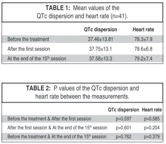
A total of 44 patients were enrolled in the study and three of them were lost at follow-up. The study was completed with 41 patients (26 females, 15 males, mean age: 53.39±10.9 years). No significant differences were noted in heart rate between the first (mean: 78.3±7.9), second (mean: 78.6±6.8), and third (mean: 79.2±7.4) measurements (Table 1 and 2). The mean QTc dispersion was 37.46 ms at the first, 37.75 ms at the second, and 37.58 ms at the last measurement (Table 1). No statistically signif-icant difference was observed between the meas-urements (first&second: P=0.597, second&third: P=0.601, first&third: P=0.762) (Table 2).
DISCUSSION
This prospective study evaluated the effects of TENS in the cardiac rhythm and ventricular recov-ery time by measuring the QT dispersion in painful patients without any cardiac disease. TENS applica-tion to the left shoulder had no significant effect on the heart rate or the QT dispersion in the ECG.
124
Mehmet AĞIRMAN et al. J PMR Sci 2018;21(3):122-5
TENS is commonly used in physical therapy practice for the treatment of acute and chronic pain, such as low-back pain, postoperative pain, neuro-pathic pain, osteoarthritis, dysmenorrhea, cancer pain, etc.8,9 Several mechanisms have been
pro-posed to explain the TENS effect following spinal, supraspinal, and peripheral administration. One of the first theories was based on the “Gate control theory” conceptualized by Melzack and Wall, which postulated that the stimulation of the large diameter fibers (A-beta) inhibits the transmission of pain signals in small diameter fibers (A-delta and C) in the dorsal horn.10 At the supraspinal sites,
TENS activates endogenous opioid, seratonergic, and muscarinic receptors, resulting in analgesia.2,4,11
King et al. demonstrated that peripherally located alpha 2 adrenergic receptors contributed to TENS-mediated antihyperalgesia in an animal study.12
Many types of TENS applications are used in ther-apy by modulating the amplitude and pulse width. The most commonly used procedure in clinical practice uses a high frequency (50-100 Hz) and low intensity, and we used this conventional TENS pro-tocol in this study.
TENS is a safe technique, but it has some pre-cautions, as well as situations where it should not be used. The contraindications of TENS are mostly relative. The manufacturers indicate pregnancy, cardiac pacemakers, and epilepsy as contraindica-tions because of the difficulty of excluding TENS as a potential risk in these situations. The Canadian
Physiotherapy Association (ACPA) has stated that TENS is contraindicated in patients with impaired sensory awareness and cognitive impairments.5The
literature is not clear for cases of patients with im-planted electronic devices (pacemakers, car-dioverter defibrillator etc.), cardiac failure, or arrhythmias; TENS can also lead to misinterpreta-tion of ECG findings.13-16Some studies have
inves-tigated TENS effects in patients with cardiac implanted devices; however, these results were mostly retrospective and obtained from case stud-ies.16,17To our knowledge, the present study is the
first prospective investigation to examine the early and late effects of TENS on ECGs.
The range of the durations, termed the “QT dispersion,” was proposed as an index of the spatial dispersion of the ventricular recovery times. The different ECG leads magnify the ECG signal of dif-ferent myocardial regions; consequently, the QTc dispersion is an almost direct measure of the het-erogeneity of myocardial repolarization. The QTc dispersion is the most frequently used parameter in the electrocardiogram for determination of malig-nant ventricular arrhythmias and the risk of sud-den cardiac death.18
Our results indicated no significant differences between the three measurements in the QTc dis-persion and heart rates. The aim of this study was to investigate the TENS effect on the cardiac rhythm in the clinical setting. TENS application to the chest wall is not recommended; therefore, we chose the left shoulder because of its proximity to the heart. We investigated the early and cumula-tive effect of the TENS by recording the ECGs just after the first and at the end of the 15thapplications.
These measurements showed no significant differ-ences in the QTc dispersion and heart rate.
The main limitations of this study are that it was conducted on healthy subjects are those who do not have any cardiac problems and do not have any additional disease that can disrupt the heart rhythm and it used a manual assessment of the QTc dispersion. However, these deficiencies can be remedied with further studies. The second limita-tion was that the pain cause (diagnosis, duralimita-tion, pain severity etc.) was not assessed.
QTc dispersion Heart rate Before the treatment 37.46±13.81 78.3±7.9 After the first session 37.75±13.1 78.6±6.8 At the end of the 15thsession 37.58±13.3 79.2±7.4
TABLE 1: Mean values of the
QTc dispersion and heart rate (n=41).
QTc dispersion Heart rate
Before the treatment & After the first session p=0.597 p=0.585 After the first session & At the end of the 15thsession p=0.601 p=0.204
Before the treatment & At the end of the 15thsession p=0.762 p=0.379
TABLE 2: P values of the QTc dispersion and
125
Mehmet AĞIRMAN et al. J PMR Sci 2018;21(3):122-5
In conclusion, conventional TENS application to the left shoulder (which is the closest area to the heart in daily practice) has no deleterious effects on the heart rhythm in painful patients without any cardiac disease. However, further studies are
required to confirm these findings in patients with cardiac arrhythmias.
A
Acckknnoowwlleeddggmmeennttss
We thank Hulya Binici for his help in ECG shots.
1. Murina F, Francesco SD. Transcutaneous electrical nerve stimulation. In: Martellucci J, ed. Electrical Stimulation for Pelvic Floor Dis-orders. 1sted. Switzerland: Springer Interna-tional Publishing; 2015. p.105-17.
2. Sluka KA, Walsh D. Transcutaneous electri-cal nerve stimulation: basic science mecha-nisms and clinical effectiveness. J Pain. 2003;4:109-21.
3. Page MJ, Green S, Kramer S, et al. Elec-trotherapy modalities for adhesive capsulitis (frozen shoulder). Cochrane Database Syst Rev. 2014;10:CD011324.
4. Khadilkar A, Odebiyi DO, Brosseau L, et al. Transcutaneous electrical nerve stimulation (TENS) versus placebo for chronic low-back pain. Cochrane Database Syst Rev. 2008;8: CD003008.
5. Canadian Physiotherapy Association. Elec-trophysical Agents-Contraindications and pre-cautions: an evidence-based approach to clinical decision making in physical therapy. Physiother Can. 2010;62:1-80.
6. Digby GC, Daubney ME, Baggs J, et al. Phys-iotherapy and cardiac rhythm devices: a
re-view of the current scope of practice. Eu-ropace. 2009;11:850-9.
7. Funada A, Hayashi K, Ino H, et al. Assess-ment of QT intervals and prevalence of short QT syndrome in Japan. Clin Cardiol. 2008;31: 270-4.
8. Johnson M. Transcutaneous electrical nerve stimulation: review of effectiveness. Nurs Stand. 2014;28:44-53.
9. Bedwell C, Dowswell T, Neilson JP, et al. The use of transcutaneous electrical nerve stimu-lation (TENS) for pain relief in labour: a review of the evidence. Midwifery. 2011;27:e141-8.
10. Melzack R, Wall PD. Pain mechanisms: a new theory. Science. 1965;150:971-9.
11. Johnson M. Transcutaneous electrical nerve stimulation: mechanisms, clinical application and evidence. Rev Pain. 2007;1:7-11. 12. King EW, Audette K, Athman GA, et al.
Transcutaneous electrical nerve stimulation activates peripherally located alpha-2A ad-renergic receptors. Pain. 2005;115:364-73.
13. Banerjee G, Johnson MI. Transcutaneous electrical nerve stimulation (TENS): a potential intervention for pain management in India? In-dian J Pain. 2013;27:132-41.
14. Johnson MI. Contraindications, precautions, and adverse events. Transcutaneous Electri-cal Nerve Stimulation (TENS): Research to Support Clinical Practice. 1sted. Oxford: Ox-ford University Press; 2014. p.93.
15. Kimberley AP, Soni N, Williams TR. Transcu-taneous nerve stimulation and the electrocar-diograph. Anaesth Intensive Care. 1987;15: 358-9.
16. Sliwa JA, Marinko MS. Transcutaneous elec-trical nerve stimulator-induced electrocardio-gram artifact. A brief report. Am J Phys Med Rehabil. 1996;75:307-9.
17. Chen D, Philip M, Philip PA, et al. Cardiac pacemaker inhibition by transcutaneous elec-trical nerve stimulation. Arch Phys Med Re-habil. 1990;71:27-30.
18. Zhang Y, Post WS, Blasco-Colmenares E, et al. Electrocardiographic QT interval and mor-tality: a meta-analysis. Epidemiology. 2011;22: 660-70.