Corresponding author: Ahmet Murat MUSLUMAN E-mail: [email protected]
Original Investigation
Published Online: 13.11.2017Ahmet Murat MUSLUMAN
1, Baris OZONER
2, Atilla KIRCELLI
3, Songul Meltem CAN
1, Adem YILMAZ
1,
Ayca KALDIRIMOGLU
1, Balkan SAHIN
11University of Health Sciences, Hamidiye Sisli Etfal SUAM, Department of Neurosurgery, Istanbul, Turkey 2Erzincan University, School of Medicine, Department of Neurosurgery, Erzincan, Turkey
3Baskent University, School of Medicine, Department of Neurosurgery, Istanbul, Turkey
This study was presented as an oral presentation at the 31st National Congress of Turkish Neurosurgical Society between 29 March-02 April 2017,
Antalya, Turkey.
Chronic Subdural Hematoma Associated with Arachnoid
Cyst of the Middle Fossa: Surgical Treatment and Mid-Term
Results in Fifteen Patients
ABSTRACT
studies (9). The reported incidence of subdural fluid collection as a complication of AC in the middle fossa is 6.6%–17.5% (4-6,13,22,25).
Chronic subdural hematoma (CSDH) generally occurs in elderly patients who had a head trauma more than 3 weeks prior to admission. Classically, CSDHs contain dark motor oil-like fluid which is different from CSF and probably arise from
█
INTRODUCTION
A
rachnoid cysts (ACs) are congenital, benign, extra-axial lesions that arise from the splitting of arachnoidal mem-branes with components similar to the cerebrospinal fluid (CSF)(1,4-7,21,24). ACs make up almost 1% of all intra-cranial masses. In the healthy population, ACs are incidentally seen in 0.8–1.7% of all magnetic resonance imaging (MRI)AIM: To report neurological and radiological features, surgical management, and mid-term outcomes of patients with chronic subdural hematoma (CSDH) associated with ipsilateral arachnoid cyst (AC) of the middle fossa.
MATERIAL and METHODS: A total of 453 patients with CSDH were treated at our clinic between August 2004 and August 2012. Of these patients, 15 had ipsilateral AC in the middle fossa. A single burr hole craniostomy was performed to drain the hematoma. The AC was left intact in 14 patients, and one patient had no surgical intervention. The follow-up period ranged from 13 to 88 months (mean 43.07 ± 23.23 months).
RESULTS: The patients with AC associated CSDH were found to be younger than the patients with CSDH alone, and the mean age of 58 patients was 13.15 ± 13.17 years, while it was 11 ± 14.22 years in the other patients. Eleven patients had experienced head trauma at 21–50 days before admission. Hematoma evacuation through a single burr hole with closed-system subdural drainage performed at 2–4 days after surgery improved the symptoms in all patients. Two patients developed subdural fluid collection, which was treated by subduroperitoneal shunt placement.
CONCLUSION: Greater prevalence of ACs in patients with CSDHs has been reported in the literature. We recommend the drainage of the hematoma via a single craniostomy and to leave the AC intact as the first choice of treatment if the associated AC is a Galassi type I or II. Additional subduroperitoneal shunting may be performed in patients with Galassi type III cyst.
acute subdural hemorrhage which evokes an inflammatory process (3,14,15).
CSHD is the most frequent hemorrhagic event associated with AC development (2,13,16-19,22,25). Parsch et al. reported 2.43% incidence of ACs in their study of 658 patients (18). However, very few studies have reported about this subject. The common practice of CSDH treatment in leading neurosurgical centers tends toward minimal invasive surgical approaches using a burr-hole craniostomy (BHC) (20).
This study aimed to investigate the neurological and radiological features and mid-term outcomes of patients with CSDHs associated with ipsilateral AC. We discussed the probable causes of these associated conditions to rationalize the surgical treatment.
█
MATERIAL and METHODS
This study was a retrospective review of all clinical records and imaging studies of a neurosurgery clinic between August 2004 and August 2012. During this period, 453 patients diagnosed with CSDH were treated. Among them, 15 patients who had an ipsilateral AC were included in this study. Table I outlines the summary of the data of this series.
MRI or computed tomography (CT) findings were noted in terms of laterality (unilateral and bilateral), thickness of the lesion, midline shift, and lesion volume. Furthermore, 14 patients were surgically treated, and one patient was observed, with clinical observation as the first treatment option. The Galassi classification system was used for classifying ACs (5,6). All patients who were surgically treated underwent early cranial CT scanning and late MRI during the follow-up period. All hematomas were evacuated and irrigated using isotonic saline solution at body temperature through a single burr hole performed at where the hematoma was wide, except in one patient. A closed-system subdural drainage was continued 2–4 days postoperatively. Figure 1 shows the treatment flow-chart we followed.
Patients were followed up through clinical examinations and cranial MRI or CT scans at 1, 6, and 12 months after surgery. The neurological status on admission and that on discharge were classified according to the grading scheme for CSDH proposed by Markwalder (15).
█
RESULTS
A total of 453 patients diagnosed with CSDH were admitted during a 9-year period. Among them, 438 patients were without AC, and the mean age of 58 patients was 11 ± 14.22 years. In 15 patients, CSDH associated with ipsilateral AC in the middle fossa was found. Moreover, 10 patients (66.7%) were male, and 5 were female (33.3%). The mean age was 13.93 ± 12.37 years, and mean follow-up was 43.07 ± 23.23 months. In addition, 8 of 15 patients were aged below 10 years. Eleven patients had experienced head trauma 21–50 days before admission, but two of them had no history of
trauma. Falls were the most frequent trauma type. The mean CSDH hematoma thickness was 11.1 ± 2.62 mm (Table I). According to the Galassi classification system, the ACs were type I in four, type II in nine, and type III in two patients. One patient who had type I AC with CSDH, which is 3 mm in diameter, was observed without surgical intervention. The other 14 patients underwent surgery. Three patients with Galassi type I AC and nine patients with Galassi type II AC demonstrated the complete resolution of CSDH on follow-up CT examinations, and they were treated using BHC irrigation and non-vacuuming drainage. One of the patients with Galassi type III AC also had a posterior fossa AC. At first, he underwent BHC with drainage. However, drainage was terminated on the third postoperative day, and he was discharged on the fifth day after surgery. One week later, he was admitted with swelling on the operation field. Cranial CT scan showed mild subdural and subcutaneous fluid collection. He recovered after the placement of a low-pressure subduroperitoneal shunt system. CT scans were performed on the fourth postoperative day in patients with type III AC, in whom drainage was terminated on the third postoperative day, and revealed subdural fluid collection. On the same day, subduroperitoneal shunting was performed. All patients returned to their daily activities within 15 days after surgery. Their neurological examinations were found to be normal (Markwalder grade 0), and they were free of headache at long-term follow-up.
Figure 1: AC-associated CSDH, suggested treatment protocol. (AC: Arachnoid cyst, BHC: burr hole craniostomy, CSDH: chronic subdural hematoma, CT: computed tomography, SDS: subduroperitoneal shunt).
Illustrative Case
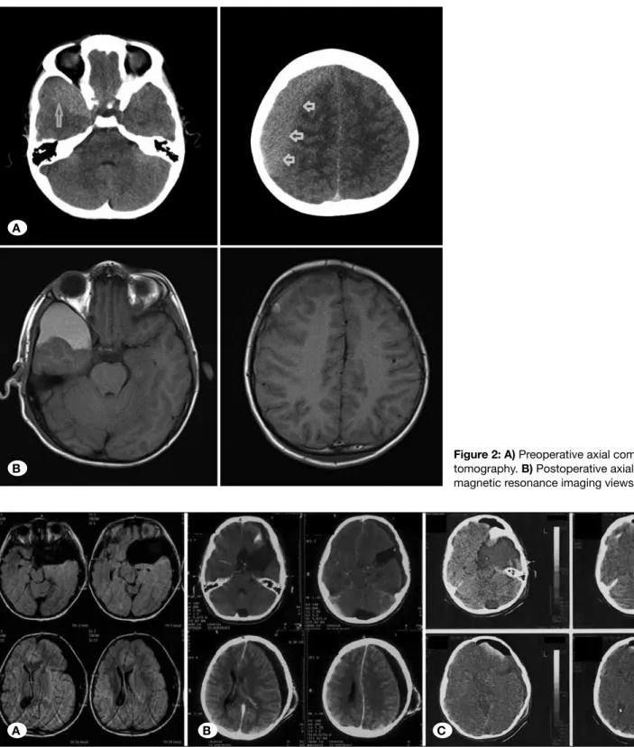
A 10-year-old male patient who had a bicycle accident 1 month ago was admitted to the emergency room. He complained of headache for 10 days, drowsiness, and left-sided weakness of the arm and leg (Case 4). Physical examination revealed no sign of acute trauma. Neurological examination showed confusion and left-sided hemiparesis. CT scans revealed subacute subdural hematoma, 25 mm in diameter, in the right frontotemporoparietal region which caused an 11-mm midline shift. An isodense image which measured 33×35 mm on the right temporal lobe was interpreted as AC (Figure 2A). The decision was to perform immediate surgery because of the patient’s neurological status. An MRI scan was avoided to not lose any time. The hematoma was evacuated and irrigated with isotonic saline solution through a single burr hole, and a closed-system subdural drainage was placed. The AC was left intact. His consciousness and hemiparesis immediately recovered after the surgery. The postoperative CT scans showed near-complete drainage of the hematoma. The drainage system was pulled out on the third postoperative day.
The MRI scans on the fourth postoperative day showed that the hematoma was totally drained, the AC was intact, and the back of the AC contained partially hemorrhagic characteristic (Figure 2B). The patient was discharged on the sixth postoperative day with perfect recovery. Figures 3A-C; 4A, B; 5A-C show other case examples.
█
DISCUSSION
The etiology of AC is not widely explained. This condition occurs when the space formed by the separation of the two layers of the arachnoid membrane is filled with cerebrospinal fluid (7,21). Basically, it is considered that the cavity caused by minor developmental anomalies is wrapped with arachnoid membranes (2,16,21). The dural border cells beneath the dura mater are attached to the arachnoid mater in tight junctions (3,8,18). The loosening of these junctions as a result of possible trauma forms cavities called subdural components. Moreover, CSDH is considered to have occurred as a tiny bleeding, which triggered an inflammatory process in these junctions (3). Developmental disorders of the arachnoid membranes will make these junctions weaker (2,16,18).
Generally, CSDHs occur in elderly patients who had a head trauma more than 3 weeks previously (14). Classically, CSDHs contain dark motor oil-like fluid which differ from CSF and probably arise from acute subdural hemorrhage, which evokes an inflammatory process. It was first described by Virchow, in 1857, as “an internal hemorrhagic pachymeningitis.” Later, in 1914, Trotter launched the theory of traumatic brain injury, and the consecutive lesion of the “bridging veins” was the cause of what he called “hemorrhagic subdural cyst” (14).
The detailed features of AC-associated CSDH remain not unelucidated. Hemorrhage is considered to emerge from the small bridging vessels surrounding the cyst wall between the dura and outer membranes of the cyst wall or floating inside the cyst. Several authors have emphasized the structures
Table I: Summary of 15 Cases
Case Age (years), Sex Injury AC-Galassi Type CSDH Treatment Follow up
1 27, F Fall L Type II R Hemispheric BHC 2y, 2m 2 16, M Accident L Type II L Hemispheric BHC 1y, 1m 3 6, F No Trauma L Type I L Hemispheric BHC 4y, 4m 4 10, M Fall R Type II R Hemispheric BHC 1y, 7m 5 1, M Fall R Type I L Temporofrontal Observation 1y, 4m 6 1, M Fall L Type I R Hemispheric BHC 7y, 4m 7 5, M MVA L Type III+P. Fossa L Hemispheric BHC+SDS 1y, 10m 8 28, M No Trauma L Type II L Temperoparietal BHC 7y, 2m 9 46, F MVA R Type II R Temporofrontal BHC 2y, 8m 10 9, M Fall R Type I R Temporofrontal BHC 3y, 9m 11 12, F Fall L Type III L Hemispheric BHC+SDS 5y, 6m 12 7, F Fall L Type II L Hemispheric BHC 4y, 1m 13 3, M Fall L Type II R Hemispheric BHC 7y, 2m 14 18, M Fall R Type I R Hemispheric BHC 2y, 1m 15 20, M Fall R Type II R Temporofrontal BHC 1y, 6m
F: Female, M: Male, MVA: Motor vehicle accident, AC: Arachnoid cyst, CSDH: Chronic subdural hematoma, L: Left, R: Right, BHC: Burr-hole
of relationships between the location of AC and CSDH on neuroimaging studies: (1) a CSDH close to an AC, (2) a CSDH apart from an AC at the ipsilateral side, and (3) a CSDH apart from an AC at the contralateral side. CDHs were found to be close to the ACs in all of our reported patients.
between the dura and arachnoid membrane (17,25,27). We consider these small vessels as the source of initial bleeding that leads to the development of CSDH in patients with an AC. Therefore, the presence of an AC may be a risk factor for the development of CSDH (18). There are three types
Figure 2: A) Preoperative axial computed tomography. B) Postoperative axial T1W magnetic resonance imaging views (case 4).
Figure 3: A) Preoperative view of Galassi type III arachnoid cyst and chronic subdural hematoma are seen on axial T1-weighted MR images. B) Fluid collection and swelling seen on computed tomography (CT) 1 week later after BHC surgery. C) Subduroperitoneal shunt is seen on CT after shunt surgery (case 7).
A
B
(10,11,17,19). In some cases, AC fenestration with craniotomy or craniectomy was chosen (10,12,21). Arai et al. reported a series of 77 cases of AC by performing arachnoidoperitoneal shunting in 1998 (1). Some patients had CSDH; hence, drainage with burr hole and shunting was performed. Page et al. reported seven patients in their series in 1987: three craniotomies, one craniotomy and shunting, two burr hole drainages, and one shunting after burr hole drainage were preferred (17). Rogers et al. in their study in 1990 with a series of six cases suggested shunting after the drainage of the hematoma (19). Recent studies have suggested that the irrigation of hematoma via a single burr hole should be chosen as an initial surgical management (23,26). Similar studies in the last decade are as follows: Domenicucci et al. reported The BHC approach has replaced the more aggressive
craniotomies that were the standard-of-care until the report by Markwalder in the early 1980s (15). Little evidence has been found in the literature on the management of CSDH. BHC has been associated with reduced morbidity compared with craniotomy (3.8% vs. 12.3%, respectively)
Several options for the treatment of AC include craniotomy and fenestration, endoscopic fenestration, and cystoperitoneal shunting. Optimal management of the AC associated with subdural fluid collection also remains controversial (2,13,16,18,19,22,27). Some studies have reported good results of hematoma drainage, leaving the AC intact (12,18,27). Some studies have advocated drainage with burr hole and arachnoidoperitoneal shunting performed simultaneously
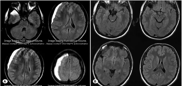
Figure 5: A) Preoperative axial CT scan of a 20-year-old patient with chronic subdural hematoma. B) Galassi type II cyst can be seen on the preoperative axial Flair sequence MRI. C) Late postoperative axial T2-weighted imaging of the same patient (case 15).
Figure 4: Flair sequence axial MR images of an 18-year-old male with chronic subdural hematoma and Galassi type 1 arachnoid cyst. A) Preoperative imaging. B) Postoperative imaging (case 14).
A B
additional subduroperitoneal shunting may be performed following BHC surgery. We recommend this minimal invasive treatment algorithm because of the shorter hospitalization duration and a relatively lower risk of infection.
█
ACKNOWLEDGEMENT
Preparation for publication of this article is partly supported by the “Turkish Neurosurgical Society”.
█
REFERENCES
1. Arai H, Sato K, Wachi A, Okuda O, Takeda N: Arachnoid cysts of the middle cranial fossa: Experience with 77 patients who were treated with cystoperitoneal shunting. Neurosurgery 39: 1108-1112; discussion 1112-1113, 1996
2. Domenicucci M, Russo N, Giugni E, Pierallini A: Relationship between supratentorial arachnoid cyst and chronic subdural hematoma: Neuroradiological evidence and surgical treat-ment. J Neurosurg 110:1250-1255, 2009
3. Foelholm R, Waltimo O: Epidemiology of chronic subdural haematoma. Acta Neurochir (Wien) 32:247-250, 1975 4. Galassi E, Gaist G, Giuliani G, Pozzati E: Arachnoid cysts of
the middle cranial fossa: Experience with 77 cases treated surgically. Acta Neurochir Suppl (Wien) 42:201-204, 1988 5. Galassi E, Piazza G, Gaist G, Frank F: Arachnoid cysts of the
middle cranial fossa: A clinical and radiological study of 25 cases treated surgically. Surg Neurol 14:211-219, 1980 6. Galassi E, Tognetti F, Gaist G, Fagioli L, Frank F, Frank G: CT
scan and metrizamide CT cisternography in arachnoid cysts of the middle cranial fossa: Classification and pathophysiological aspects. Surg Neurol 17:363-369, 1982
7. Gandy SE, Heier LA: Clinical and magnetic resonance features of primary intracranial arachnoid cysts. Ann Neurol 21:342-348, 1987
8. Haines DE, Harkey HL, Al-Mefty O: The “subdural” space: A new look at an outdated concept. Neurosurgery 32:111-120, 1993
9. Heier LA, Zimmerman RD, Amster JL, Gandy SE, Deck MD: Magnetic resonance imaging of arachnoid cysts. Clin Imaging 13:281-291, 1989
10. Huang D, Abe T, Kojima K, Tanaka N, Watauabe M, Ohkura A, Nishimura H, Hayabuchi N, Norbash AM: Intracystic hemorrhage of the middle fossa arachnoid cyst and subdural hematoma caused by ruptured middle cerebral artery aneurysm. AJNR Am J Neuroradiol 20:1284-1286, 1999 11. Ide C, De Coene B, Gilliard C, Pollo C, Hoebeke M, Godfraind
C, Trigaux JP: Hemorrhagic arachnoid cyst with third nerve paresis: CT and MR findings. AJNR Am J Neuroradiol 18: 1407-1410, 1997
12. Kulali A, von Wild K: Post-traumatic subdural hygroma as a complication of arachnoid cysts of the middle fossa. Neurosurg Rev 12 Suppl 1:508-513, 1989
13. Kwak YS, Hwang SK, Park SH, Park JY: Chronic subdural hematoma associated with the middle fossa arachnoid cyst: Pathogenesis and review of its management. Childs Nerv Syst 29:77-82, 2013
CSDH with the presence of AC in eight cases. In all the cases (2), AC was left intact, and CSDH was drained with a burr hole. Follow-up MRI showed that the view in the AC that is compatible with chronic hemorrhage disappeared with time. They concluded that fragmented blood cells diffuse through the weakened AC membrane in the presence of CSDH. Mori et al., in a series of 541 cases of CSDH, showed 12 cases of AC. Drainage and irrigation was performed in all patients, and they reported that these operations had good results (16). The most extensive series on AC-associated CSDH was that of Parsch et al., with a series of 16 cases; in 13 patients with burr hole drainage, one patient underwent hematoma drainage with fenestration, and conservative treatment was performed in two patients (18). Conservative treatment is suggested in very few cases in the literature (12). Kulali and von Wild reported one patient with a good clinical state and relatively thin subdural hematomas as in our series. Of the 15 patients, one patient had a thinner subdural hematoma (12). Huang et al. reported intracystic hemorrhage of the AC and development of SDH due to the rupture of middle cerebral artery bifurcation aneurysm (10). In addition, deeply localized hemorrhages in the AC may cause third cranial nerve paralysis (11). Domenicucci et al.(2) considered that the AC may not be the source and that destructed blood may have diffused through the weak cyst membrane in chronic hemorrhage found in the AC in some cases. The follow-up cranial MRI showed resolution of hemorrhage in cases with CSDH drainage, and the finding of this study supports their suggestion. Again, this idea supports that AC and CSDH are two different conditions and that they should be treated differently.
To our knowledge, there has been no published report on the treatment algorithm for AC-associated CSDH. We propose that the size of AC could affect the treatment course of CSDH. Therefore, we used the Galassi AC classification system. The conservative treatment in one patient with Galassi type 1 and BHC and drainage of 10 patients with grades 1–2 CSDH were found to be sufficient. Subduroperitoneal shunting besides BHC surgery was required for two patients with Galassi type III AC. In our series, CSDH and AC are considered two different conditions; 14 of 15 patients were treated with surgery. CSDH was treated by drainage, and AC was left intact in patients with Galassi type I and II AC. Additional SDS was performed on Galassi type III patients. As fenestration of the cyst wall carries some risks of morbidity, such as re-bleeding, epidural hematoma, and subdural hygroma, we assumed that treating AC simultaneously with hematoma drainage is not necessary unless AC causes symptoms (23,26).
█
CONCLUSION
We suggest a management algorithm for patients with ipsilateral AC-associated CSDH. If there is no evidence of acute bleeding in the AC, Galassi type I and type II AC-associated CSDHs can be treated with BHC drainage and irrigation of the cavity alone, leaving the AC membrane intact. In the treatment of Galassi grade III AC-associated CSDH, if there is swelling and CSF fistula on the operation site or recurrence of the hematoma or subdural effusion, then
21. Schachenmayr W, Friede RL: Fine structure of arachnoid cysts. J Neuropathol Exp Neurol 38:434-446, 1979
22. Sprung C, Armbruster B, Koeppen D, Cabraja M: Arachnoid cysts of the middle cranial fossa accompanied by subdural effusions-experience with 60 consecutive cases. Acta Neurochir (Wien) 153:75-84; discussion 84, 2011
23. Sun J, Wang W, Wang D, An S, Xue L, Wang Y, Zhu SG, Jiang RC, Yang XJ, Yue SY: Clinical analysis of 10 patients of chronic subdural hematoma associated with arachnoid cyst. Zhonghua Yi Xue Za Zhi 97(19):1502-1504, 2017 (In Chinese) 24. Weber F, Knopf H: Incidental findings in magnetic resonance
imaging of the brains of healthy young men. J Neurol Sci 240: 81-84, 2006
25. Wester K, Helland CA: How often do chronic extra-cerebral haematomas occur in patients with intracranial arachnoid cysts? J Neurol Neurosurg Psychiatry 79: 72-75, 2008 26. Wu X, Li G, Zhao J, Zhu X, Zhang Y, Hou K: Arachnoid
cyst associated chronic subdural hematoma: Report of 14 cases and a systematic literature review. World Neurosurg pii: S1878-8750(17)31622-4, 2017 (Ahead of print)
27. Zhang H, Zhang JM, Chen G: Chronic subdural hematoma associated with arachnoid cyst: Report of two cases. Chin Med J (Engl) 120:2339-2340, 2007
14. Lee KS: Natural history of chronic subdural haematoma. Brain Inj 18:351-358, 2004
15. Markwalder TM: Chronic subdural hematomas: A review. J Neurosurg 54:637-645, 1981
16. Mori K, Yamamoto T, Horinaka N, Maeda M: Arachnoid cyst is a risk factor for chronic subdural hematoma in juveniles: Twelve cases of chronic subdural hematoma associated with arachnoid cyst. J Neurotrauma 19:1017-1027, 2002
17. Page A, Paxton RM, Mohan D: A reappraisal of the relationship between arachnoid cysts of the middle fossa and chronic subdural haematoma. J Neurol Neurosurg Psychiatry 50: 1001-1007, 1987
18. Parsch CS, Krauss J, Hofmann E, Meixensberger J, Roosen K: Arachnoid cysts associated with subdural hematomas and hygromas: Analysis of 16 cases, long-term follow-up, and review of the literature. Neurosurgery 40:483-490, 1997 19. Rogers MA, Klug GL, Siu KH: Middle fossa arachnoid cysts
in association with subdural haematomas. A review and recommendations for management. Br J Neurosurg 4:497-502, 1990
20. Santarius T, Hutchinson PJ: Chronic subdural haematoma: Time to rationalize treatment? Br J Neurosurg 18:328-332, 2004