Comparison of Au(III) and Ga(III) Ions
’ Binding to Calf Thymus
DNA: Spectroscopic Characterization and Thermal Analysis
Omer Faruk Sarioglu&Refiye Tekiner-Gursacli&Ayse Ozdemir&Turgay Tekinay
Received: 3 June 2014 / Accepted: 23 June 2014 / Published online: 10 July 2014 # Springer Science+Business Media New York 2014
Abstract Metals have been studied as potential chemothera-peutic agents for cancer therapies due to their high reactivity toward a wide variety of substances. The characterization of metal ion-binding capacities is essential to understand the possible effects of metals on target biomolecules. In the pres-ent study, biochemical effects of Au(III) and Ga(III) ions on calf thymus DNA (ctDNA) were studied comparatively via bioanalytical, spectroscopic, and thermal methods. Briefly, UV-Vis absorbance spectroscopy, fluorescence spectroscopy, circular dichroism (CD) spectroscopy, and Fourier transform infrared (FT-IR) spectroscopy were utilized for spectroscopic characterization, and isothermal titration calorimetry (ITC) measurements were performed for thermal analysis. Our re-sults reveal that both Au(III) and Ga(III) ions are capable of interacting with ctDNA, and Au(III) ions display a more favorable interaction and a higher binding affinity. ITC anal-yses indicate that the Au(III)-DNA interaction displays a binding affinity (Ka) around 1.43×10
6
M−1, while a Kaaround
1.17×10⁵M−1was observed for the Ga(III)-DNA binding. It was suggested that both metal ions are unlikely to change the structural B-conformation while interacting with ctDNA.
Keywords Calf thymus DNA . B-conformation of DNA . DNA-binding studies . Au(III) and Ga(III) ions
Abbreviations
ctDNA Calf thymus DNA CD Circular dichroism FT-IR Fourier transform infrared UV-Vis Ultraviolet-visible
EB Ethidium bromide
Introduction
The investigation of novel metal-based anticancer agents has been a popular field of research since the discovery of the antitumor activity of cisplatin. While many platinum-based chemotherapeutic agents have been studied in the last 30 years [1], a limited spectrum of activity and a number of side effects associated with these drugs have triggered an increased inter-est in non-platinum metal-based agents [2,3]. To this end, various complexes of gallium, gold, ruthenium, germanium, cobalt, copper, and zinc have been studied as alternatives to platinum [3,4]. After platinum, gallium is the second metal species that has been approved for use in patients for chemo-therapeutic purposes, and gallium nitrate has been used for the treatment of hypercalcemia, lymphoma, and bladder cancer [5–7]. Ga(III) shares similar chemical properties with Fe(III), especially with respect to ionic radius, electron affinity, elec-tronegativity, coordination geometry, and Lewis bases affinity [3,6]. Because of these similarities, gallium can participate in cellular processes and interact with proteins responsible for iron metabolism [7].
Following cellular uptake, gallium binds to the enzyme ribonucleotide reductase and inhibits its catalytic activity. Ribonucleotide reductase, a key enzyme in DNA replication, catalyzes the reduction of ribonucleotides to deoxyribonucle-otides and prevents the activation of the proapoptotic factors Bax and caspase-3, thereby blocking apoptosis [1,3,8]. The O. F. Sarioglu
:
A. OzdemirUNAM-Institute of Materials Science and Nanotechnology, Bilkent University, 06800 Ankara, Turkey
T. Tekinay (*)
Department of Medical Biology and Genetics, Faculty of Medicine, Gazi University, Besevler, Ankara 06500, Turkey
e-mail: [email protected] R. Tekiner-Gursacli
:
T. TekinayLife Sciences Application and Research Center, Gazi University, Golbasi, 06830 Ankara, Turkey
inhibition of ribonucleotide reductase has been considered to be an effective means to mediate anticancer activity, and the antiproliferative and antimitotic effects of gallium are primar-ily based on its ability to interfere with the activity of this enzyme [1–3].
Au(III) compounds are isoelectronic and isostructural with Pt(II) complexes, with which they share a square-planar tetracoordinated geometry [9]. As such, Au(III) compounds are potential candidates for anticancer eval-uation and have been studied extensively for this pur-pose, with recent research focusing primarily on the remarkable cytotoxic activity displayed by Au(III) com-plexes [10,11]. Consequently, a considerable number of antineoplastic Au(III) complexes have been reported in the literature [11, 12].
DNA regulates many biochemical processes that oc-cur in cellular systems and has been established as the primary target for many antitumor agents [4, 13, 14]. These agents frequently achieve their antitumor activi-ties by directly interacting with DNA, thus interfering with the replication process and inhibiting the prolifer-ation of tumor cells [4]. Therefore, the investigation of DNA-anticancer drug-binding mechanisms is critical for both the development of new therapeutic reagents and the characterization of existing anticancer drugs [14].
In the present study, the interaction of Ga(III) and Au(III) with calf thymus DNA (ctDNA) has been ex-amined by UV-Vis absorbance spectroscopy, fluores-cence spectroscopy, circular dichroism (CD) spectrosco-py, and Fourier transform infrared (FT-IR) spectroscopy for spectroscopic characterization and isothermal titra-tion calorimetry (ITC) for thermal analysis. Overall, we describe and compare the effects of Au(III) and Ga(III) bindings on ctDNA by utilizing a wide range of analytical techniques and believe that our results will be crucial for future studies involving the antineoplastic properties of these metals.
Materials and Methods
Materials and Sample Preparation
All chemical reagents were purchased from Sigma-Aldrich (USA). ctDNA with an initial concentration of 10 mg/ml was purchased from InvitrogenTM (USA). Gold(III) chloride hydrate, gallium(III) nitrate hydrate, and ctDNA solutions were prepared in a 10 mM Tris ( t r i s ( h y d r o x y m e t h y l ) a m i n o m e t h a n e ) - H C l b u f f e r (pH 7.40) to keep the pH of the solutions stable. Stocks of ctDNA were stored at −20 °C. Fresh ctDNA solu-tions were prepared prior to each experiment.
UV-Vis Absorbance Spectroscopy
Equal concentrations of ctDNA solutions were prepared at 300 μM. Different concentrations (10, 40, 80, 160, and 320μM) of gold(III) chloride hydrate and gallium(III) nitrate hydrate solutions were mixed with ctDNA solutions, and these mixtures were incubated for 2 h. After the incubation period, absorbance spectra of the samples were read by a NanoDrop 2000 spectrophotometer (Thermo Scientific, USA). The wavelength range was 220–420 nm.
EB Displacement Assay
Equal concentrations of ctDNA solutions (10 mg/ml) were mixed with ethidium bromide at a final ratio of 10:1 (DNA/ EB) in Tris-HCl buffer (pH 7.40), to obtain a DNA/EB com-plex solution. This solution was incubated at 25 °C for 30 min in an unilluminated room and then diluted prior to mixing with different concentrations of metal solutions. The final DNA concentrations were constant for all samples (200μM), and the final concentrations of gold(III) chloride hydrate and gallium(III) nitrate hydrate solutions were set at 10, 20, 50, 100, and 200 μM. Those mixtures were also incubated for 30 min in an unilluminated room and then transferred to a microwell plate. The fluorescence values of the samples were measured by a SpectraMax M5 Microplate Reader. Excitation and emission wavelengths were 530 and 590 nm, respectively. The fluorescence intensities of the samples were evaluated by using the equation below [15]:
F ¼ I−Ið 0Þ= Ið100−I0Þ ð1Þ
where F and I are the relative fluorescence and emission intensities of EB-DNA-metal mixtures at 590 nm, respective-ly. I0 denotes the emission intensity of free EB, and I100
denotes the emission intensity of the EB-DNA mixture.
CD Spectroscopy
For CD analysis, 200μM ctDNA and 4 mM gold(III) chloride hydrate or gallium(III) nitrate hydrate stock solutions were prepared. DNA concentrations were constant for all samples (200μM), and increasing concentrations of gold(III) chloride hydrate and gallium(III) nitrate hydrate solutions (10, 20, 40, 100, and 200μM at final) were obtained by diluting the main stocks of the metal salt solutions for titration. CD spectra of ctDNA at different metal ion concentrations were recorded with a Jasco J-815 spectropolarimeter (Jasco, UK). For mea-surements within 210 to 300 nm, a quartz cell with a path length of 1 cm was utilized in nitrogen atmosphere. Three scans of accumulation were taken for each nanometer from
300 to 210 nm, with a scan speed of 100 nm per minute, and sample temperature was maintained at 25 °C.
Isothermal Titration Calorimetry (ITC)
ITC experiments for calf thymus DNA-metal ion binding were carried out at 25 °C on an iTC200 microcalorimeter
(Microcal®). Experimental setup for ITC was arranged as follows: a total of 40 injections, a syringe concentration of 4 mM (gold(III) chloride hydrate or gallium(III) nitrate hy-drate), a cell concentration of 200μM (ctDNA), and a stirring speed of 300 rpm. Injection parameters were set up using the following parameters; volume 1μl, duration 0.8 s, spacing 100 s,and filter period 2 s. Subtraction of pure buffer-metal binding was performed after data acquisition. The data anal-ysis software Origin 7.0 was used for data acquisition and manipulation.ΔG (Gibbs free energy) calculations were made by the following formula [16]:
ΔG ¼ −RT ln Ka¼ ΔH−TΔS ð2Þ
whereT is the absolute temperature in Kelvin (298 K) and R= 8.3151 J mol−1K−1.
FT-IR Spectroscopy
Solutions of ctDNA with a concentration of 200 μM and gold(III) chloride hydrate or gallium(III) nitrate hydrate at varying concentrations (50, 100, and 200μM at final) were prepared for FT-IR measurements and incubated for 2 h for the establishment of metal-DNA interactions. After the incubation
period, 20μl of final solutions were dropped and dried on a 96-well plate at 37 °C for 1 h. After drying, FT-IR transmit-tance analysis was performed by using Nicolet 6700 FT-Raman Spectrometer (Thermo Scientific, USA). The data analysis software OMNICTMwas used for FT-IR measure-ments, identification of peak locations, and basic modifica-tions such as baseline and background correcmodifica-tions. Back-ground corrections for H2O and CO2were carried out for each
analysis. Duplicate samples were utilized in each analysis, and experiments were repeated independently for at least two times.
Results and Discussion
UV-Vis Absorbance Spectroscopy Studies
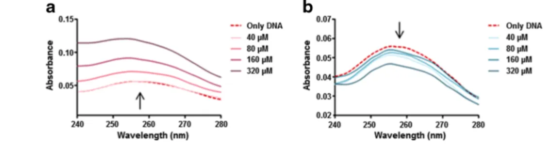
Absorption peaks of free ctDNA were observed at 258 nm and displayed altered intensities in the presence of metal ions (Fig.1). In particular, the presence of Au(III) ions was found to cause a gradual and concentration-dependent increase (hyperchromism) in the absorbance spectrum, while Ga(III) ions triggered a slight and again concentration-dependent decrease (hypochromism). This result may suggest that Au(III) and Ga(III) ions bind to DNA by different binding modes and with different binding affinities. It is reported that hypochromism is associated with charge-transfer interactions [17] and hyperchromism is related to DNA melting and de-naturation [18]. Therefore, the UV-Vis absorbance spectra suggest that while Au(III) ions lead to denaturation of ctDNA in a concentration-dependent manner, Ga(III) ions appear not to denature it. This result may indicate that Au(III) ions show stronger binding characteristics, since gold bears a higher impact on the spectral intensity of DNA and has denaturing properties that Ga(III) does not appear to display.
Competitive Binding of Au(III) and Ga(III) Ions with EB-DNA Mixture
An ethidium bromide displacement assay was performed to determine whether metal ions are capable of intercalative binding to DNA and whether the intercalative binding strengths change with the increasing metal ion concentrations. Fig. 1 UV-Vis absorbance
spectra of ctDNA within 240– 280 nm, when treated with varying molarities of a Au(III) and b Ga(III) ions (0, 40, 80, 160, and 320μM)
Table 1 Relative fluorescence ratios of EB-ctDNA mixture when treated with different concentrations of Au(III) and Ga(III) ionsa
Relative fluorescence (F) (%)
[metal ion]/[ctDNA] Au(III) Ga(III)
0.25 95±1.54 98.03±0.99
0.5 93.31±1.77 90.89±5.03
1 71.74±6.51 74.54±5.31
2 49.81±1.85 42.56±6.72
aDNA concentrations were constant for all samples (200μM), while
Both metal ions were found to trigger significant reductions in the emission intensities of EB-DNA mixtures, especially at higher concentrations (Table 1). Although, this result may suggest that both metal ions are capable of intercalating be-tween ctDNA strands, it is known that groove binders can also cause a reduction in the emission intensity of EB/DNA mix-tures, so Au(III) and Ga(III) ions may have groove-binding properties [19]. Nevertheless, UV-Vis absorbance spectrosco-py and EB displacement assays alone are not sufficient to fully evaluate the binding modes, strengths, and affinities of these metals. As such, further studies were performed to quantita-tively determine the binding affinity differences of metal-DNA interactions and the binding modes of metal ions to ctDNA.
CD Spectroscopy Studies
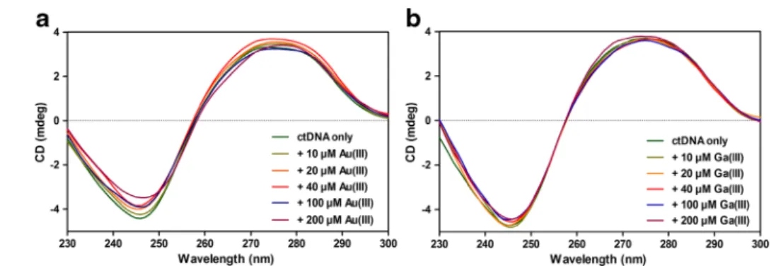
Typically, the B-conformation of ctDNA displays a CD spec-trum with two positive peaks at 220 and 275 nm and a negative peak at 245 nm [20]. The peak around 245 nm corresponds to the helicity of DNA, whereas the peak around
275 nm corresponds to the stacking of DNA bases [21], and an intensity increase at 275 nm can be attributed to B to A conformational change [22]. Our CD spectra suggest that metal-DNA bindings do not significantly alter the DNA struc-ture. As shown in Fig.2a, interactions with Au(III) ions led to decreases in the intensities of CD values at 245 nm and no consistent change at 275 nm was observed, which may indi-cate that the right-handed helicity of B-DNA is modified [21]. Since no peak shift was observed in the spectra of gold samples and no increase in the intensity at 275 nm is present, there is no indication for a B to A conformational transition in DNA structure. For gallium samples, the changes at 245 and 275 nm remained minor in the CD spectra (Fig. 2b), which may indicate that Ga(III) ions are not as influential as Au(III) ions on ctDNA. Moreover, as with Au(III) samples, Ga(III)-DNA interactions may take place without conformational transitions in DNA structure. It was previously stated that while intercalators are known to enhance the intensities of both positive and negative bands, groove binders show less or no perturbation on the helicity and base stacking bands [23]. Therefore, the lack of distinct signatures for conformational Fig. 2 a, b CD spectra within
210–300 nm region and c, d CD differences of ctDNA at the wavelengths of 220, 245, and 275 nm, when titrated with varying molarities of a Au(III) and b Ga(III) ions
Fig. 3 ITC final thermograms of a Au(III)-ctDNA and b Ga(III)-ctDNA bindings
transitions in our CD results indicates that both Au(III)-DNA and Ga(III)-DNA interactions might occur without conforma-tional changes in DNA structure. While EB displacement assay results may suggest that Au(III) and Ga(III) ions display intercalative binding properties, the lack of large perturbations and intensity enhancements in CD bands of metal-DNA com-plexes indicates that groove binding might be more responsi-ble for metal-DNA interactions. FT-IR spectroscopy was
performed to further clarify which binding modes are domi-nant for metal-DNA interactions.
Thermal Analysis
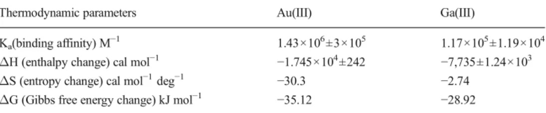
According to ITC results, both Au(III)-DNA and Ga(III)-DNA reactions were exothermic (Fig. 3, Table 2) and Au(III)-DNA interactions were observed to be much more Table 2 Thermodynamic
param-eters obtained by ITC analysis for the binding of ctDNA with Au(III) or Ga(III) ions
Thermodynamic parameters Au(III) Ga(III)
Ka(binding affinity) M−1 1.43×10⁶±3×10⁵ 1.17×10⁵±1.19×10⁴
ΔH (enthalpy change) cal mol−1 −1.745×10⁴
±242 −7,735±1.24×10³ ΔS (entropy change) cal mol−1deg−1 −30.3 −2.74
ΔG (Gibbs free energy change) kJ mol−1 −35.12 −28.92
Fig. 4 FT-IR spectra of ctDNA in the range of 600–1,800 cm−1, when treated with varying molarities of a Au(III) and b Ga(III) ions (a:0, b:100, c:200 μM of metal ions)
favorable than Ga(III)-DNA interactions in ambient condi-tions. Since the reactions were conducted under constant pressure and temperature conditions and ΔG values of Au(III)-DNA and Ga(III)-DNA reactions were negative, both reactions are exergonic and likely to occur spontaneously through forward direction at ambient conditions, and Au(III)-DNA binding seems to have a higher spontaneity over Ga(III)-DNA binding, although the homogeneity of both re-actions avoids the accurate prediction of reaction spontaneities [24]. Our Kavalues demonstrate that Au(III) ions bind with
ctDNA with a much higher affinity than Ga(III) ions. Accord-ing toΔH and ΔS values, there are considerable differences between Au(III)-DNA and Ga(III)-DNA bindings, such that Au(III)-DNA binding is enthalpically more favorable but entropically less favorable than Ga(III)-DNA binding. In ad-dition, these values were negative in both cases, and since the ΔG values were also negative, the reactions are enthalpy driven in both cases, and it reveals that non-covalent interac-tions (e.g., electrostatic, van der Waals) are dominant between Au(III)-DNA and Ga(III)-DNA bindings [25].
In summary, according to ITC analysis, binding reactions of Au(III)-DNA and Ga(III)-DNA are likely to be spontane-ous at ambient conditions, Au(III)-DNA binding was ob-served to be much more favorable and much stronger than Ga(III)-DNA binding, and this finding is parallel with the UV-Vis spectroscopy analysis results.
FT-IR Spectroscopy Studies
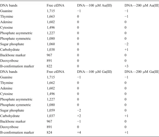
FT-IR spectroscopy was performed to determine the binding modes of Au(III) and Ga(III) ions to ctDNA within 600– 1,800 cm−1 region (Fig. 4) and to monitor whether the B-conformation of ctDNA is altered during Au(III) or Ga(III) binding [26]. The marker bands were chosen for different chemical groups: 1,715 cm−1 for guanine, 1,663 cm−1 for thymine, 1,602 cm−1for adenine, 1,496 cm−1 for cytosine, 1,227 cm−1for phosphate asymmetric, 1,080 cm−1for phos-phate symmetric, 1,060 cm−1for sugar phosphate, 1,038 cm−1 for carbohydrate, 967 cm−1for backbone marker, 891 cm−1 for deoxyribose, and 818 cm−1 for B-conformation marker [21,27–29]. For the purposes of this manuscript, shifts up to 5 cm−1were assumed as minor. In the free DNA spectrum, the marker bands at 1,710–1,717 cm−1 (guanine), 1,222– 1,227 cm−1(asymmetric PO2stretching), and 825 cm−1
(B-conformation marker) are signatures of the B-DNA confor-mation [27]. For both gold and gallium samples (Table 3), minor spectral shifts were observed for these B-conformation markers. Minor spectral shifts were observed for nucleotide base markers as well (guanine, thymine, adenine, and cyto-sine), which may indicate that the metal-DNA bindings occur via non-intercalative interactions. No major shifts occurred in phosphate-associated bands, and the marker bands exhibited no major shifts in the spectra of metal-DNA complexes, so the
Table 3 Peak positions and spectral shifts of the Au(III)-ctDNA and Ga(III)-Au(III)-ctDNA com-plexes at different molar ratios
DNA bands Free ctDNA DNA—100 μM Au(III) DNA—200 μM Au(III)
Guanine 1,715 −1 −1 Thymine 1,663 0 −1 Adenine 1,602 0 0 Cytosine 1,496 0 0 Phosphate asymmetric 1,227 0 0 Phosphate symmetric 1,080 0 0 Sugar phosphate 1,060 0 −2 Carbohydrate 1,038 0 +1 Backbone marker 967 0 0 Deoxyribose 891 0 0 B-conformation marker 822 0 +3
DNA bands Free ctDNA DNA—100 μM Ga(III) DNA—200 μM Ga(III)
Guanine 1,715 −1 −1 Thymine 1,662 0 0 Adenine 1,602 0 0 Cytosine 1,496 0 0 Phosphate asymmetric 1,227 0 0 Phosphate symmetric 1,080 0 0 Sugar phosphate 1,059 −2 0 Carbohydrate 1,037 +2 +1 Backbone marker 967 −1 0 Deoxyribose 891 0 0 B-conformation marker 824 +4 +1
minor spectral changes might occur as a result of alterations in the sugar-phosphate geometry. Hence, ctDNA likely remained in B-family structure following both Au(III) and Ga(III) ions’ binding y.
In conclusion, since minor shifts were observed in nucleo-tide base peaks and no apparent alterations occurred on the conformation of ctDNA, both Au(III) and Ga(III) ions may bind to DNA mainly through groove binding and non-covalent interactions instead of intercalation.
Conclusions
In this paper, the binding properties of Au(III)-DNA and Ga(III)-DNA were investigated by a variety of physicochem-ical characterization tools. While EB displacement assay re-sults demonstrated that an intercalative type of binding might be possible between both metal ions and ctDNA, the other analysis results indicate that groove binding and non-covalent interactions are more meaningful for Au(III)-DNA and Ga(III)-DNA interactions. Our results overall suggest that the B-family structure of ctDNA is preserved during both metal ions’ binding. Finally, thermal analysis of metal-DNA interactions revealed that both reactions were exothermic at ambient conditions, and Au(III)-DNA binding is more favor-able and has a higher affinity than Ga(III)-DNA binding. While the in vitro characterizations of Au(III)-DNA and Ga(III)-DNA interactions detailed in this manuscript are im-portant to elucidate the DNA-binding potential of Au(III) and Ga(III) ions, cellular uptake and intracellular localization/ activity of these metals should also be studied to determine their ability to access the nucleus and genomic DNA.
Acknowledgments This work was supported by grants from Scientific and Technological Research Council of Turkey (TUBITAK), European Cooperation in Science and Technology (COST) action-112S047. The authors thank Rengin Erdem for her great contributions in UV-Vis absorbance spectroscopy and fluorescence spectroscopy sessions and Alper Devrim Ozkan for his fruitful discussions.
References
1. Gómez-Ruiz S, Gallego B, Kaluderović MR, Kommera H, Hey-Hawkins E, Paschke R, Kaluderović GN (2009) Novel gallium(III) complexes containing phthaloyl derivatives of neutral aminoacids with apoptotic activity in cancer cells. J Organomet Chem 694: 2191–2197
2. Samari F, Hemmateenejad B, Shamsipur M, Rashidi M, Samouei H (2012) Affinity of two novel five-coordinated anticancer Pt(II) com-plexes to human and bovine serum albumins: a spectroscopic ap-proach. Inorg Chem 51:3454–3564
3. Kaluderović MR, Gómez-Ruiz S, Gallego B, Hey-Hawkins E, Paschke R, Kaluderović GN (2010) Anticancer activity of dinuclear gallium(III) carboxylate complexes. Eur J Med Chem 45:519–525
4. Tabassum S, Asim A, Arjmand F, Afzal M, Bagchi V (2012) Synthesis and characterization of copper(II) and zinc(II)-based po-tential chemotherapeutic compounds: their biological evaluation viz. DNA binding profile, cleavage and antimicrobial activity. Eur J Med Chem 58:308–316
5. Ortega R, Suda A, Devès G (2003) Nuclear microprobe imaging of gallium nitrate in cancer cells. Nucl Instrum Methods Phys Res Sect B 210:364–367
6. Enyedy ÉA, Dömötör O, Varga E, Kiss T, Trondl R, Hartinger CG, Keppler BK (2012) Comparative solution equilibrium studies of anticancer gallium(III) complexes of 8-hydroxyquinoline and hydroxy(thio)pyrone ligands. J Inorg Biochem 117:189–197
7. Lessa JA, Parrilha GL, Beraldo H (2012) Gallium complexes as new promising metallodrug candidates. Inorg Chim Acta 393:53–63 8. Despaigne AAR, Parrilha GL, Izidoro JB, da Costa PR, dos Santos
RG, Piro OE, Castellano EE, Rocha WR, Beraldo H (2012) 2-Acetylpyridine- and 2-benzoylpyridine-derived hydrazones and their gallium(III) complexes are highly cytotoxic to glioma cells. Eur J Med Chem 50:163–172
9. Gabbiani C, Casini A, Messori L (2007) Gold (III) compounds as anticancer drugs. Gold Bull 40:73–88
10. Aldinucci D, Ronconi L, Fregona D (2009) Groundbreaking gold(III) anticancer agents. Drug Discov Today 14:1075–1076
11. Casini A, Hartinger C, Gabbiani C, Mini E, Dyson PJ, Keppler BK, Messori L (2008) Gold(III) compounds as anticancer agents: rele-vance of gold-protein interactions for their mechanism of action. J Inorg Biochem 102:564–575
12. Maiore L, Cinellu MA, Nobili S, Landini I, Mini E, Gabbiani C, Messori L (2012) Gold(III) complexes with 2-substituted pyridines as experimental anticancer agents: solution behavior, reactions with model proteins, antiproliferative properties. J Inorg Biochem 108: 123–127
13. Arjmand F, Parveen S, Afzal M, Shahid M (2012) Synthesis, characterization, biological studies (DNA binding, cleavage, antibacterial and topoisomerase I) and molecular docking of copper(II) benzimidazole complexes. J Photochem Photobiol B 114:15–26
14. Wu SS, Yuan WB, Wang HY, Zhang Q, Liu M, Yu KB (2008) Synthesis, crystal structure and interaction with DNA and HSA of (N, N’-dibenzylethane-1,2-diamine) transition metal complexes. J Inorg Biochem 102:2026–2034
15. Sheng R, Luo T, Zhu Y, Li H, Sun J, Chen S, Sun W, Cao A (2011) The intracellular plasmid DNA localization of cationic reducible cholesterol-disulfide lipids. Biomaterials 32:3507– 3519
16. Chatterjee T, Pal A, Dey S, Chatterjee BK, Chakrabarti P (2012) Interaction of virstatin with human serum albumin: spectroscopic analysis and molecular modeling. PLoS One 7:e37468
17. Markovitsi D (2009) Interaction of UV radiation with DNA helices. Pure Appl Chem 81:1635–1644
18. Eshkourfu R,Čobeljić B, Vujčić M, Turel I, Pevec A, Sepčić K, Zec M, Radulović S, Srdić-Radić T, Mitić D, Andjelković K, Sladić D (2011) Synthesis, characterization, cytotoxic activity and DNA bind-ing properties of the novel dinuclear cobalt(III) complex with the condensation product of 2-acetylpyridine and malonic acid dihydrazide. J Inorg Biochem 105:1196–1203
19. Ju CC, Zhang AG, Yuan CL, Zhao XL, Wang KZ (2011) The interesting DNA-binding properties of three novel dinuclear Ru(II) complexes with varied lengths of flexible bridges. J Inorg Biochem 105:435–443
20. Ivanov VI, Minchenkova LE, Schyolkina AK, Poletayer AI (1973) Different conformations of double-stranded nucleic acid in solution as revealed by circular dichroism. Biopolymers 12:89–110 21. Sun J, Lu Y, Wang L, Cheng D, Sun Y, Zeng X (2013) Polym Chem
22. Jangir DK, Charak S, Mehrotra R, Kundu S (2011) FTIR and circular dichroism spectroscopic study of interaction of 5-fluorouracil with DNA. J Photochem Photobiol B 105:143– 148
23. Uma V, Elango M, Nair BU (2007) Copper(II) terpyridine com-plexes: effect of substituent on DNA binding and nuclease activity. Eur J Inorg Chem 22:3484–3490
24. McNaught AD, Wilkinson A (1997) Compendium of chemical ter-minology. Blackwell, Oxford
25. Sikora C, Turner R (2005) Investigation of ligand binding to the multidrug resistance protein EmrE by isothermal titration calorimetry. Biophys J 88:475–482
26. Keller PB, Hartman KA (1986) The effect of ionic environment and mercury(II) binding on the alternative structures of DNA. An infrared spectroscopic study. Spectrochim Acta A 42:299–306
27. Tyagi G, Charak S, Mehrotra R (2012) Binding of an indole alkaloid, vinblastine to double stranded DNA: a spectroscopic insight in to nature and strength of interaction. J Photochem Photobiol B 108:48–52 28. Nafisi S, Sobhanmanesh A, Esm-Hosseini M, Alimoghaddam K, Tajmir-Riahi HA (2005) Interaction of antitumor drug Sn(CH3)2Cl2 with DNA and RNA. J Mol Struct 750:22–27 29. Ozdemir A, Tekiner-Gursacli R, Tekinay T (2014) Non-intercalative,
deoxyribose binding of boric acid to calf thymus DNA. Biol Trace Elem Res 158:268–274