Abstract. – OBJECTIVE: Studies in animals have provided key evidence that antagonizing TNF-αα is a viable therapeutic strategy for diffuse severe brain injury. This study is planned to pre-vent post-traumatic secondary tissue damages in rat diffuse severe brain injury model, which is induced by alone or combined administration of Etanercept and lithium chloride (LiCl).
MATERIALS AND METHODS: Male Sprague-Dawley rats were used in the current study. Rats were divided into 5 groups. Trauma was not induced and treatment was not applied to rats of Sham group. For rats of Trauma+Saline group, saline 0.9% was administered via in-traperitoneal (i.p.) route at dose of 1 mg/100 g body weight 1 hour after trauma. For rats of Trauma+Etanercept group, Etanercept was ad-ministered via i.p. route at dose of 5 mg/kg body weight 1 hour after trauma. For rats of Trauma+LiCl group, LiCl was administered via i.p. route at dose of 50 mg/kg body weight 1 hour after trauma. For rats of Etanercept+LiCl group, Etanercept and LiCl were administered via i.p. route at dose of 5 mg/kg body weight and 50 mg/kg body weight, respectively, 1 hour after trauma. Serum glial fibrillary acidic protein (GFAP) and Tau levels were analyzed with ELISA. For analyses H&E, TUNEL, GFAP and TNF-αα staining methods were used.
RESULTS: We demonstrate that Etanercept treatment reduced the TBI-induced brain tis-sues alteration, reduced the expression of TNF-αα and improve edema and axonal swelling. We observed a significant decrease in TNF-αα and GFAP positivity after LiCl was administered.
CONCLUSIONS: The findings obtained in this study suggest that the combination therapy with Etanercept and LiCl decreased neuronal
Effect of etanercept and lithium chloride on
preventing secondary tissue damage in rats with
experimental diffuse severe brain injury
M.A. EKICI
1, O. UYSAL
2, H.I. CIKRIKLAR
3, Z. ÖZBEK
4, D. TURGUT CO
S
AN
5,
C. BAYDEMIR
6, B. KAZANCI
7, D. HAFIZO
Ğ
LU
81Neurosurgery Clinic, Ministry of Health, Sevket Yilmaz Research and Training Hospital, Bursa, Turkey 2Vocational School of Health Services, Eskisehir Osmangazi University, Eskisehir, Turkey
3Emergency Clinic, Ministry of Health, Sevket Yilmaz Research and Training Hospital, Bursa, Turkey 4Department of Neurosurgery, Eskisehir Osmangazi University, Medical Faculty, Eskisehir, Turkey 5Department of Medical Biology, Eskisehir Osmangazi University, Medical Faculty, Eskisehir, Turkey 6Department of Biostatistics, Eskisehir Osmangazi University, Medical Faculty, Eskisehir, Turkey 7Department of Neurosurgery, Ufuk University, Medical Faculty, Ankara, Turkey
8Department of Pediatric Immunology, Uludag University, Medical Faculty, Bursa, Turkey
degeneration and alleviated secondary tissue damage in post-traumatic period.
Key Words:
Traumatic brain injury, Etanercept, Lithium chlo-ride, TNF-α.
Introduction
Traumatic brain injury (TBI), the leading cause of morbidity and mortality in young adults and children, is a major public health problem globally1-5. Depending on severity and duration,
brain injury may cause either infarction or selec-tive neuronal death6.
Injury to the cerebral vasculature breaks the blood-brain barrier (BBB) is accompanied by an inflammatory response marked by infiltration of neutrophils and macrophages, activation of glial cells and up-regulated expression of pro-inflam-matory cytokines7-9. The molecular events result
in apoptosis, reactive astrogliosis, microglial ac-tivation, infiltration of immune cells in the cen-tral nervous system (CNS), neurodegeneration, altered plasticity and neuronal regeneration4,10-12.
Diffuse brain injury includes diffuse axonal injury (DAI), diffuse vascular injury and diffuse hypoxia/ischaemia. One of the major pathologi-cal outcomes of these mechanisms is DAI, the main clinical feature of human TBI, leading to diffuse degeneration of cerebral white matter13,14.
that is primarily localized in the axonal compart-ment of neurons. Loss of axonal microtubules following injury releases intracellular micro-tubule binding proteins, such as Tau, into the ex-tracellular space, where they are transported to the cerebral spinal fluid (CSF). CSF levels of a cleaved form of Tau (C-tau) reflect axonal dam-age after head injury, Alzheimer’s disease and meningitis5,15,16.
Astrocytes are the most numerous non-neu-ronal cell type in the CNS and make up about 50% of human brain volume. Within a few hours of virtually any type of brain injury, surviving as-trocytes in the affected region begin to exhibit hy-pertrophy and proliferation, termed “reactive as-trogliosis”. This response is fortified by the mi-gration of microglia and macrophages to the dam-aged area. Reactive astrocytes increase the ex-pression of their structural protein, Glial fibrillary acidic protein (GFAP), which is commonly used as astrocyte markers2,6,17,18. Increasing numbers of
GFAP(+) astroglial cells following TBI have been described in several experimental studies in animals19,20. Astrocytes are known to be a source
of pro-inflammatory cytokines and in the ad-vanced stages of injury progression form a glial scar inhibitory to neural regeneration. These ob-servations suggest that conversion of glial cells to their “reactive” state, and the associated increase in expression of cytokines and chemokines, may play a role in neurodegeneration3.
TNF-α is 17 Kda major pro-inflammatory cy-tokine involved with development of cerebral edema and secondary neuronal loss after TBI. TNF-α may contribute to ischemic brain damage through a variety of pro-inflammatory effects, such as alteration of BBB permeability, activa-tion of microglia and astrocytes, inducactiva-tion of cel-lular adhesion molecule expression and recruit-ment of neutrophils21,22. Clinically, elevated
lev-els of TNF-α has been detected in CSF and serum from patients with head trauma1,23.
Etanercept is a TNF-α antagonist that repre-sents a dimeric fusion protein of the extracellular ligand-binding portion of the soluble 75-kDa TNF-α receptor II (p75-TNFR, or TNFRII) and the fragment-crystallizable (Fc) portion of hu-man immunoglobulin (IgG)24-27. More recent
studies showed that Etanercept was able to inter-vene in the brain inflammatory response and to protect brain and spinal cord from secondary damage caused by infiltrating leukocytes. This raised the possibility that experimental TBI could be affected by Etanercept therapy1,28.
Lithium has now been shown to be neuropro-tective in animal models of stroke, Huntington’s disease, and other neurodegenerative diseases29.
Su et al30 reported that Lithium can increase the
proliferation and neuronal differentiation of spinal cord-derived neural progenitor cells (NPCs) after transplantation into the intact spinal cord of adult rats, indicating that Lithium may have a therapeutic potential in cell transplanta-tion strategies to treat neurological disorders. However, the precise mechanisms of how Lithi-um exerts its promoting effects on the prolifera-tion and neuronal generaprolifera-tion of NPCs in vitro and in vivo remain unknown.
This study is planned to prevent post-traumat-ic secondary tissue damages in rat diffuse severe brain injury model, which is induced by alone or combined administration of Etanercept and LiCl (lithium chloride).
Materials and Methods
Animals
In this study Male Sprague-Dawley rats (n=40) weighing 250 ± 50 g were obtained from the Eskisehir Osmangazi University Experimen-tal Research Center. They were used after 2 weeks of adaptation. The rats were housed in polycarbonate cages in a temperature (21 ± 10C)
and humiditiy (45-55%) controlled room which was maintained on a 12/12 reversed light-dark cycle and were fed with a standard rat chow (Oguzlar Yem, Eskisehir, Turkey) and allowed to drink ad libitum. The procedures involving ani-mal care, surgery and sample preparation were approved by the Institutional Animal Care and Experiments Committee of Eskisehir Osmangazi University Faculty of Medicine, and the Regula-tions and Guidelines for the Care and Use of Laboratory Animals of Institutional Animal Care and Experiments Committee of Eskisehir Os-mangazi University Faculty of Medicine (23.02.2012/45-262), and the Guidelines laid down by the NIH in the US regarding the care and use of animals for experimental procedures were observed.
Experimental Groups
In this experimental study, experimental dif-fuse severe head injury was examined in five groups (n=8). Animals were sedatized with intra-muscular (i.m.) xylazine 10 mg/kg (Rompun, Bayer Ilaç San, Istanbul) and anesthesia was
in-from the tube immediately following the initial impact. The rats with post-traumatic fracture of skull or died secondary to trauma were excluded.
Detection of GFAP Levels
Rat GFAP levels were detected in the serum by Rat GFAP ELISA kit (Eastbiopharm, Hangzhou). Firstly samples and standards were added into the well. Antibodies were labeled with enzyme and the plate was incubated 60 minutes at 37ºC. Then plate was washed five times and chromogen solutions were added. It was incubated 10 minutes at 37ºC and stop solution added into wells. Optical density (OD) was measured under 450 nm wavelengths with a microplate reader (LabSystems, UV/Vis. Spectrum Finstruments™ Multiskan Model 347 Finland). According to standards concentration and the corresponding OD values were calculated out the standard curve linear regression equation to cal-culate the corresponding sample’s concentration.
Detection of Tau Protein Levels
The protein levels rat Tau protein in the serum were quantified using Enzyme Linked Im-munosorbent Assay Kit (ELISA) for microtubule associated protein Tau (Uscn, PR China). Sam-ples, standards and blank were added into the ap-propriate wells. Then detection reagents were added and plate was incubated 60 minutes at 37ºC. Plate was washed five times. Substrate so-lution was added to each well and it was incubat-ed 20 minutes at 37ºC. Finally stop solution was added into the well. Measurement was conducted using ELISA reader (LabSystems, UV/Vis. Spectrum Finstruments™ Multiskan Model 347 Finland) at 450 nm.
Histological Examinations
H&E Staining
Brain tissue was carefully excited, fixed in neutral buffered formalin for histologic analyses. After the fixation, the tissues were embedded in paraffin and serial sections (4 µm) were prepared for each of the paraffin blocks. On average, 50 sections were collected per rat. Sections were stained for histological analyses with Hema-toxylin and Eosin (H&E). Digital images were obtained by an Olympus BX-61 (Olympus America Inc., Hauppauge, NY, USA) micro-scope with a DP70 digital camera. The histologi-cal evaluation of the brain injury was semi-quan-titatively scored by two independent observers duced with i.m. ketamine hydrochloride 50
mg/kg (Ketalar, with licence Parke-Davis lisansi ile Eczacibasi Ilaç San, Istanbul).
Trauma was not induced and treatment was not applied to rats of Sham group.
For rats of Trauma+Saline group, saline 0.9% was administered via intraperitoneal (i.p.) route at dose of 1 mg/100 g body weight 1 hour after trauma.
For rats of Trauma+Etanercept group, Etaner-cept was administered via i.p. route at dose of 5 mg/kg body weight 1 hour after trauma.
For rats of Trauma+LiCl group, LiCl was ad-ministered via i.p. route at dose of 50 mg/kg body weight 1 hour after trauma.
For rats of Etanercept+LiCl group, Etanercept and LiCl were administered via i.p. route at dose of 5 mg/kg body weight and 50 mg/kg body weight, respectively, 1 hour after trauma.
Blood was drawn and cerebral tissue speci-mens were biopsied for biochemical and histo-logical analyses, respectively, at 24 hours follow-ing the induction of trauma, and all animals were sacrificed.
Induction of Head Trauma
Diffuse severe head injury was induced using the modified version of the model which was de-scribed by Marmarou et al31. Briefly, after
anes-thesia was induced, rats of all groups were placed prone position; scalp was shaved and dis-infected with betadine. Following vertical skin incision, periosteum was dissected. The trauma device consists of a column of brass weights falling freely by gravity onto a metallic helmet fixed to the skull vertex of the rat by dental acrylic. The brass weights were threaded so that they could be connected to produce a falling weight 100 g. From a designated height, the weight falls through 100 cm vertical section of a transparent Plexiglas tube held in place with a ring stand. The helmet is a stainless-steel disc 10 mm in diameter and 3 mm thick. The contact side of the disc is grooved concentrically to ac-cept acrylic and firm the contact. The animal was placed in a prone position on a foam bed of known spring constant contained within a Plexi-glas frame and secured in place with two belts. The lower end of the Plexiglas tube was then po-sitioned directly above the helmet. Diffuse se-vere brain injury was delise-vered by dropping the weight from a predetermined height. Rebound impact was prevented simply by sliding the Plex-iglas box (foam bed) containing the animal away
evaluated 25 optical fields for each 50 sections per animal chosen randomly using a x40 ocular micrometer by light microscopy and median 25%-75% percentiles were calculated35,37,39.
GFAP Immunohistochemistry (Avidin-biotin method)
To identify protoplasmic astrocytosis, the GFAP immunohistochemical staining was per-formed on formalin-fixed, paraffin-embedded brain tissue. Three sections from each brain were examined. In each case, 4 µm thick serial paraffin sections were obtained on poly-L-lysine coated slides. The sections were dewaxed, put in 0.3% H2O2in methanol for 30 min for antigen retrieval,
and incubated with 1% horse serum and 1% bovine serum albumin (BSA) mixture for 30 min to reduce nonspecific binding. Monoclonal mouse anti-human primary antibody to GFAP (Novocas-tra Laboratories, Ltd., Newcastle-upon-Tyne, UK), was applied at a concentration of 1:200 and incubated at room temperature for 90 min. After three washes in 0.01 M PBS for 10 min each, bi-otinylated horse anti-mouse secondary antibody (1:100, Vectastain Elite ABC kit; Vector Labora-tories, Inc., Burlingame, CA, USA) was applied for 30 min and followed by avidin-biotin complex for another 30 min. DAB (DAB kit; Vector Labo-ratories, Inc.) was used for substrate chromogen staining for 2 to 5 min. The sections were counter-stained with 0.1% Mayer’s hematoxylin (Sigma Diagnostics, St. Louis, MO, USA) and mounted. For analysis of GFAP(+) protoplasmic astrocytes located at perilesional region of the cortex, two in-dependent observers evaluated 25 optical fields for each 50 sections per animal chosen randomly using a x40 ocular micrometer by light mi-croscopy and median 25%-75% percentiles were calculated18,33-35,38,40-43.
TNF-αα Immunohistochemistry
The TNF-α immunohistochemical staining was performed on formalin-fixed, paraffin-em-bedded brain tissue. Three sections from each brain were examined. In each case, 4 µm thick serial paraffin sections were obtained on Poly-L-Lysine coated slides and deparaffinized in histo-clear, rehydrated through graded ethanol, treated with antigen unmasking solution and 0.3% H2O2
at room temperature for 20 min. Sections were blocked with Clean Vision™ blocking solution (Immuno Vision Technologies Co, Hillsborough, CA, USA) at room temperature for 2 h and then incubated overnight at 4°C with goat polyclonal who undertook the evaluation using an ocular
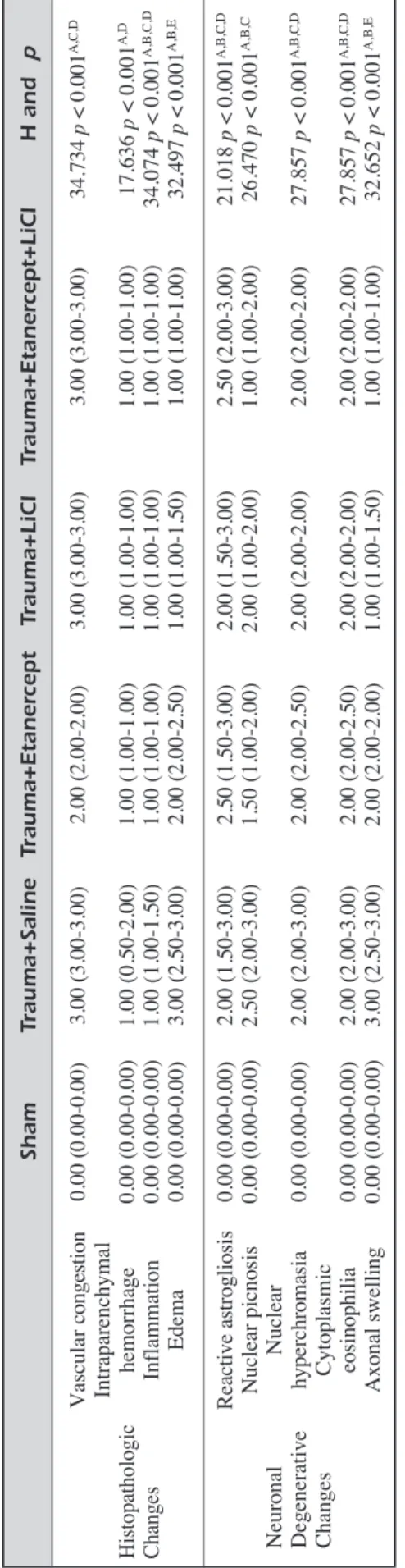
micrometer by light microscopy. The severity of the histopathologic changes and neuronal degen-erative changes was scored viz. (0), no injury; (1), mild; (2), moderate and (3) dense/common. The examination of the histologic changes con-sisted of the evaluation of the following: vascular congestion, intraparenchymal hemorrhage, in-flammation, edema and gliosis. The examination of the neuronal degenerative changes consisted of the evaluation of the following: nuclear py-knosis, nuclear hyperchromasia, cytoplasmic eosinophilia, and axonal swelling18,32-38.
In situ Apoptosis Detection
Cleavage of genomic DNA during apoptosis may yield double-stranded, low molecular-weight DNA strand breaks that can be labeled by terminal deoxynucleotidyl transferase (TdT) which catalyzes polymerization of labeled nu-cleotides to free 3’-OH ends in an template-inde-pendent manner (Terminal deoxynucleotidyl transferase mediated dUTP-biotin nick end label-ing, TUNEL reaction). The quantity of apoptotic cell death was evaluated using an in situ cell death detection kit (Cat. no.S7101; Chemicon In-ternational-ApopTag Plus Peroxidase Kits, Temecula, CA, USA). Firstly, the serial sections were deparaffinized three times in xylene. The sections were then rehydrated through a series of decreasing concentrations of ethanol before the slides were washed in phosphate-buffered saline (PBS). After that, the sections were subjected to partial digestion with proteinase K (20 µg/ml) (Merck) at room temperature for 15 min and were washed 2 times in PBS. Thereafter, the sec-tions were incubated at room temperature in an equilibration buffer. The tissue sections were in-cubated at 37°C for 60 min with the TUNEL re-action mixture (70% rere-action buffer, 30% TdT enzyme) in a humidified chamber in the dark. The slides were agitated and incubated in the stop/wash buffer for 10 min at room temperature. After the incubation, the slides were washed 3 times with PBS (phosphate buffered saline). Then, anti-digoxignenin conjugate was applied to the slides and incubated in a humidified chamber for 30 min at room temperature. The slides were washed 4 times with PBS. 3-3 diaminobenzidine (DAB) was then added onto the slides for 10 min, and the slides were washed in distilled wa-ter three times before drying. For analysis of TUNEL(+) motor neurons located at perilesional region of the cortex, two independent observers
Results
Serum GFAP and Tau Levels
Mean values of venous blood GFAP concen-tration was 0.292 ± 0.0752 for Sham group, 1.042 ± 0.561 for Trauma+Saline group, 0.795 ± 0.165 for Trauma+Etanercept group, 0.961 ± 0.222 for Trauma+LiCl group, 0.856 ± 0.179 for Trauma+Etanercept+LiCl group. Mean values of venous blood GFAP concentration was statisti-cally significantly between groups (p < 0.001) (Table I).
Median (25%-75% percentiles) values of ve-nous blood TAU concentration was 0.00550 (0.00385-0.0152) for Sham group, 0.0533 (0.0388-0.0650) for Trauma+Saline group, 0.0232 (0.0145-0.0462) for Trauma+Etanercept group, 0.0392 (0.0230-0.0447) for Trauma+LiCl group, 0.0439 (0.0240-0.0690) for Trauma+Etan-ercept/LiCl group. Median (25%-75% per-centiles) values of venous blood GFAP concen-tration was statistically significantly between groups (p = 0.009) (Table II).
Histological Results
Etanercept Decreased Edema Axonal Swelling During TBI
Based on H&E staining, histopathological changes were examined on the perilesional re-gion of the cerebral cortex, and a statistically sig-nificant difference was observed between study groups in comparison with Trauma+Saline group with respect to the vascular congestion, intra-parenchymal hemorrhage, inflammation and gliosis. Etanercept remarkably suppressed edema in post-traumatic period (p < 0.001). This finding anti-TNF-α antibody (Santa Cruz Biotechnology,
Santa Cruz, CA, USA; 1:200). After washes in 0.05 M PBS, sections were incubated with bi-otinylated donkey anti-goat (1:200) for 1 h, and avidin-biotin complex (Vector Laboratories Inc., Burlingame, CA, USA) for 1 h. Immunoreactivi-ty was detected with 0.05% DAB and 0.03% H2O2. As negative controls, alternate sections
were processed in parallel without primary anti-body. The specificity of TNF-α antibody was verified using the relative blocking peptides (Santa Cruz Biotechnology) according to the manufacturer’s instruction. Simply, anti-TNF-α 1 µg/ml combined with TNF-α blocking peptides 5 µg/ml were incubated at room temperature for 2 h. Following blocking, the antibody/peptide mixture was applied for immunostaining to veri-fy the specificity of TNF-α antibodies.
Neuronal immunoreactivity was analyzed by counting 5 fields per sections from 4-6 sections for each subject Because of less frequent and more heterogenous immunoreactivity in neu-ronal, glial, and endothelial cells, their TNF-α expression was graded as follows: (0), no im-munoreactivity; (1), trace of immunoreactivity, and (2) increased TNF-α immunoreactivity44.
Statistical Analysis
SPSS version 15.0 (SPSS for Windows Inc., Chicago, IL, USA) was used for statistical analy-sis. Normal distribution of the data was evaluated with Kolmogorov-Smirnov test. Kruskal Wallis and ANOVA Tests with Tukey Multiple Com-parison Tests and Holm Sidak Multiple Compari-son Tests were used to compare the groups. The results were expressed as mean ± standard devia-tion and Median (25-75% percentiles). p < 0.05 was considered statistically significant.
Groups Serum GFAP F and p Sham 0.292 ± 0.0752 Trauma+Saline 1.042 ± 0.5610 Trauma+Etanercept 0.795 ± 0.1650 8.038 p < 0.001A,B,C,D Trauma+LiCl 0.961 ± 0.2220 Trauma+Etanercept +LiCl 0.856 ± 0.1790
Table I.Assessment of serum GFAP concentration (ng/ml) (Mean ± St. Deviation) for all groups.
The statistical difference resulting from the comparison of the Trauma+Saline vs. Sham group (A), Trauma+LiCl vs. Sham group (B), Trauma+Etanercept+LiCl vs. Sham group (C) and Trauma+Etanercept vs. Sham group (D).
Groups Serum TAU F and p Sham 0.0055 (0.0038-0.0152) Trauma+Saline 0.0533 (0.0388-0.0650) Trauma+ Etanercept 0.0232 (0.0145-0.0462) 13.451 p = Trauma+LiCl 0.0392 (0.0230-0.0447) 0.009 A Trauma+ Etanercept+LiCl 0.0439 (0.0240-0.0690)
Table II.Assessment of median serum TAU concentrations (ng/ml; 25%-75% percentiles) for all groups.
The statistical difference resulting from the comparison of the Trauma+Saline vs. Sham group (A)
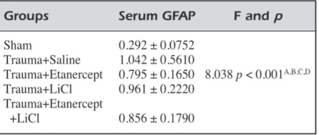
provides further support to anti-inflammatory ef-fect of Etanercept (Figure 1 a-c) (Table III).
When neurodegenerative changes are exam-ined, it was found that nuclear pyknosis, nuclear hyperchromacia and cytoplasmic eosinophilia sig-nificantly increased. However, in comparison with Trauma+Saline group, no therapeutic finding was found in experiment groups. Significant improve-ment was observed only for axonal swelling in Trauma+Etanercept and Trauma+Saline groups (p
< 0.001). This finding also provides further sup-port to neuronal injury relieving effect of Etaner-cept (Figure 1 d, e) (Table III).
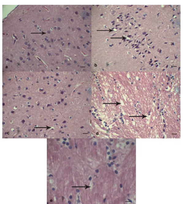
Etanercept and LiCl Decreased Neuronal and Glial Apoptosis During TBI
When TUNEL(+) motor neurons are counted in perilesional region of cortex, a significant in-crease in observed in number of apoptotic motor neurons in Trauma+Saline group. Although
de-Figure 1. Neuronal morphology indicated by hematoxylin-eosin staining. A, Histological appearances of normal brain parenchyma in Sham group. Neurons have a large, round nucleus with a single prominent nucleolus (arrow). H&E, Scale Bar 20 µm. B, Edema and pyknosis in Trauma+Saline group. Scattered clusters of angular ischemic neurons with pyknotic nuclei and vacuolization (edema) in the neuropil (arrow). H&E, Scale Bar 20 µm. C, Decrease in edema (arrow) and pyknosis in Trauma+Etanercept group. H&E, Scale Bar 20 µm. D, Dense axonal swelling (arrow) in Trauma+Saline group. H&E, Scale Bar 20 µm. E, Moderate axonal swelling (arrow) in Trauma+Etanercept group. H&E, Scale Bar 20 µm.
crease was observed in number of TUNEL(+) motor neurons in Trauma+Etanercept group, it could not reach statistically significance level in comparison with Trauma+Saline group. In LiCl alone or combination of LiCl and Etanercept in post-traumatic period, a slight decrease is ob-served in number of apoptotic motor neurons in comparison with Trauma+Saline group. Findings were not statistically significant although we ob-served anti-apoptosis effect of LiCl (Figure 2 a-e) (Table IV).
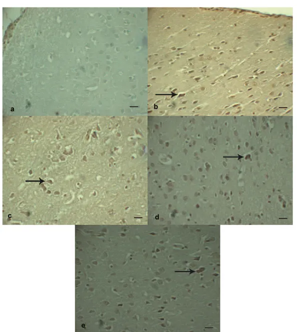
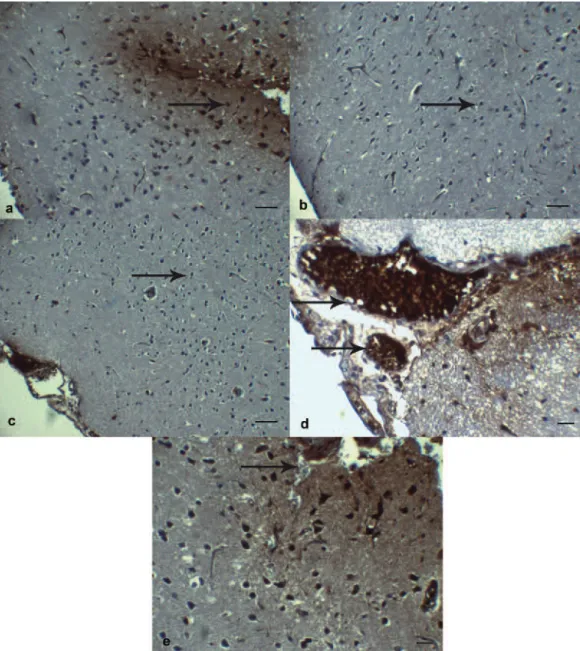
Etanercept and LiCl Decreased GFAP Expression During TBI
When the GFAP(+) protoplasmic astrocytes were counted in the perilesional region of the cortex, GFAP(+) cell number was seriously high in Trauma+Saline group and statistically signifi-cant increase was observed in comparison with Sham group (p < 0.001). In comparison with Trauma+Saline group, a statistically significant decrease was present in GFAP(+) cell count of Trauma+Etanercept, Trauma+LiCl and Trau-ma+Etanercept+LiCl groups (p < 0.001) (Figure 3 a-e) (Table IV).
Etanercept ve LiCl Decreased TNF Activity During TBI
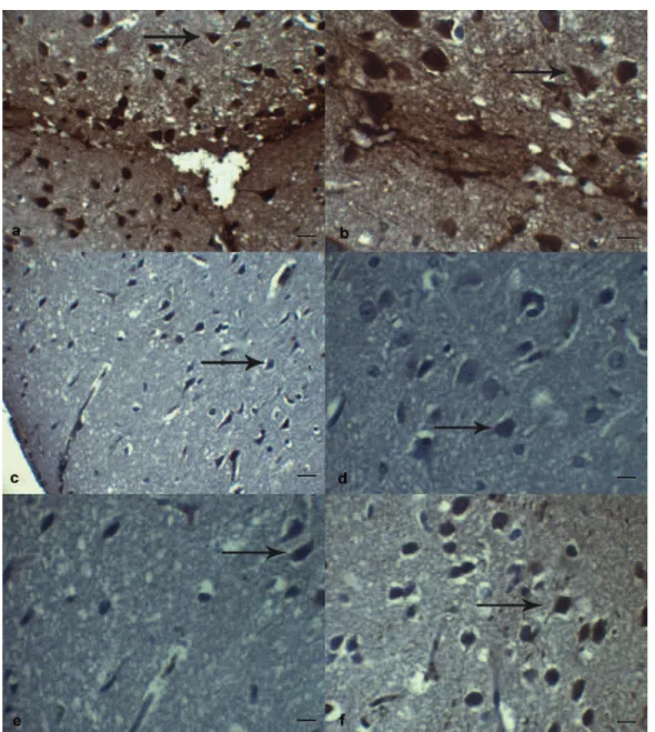
When TNF-α immunoreactivity was evaluat-ed, a statistically significant decrease was noted in TNF-α immunoreactivity of motor neurons in Travma+Etanercept group in comparison with Trauma+Saline group (p < 0.001) (Figure 4 a-d). When Trauma+Etanercept group was compared with combination group, the group of Etanercept alone had significant decrease in TNF-α im-munoreactivity (p < 0.001) (Figure 4 e, f). Such effect of Etanercept, a TNF-α antagonist, is an expected finding.
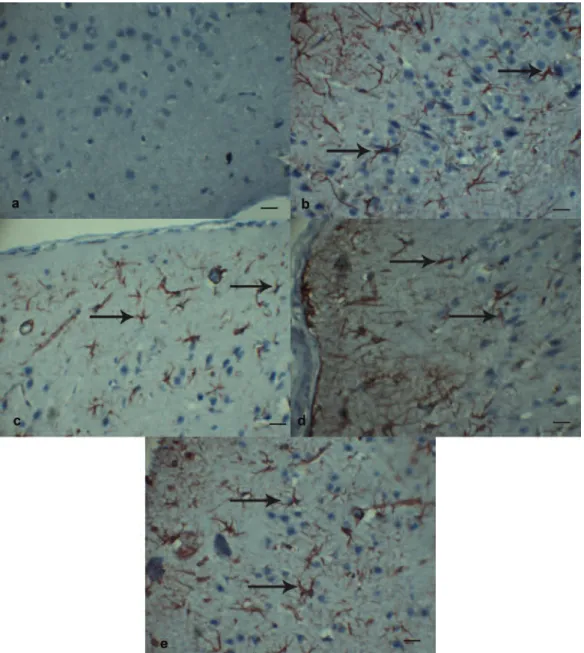
When glial immunoreactivity was examine, glial immunoreactivity significantly decreased in Trama+Etanercept group in comparison with Trauma+Saline group (p < 0.001). LiCl did not have any suppressive effect on TNF-α im-munoreactivity. In comparison with Trauma+Saline group, statistically significant de-crease was observed again in Trauma+Etaner-cept+LiCl group (p < 0.001) (Figure 5 a-c).
When endothelial cells were taken into consid-eration, there was decrease in TNF-α immunore-activity in Trauma+Etanercept group in son with Trauma+Saline group. When compari-son was made between Trauma+LiCl group and Trauma+Etanercept+LiCl group, a significant
S h a m T ra u m a + S a li n e T ra u m a + E ta n e rc e p t T ra u m a + L iC l T ra u m a + E ta n e rc e p t+ L iC l H a n d p Va sc ul ar co ng es tio n 0.0 0 ( 0.0 0-0.0 0) 3.0 0 ( 3.0 0-3.0 0) 2.0 0 ( 2.0 0-2.0 0) 3.0 0 ( 3.0 0-3.0 0) 3.0 0 ( 3.0 0-3.0 0) 34 .73 4 p < 0.0 01 A, C, D In tra pa re nc hy m al Hi sto pa th ol og ic he m or rh ag e 0. 00 (0 .0 0-0. 00 ) 1. 00 (0 .5 0-2. 00 ) 1. 00 (1 .0 0-1. 00 ) 1. 00 (1 .0 0-1. 00 ) 1. 00 (1 .0 0-1. 00 ) 17 .6 36 p < 0. 00 1 A, D Ch an ge s In fla m m ati on 0. 00 (0 .0 0-0. 00 ) 1. 00 (1 .0 0-1. 50 ) 1. 00 (1 .0 0-1. 00 ) 1. 00 (1 .0 0-1. 00 ) 1. 00 (1 .0 0-1. 00 ) 34 .0 74 p < 0. 00 1 A, B, C, D Ed em a 0. 00 (0 .0 0-0. 00 ) 3. 00 (2 .5 0-3. 00 ) 2. 00 (2 .0 0-2. 50 ) 1. 00 (1 .0 0-1. 50 ) 1. 00 (1 .0 0-1. 00 ) 32 .4 97 p < 0. 00 1 A, B, E Re ac tiv e a str og lio sis 0. 00 (0 .0 0-0. 00 ) 2. 00 (1 .5 0-3. 00 ) 2. 50 (1 .5 0-3. 00 ) 2. 00 (1 .5 0-3. 00 ) 2. 50 (2 .0 0-3. 00 ) 21 .0 18 p < 0. 00 1 A, B, C, D Nu cle ar p icn os is 0. 00 (0 .0 0-0. 00 ) 2. 50 (2 .0 0-3. 00 ) 1. 50 (1 .0 0-2. 00 ) 2. 00 (1 .0 0-2. 00 ) 1. 00 (1 .0 0-2. 00 ) 26 .4 70 p < 0. 00 1 A, B, C Ne ur on al Nu cle ar De ge ne ra tiv e hy pe rc hr om as ia 0. 00 (0 .0 0-0. 00 ) 2. 00 (2 .0 0-3. 00 ) 2. 00 (2 .0 0-2. 50 ) 2. 00 (2 .0 0-2. 00 ) 2. 00 (2 .0 0-2. 00 ) 27 .8 57 p < 0. 00 1 A, B, C, D Ch an ge s Cy to pl as m ic eo sin op hi lia 0. 00 (0 .0 0-0. 00 ) 2. 00 (2 .0 0-3. 00 ) 2. 00 (2 .0 0-2. 50 ) 2. 00 (2 .0 0-2. 00 ) 2. 00 (2 .0 0-2. 00 ) 27 .8 57 p < 0. 00 1 A, B, C, D Ax on al sw ell in g 0. 00 (0 .0 0-0. 00 ) 3. 00 (2 .5 0-3. 00 ) 2. 00 (2 .0 0-2. 00 ) 1. 00 (1 .0 0-1. 50 ) 1. 00 (1 .0 0-1. 00 ) 32 .6 52 p < 0. 00 1 A, B, E T a b le I II .H ist ol og ica l a ss es sm en t o f m ot or n eu ro ns in p er ile sio na l c or tex an d m ed ian v alu es (2 5% -7 5% p er ce nt ile s) fo r a ll gr ou ps . no in ju ry : 0 , m ild :1 , m od er ate :2 , d en se /co m m on :3 Th e sta tis tic al di ffe re nc e re su lti ng fr om th e co m pa ris on o f t he T ra um a+ Sa lin e vs . S ha m g ro up (A ), Tr au m a+ Et an er ce pt v s. Sh am g ro up (B ), Tr au m a+ Li Cl v s. Sh am g ro up (C ), Tr au m a+ Et an er ce pt +L iC l v s. Sh am g ro up (D ) a nd T ra um a+ Sa lin e v s. Tr au m a+ Et an er ce pt g ro up (E ).
decrease was observed in TNF-α immunoreac-tivity in combined group (p < 0.001). We believe that decrease was secondary to effect of Etaner-cept. When comparison was made between Trau-ma+Saline group and Trauma+Etanercept+LiCl group, statistically significant decrease was ob-served again in TNF-α immunoreactivity in combined group (p < 0.001) (Figure 5 d, e). When comparison was made between Trau-ma+Etanercept group and Trauma+LiCl group,
statistically significant decrease was observed again in TNF-α immunoreactivity in Trauma+Li-Cl group (p < 0.001).
In general terms, when results are examined, we found that TNF-α immunoreactivity increase in perilesional cortex 24 hours after TBI. TNF-α activity decreased after Etanercept and LiCl were administered, and tissue edema and axonal swelling were also decreased, resulting with fa-cilitation of alleviating brain damage.
Figure 2. TUNEL immunohistochemistry staining in the perilesional region of the cortex of the brain following TBI. A, Sham group rats showing few/not TUNEL(+) apoptotic cells. TUNEL, Scale Bar 20 µm. B, Trauma+Saline group rats showing more TUNEL(+) apoptotic cells (arrow) than Sham and other groups. TUNEL, Scale Bar 20 µm. C, TUNEL(+) apoptotic cells (arrow), in Trauma+Etanercept group. TUNEL, Scale Bar 20 µm. D, TUNEL(+) apoptotic cells (arrow), in Trauma+LiCl group. TUNEL, Scale Bar 20 µm. E, TUNEL(+) apoptotic cells (arrow), in Trauma+Etanercept+LiCl group. TUNEL, Scale Bar 20 µm.
Discussion
TBI is a leading cause of death and disability worldwide14. Immediately following TBI, the
di-rect trauma and lack of blood flow cause necrotic neuronal death4,10,23. Neuronal cell death,
includ-ing apoptosis, in general peaks after 24 to 48 h
after transient middle cerebral artery occlusion44.
Apoptosis in the traumatically injured brain oc-curs not only at the impact site but also as a re-sult of secondary brain inre-sults such as intracra-nial hypertension, hypoxia, or disturbances of microcirculation3. Genovese et al45 using the
TUNEL coloration they have clearly confirmed
Figure 3. Immunohistochemistry for GFAP in the perilesional region of the cortex of the brain following TBI. A, Appearance of protoplasmic astrocytes in Sham group. Note that the territories of astrocyte processes do not overlap and that many astrocytes do not express detectable levels of GFAP. GFAP, Scale Bar 20 µm. B, Severe diffuse reactive astrogliosis with pronounced up regu-lation of GFAP expression, astrocyte hypertrophy, astrocyte proliferation and pronounced overlap of astrocyte processes resulting in disruption of individual astrocyte domains in Trauma+Saline group (arrow). GFAP, Scale Bar 20 µm. C, Mildly reactive as-trogliosis in Trauma+Etanercept group. Note that the territories of protoplasmic astrocyte processes do not overlap (arrow) and that many astrocytes do not express detectable levels of GFAP. GFAP, Scale Bar 20 µm. D, Moderately reactive astrogliosis in which most (if not all) protoplasmic astrocytes have up regulated expression of GFAP and exhibit cellular hypertrophy, but with preservation of individual astrocyte domains and without pronounced overlap of astrocyte processes in Trauma+LiCl group (ar-row). GFAP, Scale Bar 20 µm. E, GFAP(+) protoplasmic astrocytes in Trauma+Etanercept+LiCl group. GFAP, Scale Bar 20 µm.
that apoptosis is present in the perilesional area after spinal cord injury (SCI) and that the combi-nation treatment with etanercept and dexametha-sone (DEX) attenuates the degree of apoptosis. Moreover, this attenuation is due to the preven-tion of loss of the anti-apoptotic way and reduc-tion of pro-apoptotic pathway activareduc-tion with a mechanism still to discover. In the current study, we determined apoptosis in perilesional cortical region of brain tissue with TUNEL staining method following TBI. Our findings were not statistically significant in comparison with Trau-ma+Saline group, although severity of apoptosis decreased in Etanercept, LiCl and Etanercept+Li-Cl groups.
Neutrophils, macrophages and activated mi-croglia may act as scavenger cells to remove cel-lular debris and release cytotoxic or neurotrophic molecules into the injured tissue7-9. However,
even greater apoptotic neuronal loss occurs hours and days later, caused by secondary injury from cerebral ischemia/hypoxia as well as inflamma-tory and oxidative stress. Inhibition of cytokine production by anti-inflammatory agents, includ-ing minocycline and statins, has been shown to reduce TBI. Therefore, the major focus of TBI research should be protection of neurons from apoptotic cell death by reducing the secondary injury of inflammation and oxidative stress12.
Brain inflammation represents only one of the numerous processes activated after TBI. The ma-jor effectors in this cascade are the pro-inflam-matory cytokines that are usually released within minutes after challenge because they are stored intracellular as precursor proteins eventually modified into active molecules3. Our study
showed that cortical levels of TNF-α was signifi-cantly induced by TBI at 24 h after trauma. We have clearly demonstrated, by immunohisto-chemistry, a significant increase of positive
staining for TNF-α in Trauma+Saline group compared with Sham group animals. On the con-trary, no significant expression of TNF-α was observed in the brain tissue sections obtained from Etanercept-treated rats. In this study, we observe that Etanercept treatment reduces the ex-pression of TNF-α. Finally, in this work, we demonstrate that Etanercept treatment signifi-cantly reduced the TBI-induced brain tissues al-teration and improve edema and axonal swelling. Histopathological studies following severe closed head injury in our model demonstrated a diffuse injury that affected the perilesional cor-tex. Vascular congestion was noted throughout the brain. The presence and pattern of diffuse vascular congestion, intraparenchymal hemor-rhage, inflammation, edema, and reactive as-trogliosis were similar at the high levels of in-jury. In our diffuse injury model, rats in the Trauma+Saline group had appreciable cerebral edema noted histologically 24 hours after injury.
Axonal injury is one of the most common pathologies resulting from brain trauma, where the extent of axonal damage is thought to play a major role in outcome. Initial swelling and further dam-age of injured axons may result from a change in ionic homeostasis. As a result of these ionic changes, osmotic swelling of axons may occur shortly following injury due to sodium influx, while the increase in intracellular calcium may in-duce the deleterious activation of proteases lead-ing to additional cytoskeletal damage. Although these processes are thought to play important roles in the ultimate demise of injured axons, swelling alone is not considered the principle feature of traumatically injured axons. Rather, accumulation of axonal transport proteins in swollen regions of axons is the clear signature of catastrophic dam-age13. Beginning from only hours following
trau-ma, both disconnected axonal bulbs and
still-con-Groups Tunel(+) GFAP(+)
Sham 90.00 (81.00-134.00) 276.00 (248.50-276.00)
Trauma+Saline 1286.50 (479.00-2289.00) 2021.00 (1569.00-2632.00) Trauma+Etanercept 1107.50 (879.00-1369.50) 930.00 (770.50-1017.00) Trauma+LiCl 795.50 (663.00-968.50) 996.50 (891.00-1182.00) Trauma+Etanercept+LiCl 781.00 (541.50-961.50) 1060.00 (693.00-1170.50) H and p 21.035 p < 0.001A,B,C,D 18.941 p < 0.001A,B,C,D,E,F,G
Table IV.Median values of total TUNEL(+) motor neurons in perilesional cortex and total GFAP(+) protoplasmic astrocytes for all groups (25%-75% percentiles).
The statistical difference resulting from the comparison of the Trauma+Saline vs. Sham group (A), Trauma+Etanercept vs. Sham group (B), Trauma+LiCl vs. Sham group (C), Trauma+Etanercept+LiCl vs. Sham group (D), Trauma+Saline vs. Trauma+Etaner-cept group (E), Trauma+Saline vs. Trauma+LiCl group (F), and Trauma+Saline vs. Trauma+EtanerTrauma+Etaner-cept+LiCl group (G).
nected axonal swellings can be identified13,14 In
the current study, we observed axonal swelling in Trauma+Saline group. The finding resulted from change in ionic homeostasis following TBI. In Trauma+Etanercept group, we observed that ax-onal swelling significantly decreased.
Microtubule-associated protein Tau is local-ized primarily in the axonal compartment of neu-rons. Functionally, Tau binds to axonal micro-tubules resulting in the formation of microtubule bundles. These bundles form important structural elements of the axonal cytoskeleton and are criti-cal elements in the axoplasmic flow of proteins between the nerve terminal and neuronal cell body15. Tau protein that can be measured in
serum is released from CNS neurons after TBI. Liliang et al5 show that Tau protein levels also
rise slightly in Sham group rats in the first 6 h af-ter TBI. However, the rise in Sham group did not reach statistical significance. As with any surgery, there was possible some additional in-jury to brain and Tau protein levels increased. This increase in total Tau is consistent with stud-ies employing other biomarkers such as c-Tau, S-100 and neuron-specific enolase, where in-creased levels were observed within 6 h after TBI in rats5. Wunderlich et al16reported that Tau
protein concentrations in serum showed a contin-uous increase from admission to last measure-ment 120 h after stroke and were elevated in 27% of their patients within the first day and in 41% until day 5. Tau protein levels rapidly ele-vate in the CSF after TBI. The peak has been re-ported after 2 days or 1 week. Although serum Tau protein can be measured following TBI, the peak and progression are unclear5,15,16. In the
cur-rent study, statistically significant increase in serum Tau level 24 hours after TBI reflects both extensive neuronal damage in the brain as well as a compromised BBB. However, we determined that post-TBI Etanercept and LiCl administration did not significantly decrease serum Tau level.
Hypertrophic astrocytes with prominent im-munoreactive processes were scattered diffusely throughout the region of cell body injury. These changes, which appeared to involve all astrocytes in the area, are consistent with classic astrocytic hypertrophy in response to local injury46. At the
immediate vicinity of injury, reactive astrocytes interweave their processes to form a barrier termed “anisomorphic gliosis”. This glial scar can be an impediment to regenerating axons4,7-12.
Al-though the presence of glial activation in the in-jured brain certainly represents a disturbance of
S h a m T ra u m a + S a li n e T ra u m a + E ta n e rc e p t T ra u m a + L iC l T ra u m a + E ta n e rc e p t+ L iC l H a n d p Ne ur on 0 47 .2 0 (4 1. 60 -5 2. 95 ) 18 .2 0 (1 4. 85 -4 6. 80 ) 57 .4 0 (5 3. 25 -6 5. 75 ) 27 .4 0 (2 3. 90 -9 .4 0) 45 .8 0 (4 2. 85 -5 4. 70 ) 7. 67 8 p = 0. 10 4 1 2. 00 (1 .3 0-2. 65 ) 3. 40 (0 .0 0-6. 25 ) 1. 40 (0 .8 5-3. 05 ) 4. 80 (4 .6 5-12 .1 5) 18 .2 0 (1 4. 85 -4 6. 80 ) 15 .2 43 p = 0. 00 4 D, F 2 0. 00 (0 .0 0-0. 45 ) 26 .0 0 (1 8. 45 -2 6. 65 ) 0. 40 (0 .0 0-5. 90 ) 6. 00 (5 .8 0-12 .0 5) 3. 80 (1 .2 0-8. 15 ) 16 .1 79 p = 0. 00 3 A, E Gl ia 0 20 .6 0 (1 9. 05 -2 9. 45 ) 8. 80 (5 .6 5-9. 75 ) 14 .4 0 (1 2. 20 -1 7. 40 ) 15 .0 0 (1 1. 10 -1 7. 00 ) 16 .4 0 (1 3. 75 -1 6. 95 ) 9. 02 7 p < 0. 00 1 A, B, C, D 1 4. 20 (3 .3 0-5. 05 ) 0. 00 (0 .0 0-0. 00 ) 3. 60 (2 .6 0-4. 55 ) 0. 00 (0 .0 0-0. 05 0) 0. 00 (0 .0 0-0. 40 ) 19 .8 70 p < 0. 00 1 A 2 1. 80 (0 .0 0-6. 05 ) 13 .2 0 (1 1. 60 -1 5. 40 ) 5. 40 (2 .5 0-9. 10 ) 10 .0 0 (8 .0 5-15 .9 0) 6. 40 (5 .8 0-6. 95 ) 8. 38 7 p < 0. 00 1 A, C, E, H En do th eli um 0 3. 80 (2 .7 0-6. 15 ) 1. 00 (0 .0 0-2. 00 ) 2. 20 (1 .9 5-4. 25 ) 0. 40 (0 .1 5-0. 70 ) 1. 60 (0 .7 0-2. 60 ) 14 .0 09 p = 0. 00 7 C 1 0. 00 (0 .0 0-2. 15 ) 0. 00 (0 .0 0-0. 00 ) 7. 40 (5 .7 5-11 .5 5) 1. 80 (0 .0 0-1. 85 ) 8. 20 (6 .3 5-9. 35 ) 17 .7 10 p < 0. 00 1 B, D, E, G, H, J 2 0. 00 (0 .0 0-0. 45 ) 3. 20 (1 .2 0-6. 60 ) 1. 40 (0 .0 0-2. 50 ) 6. 60 (6 .2 0-7. 00 ) 0. 00 (0 .0 0-1. 80 ) 14 .2 08 p =0 .0 07 C, G T a b le V .M ed ian v alu es o f T NF -α (+ ) i m m un or ea cti vi ty in m ot or n eu ro ns in p er ile sio na l c or tex , g lia an d en do th eli um fo r a ll gr ou ps (2 5% -7 5% p er ce nt ile s). 0: n o im m un or ea cti vi ty ; 1 : t ra ce im m un or ea cti vi ty ; 2 : i nc re as ed im m un or ea cti vi ty Th e sta tis tic al di ffe re nc e re su lti ng fr om th e co m pa ris on o f t he T ra um a+ Sa lin e vs . S ha m g ro up (A ), Tr au m a+ E tan er ce pt v s. Sh am g ro up (B ), Tr au m a+ Li Cl v s. Sh am g ro up (C ), Tr au m a+ Et an er ce pt +L iC l v s. Sh am g ro up (D ), Tr au m a+ Et an er ce pt v s. Tr au m a+ Sa lin e gr ou p (E ), Tr au m a+ Et an er ce pt v s. Tr au m a+ Et an er ce pt +L iC l g ro up (F ), Tr au m a+ Li Cl v s. Tr au m a+ Et an er ce pt +L iC l g ro up (G ), Tr au m a+ Et an er ce pt +L iC l v s. Tr au m a+ Sa lin e g ro up (H ) a nd T ra um a+ Et an er ce pt v s. Tr au m a+ Li Cl g ro up .
normal brain physiology or predictor of a patho-logical condition, it is still controversial as to whether reactive gliosis is harmful or beneficial to the acutely injured brain18. Reactive astrocytes
perform roles essential for the preservation of neural tissue and restriction of inflammation after mild or moderate focal brain injury, and thus play
a major part in determining outcome after TBI. The roles played by reactive astrocytes after TBI are not well understood. Because reactive astro-cytes are ubiquitous in damaged CNS tissue, they are often regarded as uniformly harmful, causing toxic edema, provoking inflammation, releasing cytotoxins and forming scars that serve no
pur-Figure 4. Photomicrographs showing TNF-α immunoreactivity at motor neurons in the perilesionel cortex. A, Note that there was a marked rise in the number of TNF-α positive cells at motor neurons (arrow) in Trauma+Saline group. TNF-α, Scale Bar 20 µm. B, A substantial increase in TNF-α production was found at motor neurons (arrow) in Trauma+Saline group. TNF-α, Scale Bar 10 µm. C, Treatment with Etanercept significantly reduced in TNF-α immunoreactivity at motor neurons (arrow) in Trauma+Etanercept group. TNF-α, Scale Bar 20 µm. D,, The number of TNF-α positive cells in motor neurons (arrow) was significantly reduced in Trauma+Etanercept group. TNF-α, Scale Bar 10 µm. E,, The number of TNF-α positive cells at motor neurons (arrow) was significantly reduced in Trauma+Etanercept group compared with Trauma+Etanercept+LiCl group TNF-α, Scale Bar 10 µm. F, Significantly reduced TNF-α immunoreactivity at motor neurons (arrow) in Trauma+Etanercept+LiCl group. TNF-α, Scale Bar 10 µm.
pose but to inhibit axonal regeneration. In the context of moderate contusive brain injury reac-tive astrocytes were not harmful, but rather played roles essential for preserving cortical neu-rons and tissue integrity, and for restricting post-injury inflammation. Reactive astrocytes play im-portant roles in restricting the infiltration of in-flammatory cells into viable neural tissue after CNS injuries in vivo. In this regard it is of interest
that astrocytes may be lost early after TBI2. TBI
is accompanied by secondary or delayed brain edema and these edema processes are also be-lieved to participate in the pathogenesis of TBI47.
Our findings demonstrated that reactive astro-cytes played major role in post-TBI period. Post-TBI enhanced reactive astrogliosis antagonized edema and inflammation in all groups and it had protective effect on cortical neurons.
Figure 4. α immunoreactivities at glial and endothelial cells in the perilesionel cortex. A, A substantial increased TNF-α expression was found at glial cells (arrow) in Trauma+Saline group.TNF-TNF-α, Scale Bar 50 µm. B, Treatment with Etanercept significantly reduced in TNF-α immunoreactivity at glial cells (arrow) in Trauma+Etanercept group.TNF-α, Scale Bar 50 µm. C,The combination therapy with Etanercept and LiCl significantly reduced the TNF-α immunreactivity at glial cells (arrow). TNF-α, Scale Bar 50 µm. D, Significant increased TNF-α expression was found at endothelial cells (arrow) in Trauma+Saline group.TNF-α, Scale Bar 20 µm. E, TNF-α immunoreactivity significantly reduced at endothelial cells (arrow) in Trauma+Etanercept+LiCl group.TNF-α, Scale Bar 20 µm.
The astrocytic reaction described above may play a role in the protection of the cortex after in-jury. It is known that astrocytes can synthesize trophic factors. These neurotrophins in turn may protect the surrounding neurons. Thus, it is likely that the phenomenon depicted here could be part of a series of cellular events that may result in neuronal protection48. Under normal condition,
the BBB gates the entrance of immune cells into the CNS. Barrier disruption is associated with uncontrolled inflammatory cell entry into the CNS parenchyma. Astrocytes are considered to play a prominent role in controlling this process, since their end-feet processes are in intimate con-tact with the brain micro-vessels and are known to form an essential functional component of the BBB22. In the current study, we observed that
glial cells, which are located around micro-ves-sels, intensely expressed TNF-α 24 hours after TBI and number of glial cells rapidly increased. We believed that this finding may play a signifi-cant role in impairment of BBB and inducing cerebral inflammatory response after TBI.
GFAP is an astrocyte specific intermediate fil-ament protein whose expression is required for fibrous astrocyte normal function including maintenance of CNS white matter and BBB in-tegrity. Increased GFAP expression is a hallmark of reactive astrocytes and this cytoskeletal pro-tein contributes to a barrier effect of the glial scar for axonal extension2,6,17,18. Though not all
astro-cytes express GFAP, those reacting to injury show a rise in the content of these filaments. This makes GFAP immunohistochemistry a very use-ful marker to study gliosis. Different models of brain injury have been studied using this mark-er48. These data correlate with an elevation in
GFAP gene expression (mRNA) which could be detected in response to mild cortical contusions in animals19,20. In this report, serum GFAP level
significantly increased in Trauma+Saline group. As indicated by tissue immunohistochemistry, GFAP(+) finding of Trauma+Saline group sig-nificantly increased in Etanercept, LiCl and Etanercept+LiCl groups in comparison with Trauma+Saline group.
The brain has been traditionally described as an “immune-privileged” organ due to its isola-tion from the peripheral immune system by the BBB. However, recent brain injury studies18have
indicated that resident brain cells are capable of synthesizing a wide variety of pro-inflammatory mediators necessary for mounting a neuroinflam-matory response. The inflamneuroinflam-matory response in
the CNS involves the participation of different cellular types of the immune system and resident cells of the CNS, adhesion molecules, cytokines, and chemokines among other proteic compo-nents. During neuroinflammation, chemotaxis is an important event in the recruitment of cells to the CNS. If this process is not controlled but is prolonged, inflammation loses its repairing func-tion and can be the cause of damage28.
In addition to chemokines, various cytokines have also been reported to be expressed follow-ing TBI, includfollow-ing TNF-α associated with acti-vated microglia and astrocytes that may initiate the inflammatory process and are thought to be the primary sources of cytokines and chemokines that play a role in brain inflammatory respons-es4,22. TNF-α is known to be bioactive both as a
soluble and as a transmembrane protein. TNF-α is released during and involved in the inflamma-tory response following TBI. Elevated levels of TNF-α have been associated with the pathologi-cal effects of a variety of infectious, neurologi-cal, neurodegenerative, and neurotoxic condi-tions. In contrast to these findings, however, some evidence suggests that TNF-α also plays a neuroprotective role following TBI. TNF-α has also been reported to participate actively in BBB breakdown as well as acute post-traumatic apop-totic and necrotic cell death1,6,17,22,23,47,49.
Both necrotic and apoptotic mechanisms of cell death after SCI have been well and exten-sively characterized in animal SCI models. It is has been thought that microglial cells might be the source of cytotoxic cytokines, such as TNF-α, that kill oligodendrocytes. Within 1 h after SCI, increased synthesis and/or secretion of TNF-α is detectable at the injury site28. Pettigrew
et al50 emphasized TNF-α is frequently
unde-tectable in quiescent neural cell populations, be-coming up-regulated in neurons and astroglial cells only after ischemic or post-traumatic stress. They speculate that the greater infarct volume observed in the transgenic animal resulted from chronic overexpression of TNF-α protein within the brain, causing receptor-mediated caspase ac-tivation that promoted neuronal apoptosis. Mao et al51 found that TNF-α expression is increased
in neonatal stroke and may be involved in neu-ronal apoptosis. TNF-α expression is increased in a wide range of CNS disorders such as trauma and ischemia. In this study, TNF-α expression increased in the ischemic cortex after stroke. In the current study, paralleled increase in remark-able TNF-α(+) and TUNEL(+) staining in
Trau-ma+Saline group indicated that TNF-α was in-volved in apoptotic mechanisms. After Etaner-cept, a α antagonist, is administered, TNF-α(+) staining and decreased apoptosis had inter-correlating results.
In particular, both IL-1β and TNF-α mRNA levels have been shown to peak 1-3 h following experimental TBI in rats18. In animal models of
cerebral ischemia, high levels of TNF-α have been found after global and focal ischemic injury50. Hang et al52showed that TNF-α and IL-6
levels were significantly increased in the sur-rounding area of injured brain by 3 h, maximal at 24 h and lasted for 7 days following TBI. Chen et al. demonstrated that pre-administration of lovas-tatin significantly reduced post-CCI TNF-α and IL-1β mRNA and protein levels. The decrease in cytokine levels was paralleled by a reduction of brain damage and neurological deficits. TNF-α di-rectly disturbs BBB integrity, leading to cerebral edema and leukocyte infiltration whereas inhibit-ing TNF-α synthesis or neutralizinhibit-ing TNF-α activi-ty reduces development of edema and neuronal degeneration and improves neurological outcome in various brain injury models. Our results are consistent with that of above specified study. In the current study, after Etanercept and LiCl were administered, decreased TNF-α activity was asso-ciated with tissue edema and axonal swelling and it facilitated regression of cerebral damage.
Kita et al47indicated that TNF-α was detected
1 h after impact at the glial cells such as astro-cytes, and inflammatory cells such as intravascu-lar leukocyte and perivascuintravascu-lar monocyte-derived cells in the hippocampus and parasagittal cortex. In addition immunoreactions for TNF-α were de-tected at the apical plasma membrane of the cap-illary endothelium and lysosomes of the edema-tous changes occurred in the vascular feet of as-trocytes 1 h after impact. In the current study, TNF-α activity of glial and endothelial cells is consistent with results of this study. Histopatho-logically remarkable vascular congestion, intra-parenchymal hemorrhage, inflammation, neu-ronal necrosis, reactive astrogliosis and edema were observed 24 hours after TBI. In addition, neuronal cell bodies were shrunken and eosinophilic, and nuclei were pyknotic. Astro-cytes increase in number and size in response to injury and swell after brain injuries after TBI. GFAP of immunohistochemical analysis demon-strated astrocyte enlargement and increased num-ber in the perilesional cortex. TBI activates an inflammatory reaction initiated by the release of
TNF-α has been observed in the rat brain. The present study also demonstrates that TNF-α con-tribute to brain inflammation following the me-chanical traumatic damage in the rat brain. Etan-ercept achieved a significant decrease in edema and axonal swelling. LiCl alone and combination of LiCl and Etanercept maintained the decrease.
Clinical investigations have supported that ele-vated TNF-α levels in human serum and CSF af-ter TBI. These studies have also provided evi-dence that the pharmacological inhibition of TNF-α after head injury mediates neuroprotec-tive effects. Studies in animals have provided key evidence that antagonizing TNF-α is a viable therapeutic strategy. Biological agents that are currently available include three agents that de-crease the activity of TNF-α (infliximab, adali-mumab, and etanercept). The major therapeutic goal when administering TNF-α antagonists is to eliminate the surplus of TNF-α from the circula-tion and from sites of inflammacircula-tion28,45. There
are few reports on the head truma with Etaner-cept1,12,53. These studies are promising, but some
author criticized that this drug has a limited transportation to the neural tissue due to the BBB54. However, the BBB disruption appear
af-ter the head trauma but it can be used differently for poorly transporting drugs55-57. Marchand et
al58 show that immediate treatment with
Etaner-cept reduces microglial activation. Genovese et al45 have demonstrated that treatment with
Etan-ercept at a dose of 5 mg/kg reduced the post-in-flammatory reaction as well as the motor dys-function associated with SCI. They have clearly demonstrated a significant increase of TNF-α and IL-1β in SCI. On the contrary, no significant expression of TNF-α and IL-1β was observed in the spinal cord sections obtained from SCI-oper-ated mice which received the combination treat-ment with Etanercept and DEX. In the current study, TNF-α expression intensely increased in neuron, glial and endothelium in Trauma+Saline group 24 hours after TBI. In addition, similar to results of other investigators, we observed that TNF-α positivity significantly decreased in en-dothelial cells after Etanercept, TNF-α antago-nist, was administered. Significant decrease in TNF-α positivity was also observed in neurons and glial cells. We believe that TNF-α accompa-nies pathogenesis of secondary brain damage, which involves astrogliosis and apoptosis. These results suggest that TNF-α might play a central role in the inflammatory response that leads to secondary insults after TBI.
Lithium have neuroprotective and neuroregen-erative properties59. Lithium has emerged as a
ro-bust neuroprotective agent that prevents apoptosis of neurons through multiple mechanisms. Lithium up-regulates anti-apoptotic Bcl-2 and down-regu-lates pro-apoptotic p53 and Bax. Lithium has an astonishing range of activity on the cells. In fact, it appears almost designed to turn on multiple cellu-lar mechanisms and genes that protect the cells against toxins, stress, ischemia, and injury. At the same time that it protects cells, it also turns genes on for growth factors, cellular repair, and regener-ation29. Su et al30showed that lithium treatment at
the therapeutic concentration (1 mM) is able to en-hance the neuronal differentiation of NPCs in
vit-ro and after transplantation into the avulsed
ven-tral horn of adult rats. They revealed the enhanc-ing effect of Lithium on proliferation and neuronal differentiation of NPCs in the adult rat spinal cord. In this paper, we observed a sigificant decrease in TNF-α positivity and GFAP positivity after LiCl was administered. Those results suggest that LiCl acts like neuroprotective and neuro-regenetative agents and it accompanies decreased apoptosis in LiCl group.
Conclusions
This study is planned to prevent post-trau-matic secondary tissue damages in rat diffuse severe brain injury model, which is induced by alone or combined administration of Etanercept and LiCl. Studies in animals have provided key evidence that antagonizing TNF-α is a viable therapeutic strategy. Biological agents that are currently available include three agents that de-crease the activity of TNF-α (infliximab, adali-mumab, and etanercept). The major therapeutic goal when administering TNF-α antagonists is to eliminate the surplus of TNF-α from the cir-culation and from sites of inflammation. In this study, we demonstrate that Etanercept treatment significantly reduced the TBI-induced brain tis-sues alteration, reduced the expression of TNF-α and improve edema and axonal swelling. Lithium have neuroprotective and neuroregen-erative properties. In this study, we observed a significant decrease in TNF-α and GFAP posi-tivity after LiCl was administered. Those find-ings suggest that LiCl acts like neuroprotective and neuro-regenetative agents and it accompa-nies decreased apoptosis in LiCl group. Apopto-sis continued decreasing when LiCl was
admin-istered alone or in combination with Etanercept. In conclusion, the combination therapy with Etanercept and LiCl decreased neuronal degen-eration and alleviated secondary tissue damage in post-traumatic period.
–––––––––––––––––-––––
Conflict of Interest
The Authors declare that there are no conflicts of interest.
References
1) CHIOCC, LINJW, CHANGMW, WANGCC, KUOJR, YANG CZ, CHANG CP. Therapeutic evaluation of
etaner-cept in a model of traumatic brain injury. J Neu-rochem 2010; 115: 921-929.
2) MY E R DJ, GU R K O F F GG, LE E SM, HO V D A DA,
SOFRONIEW MV. Essential protective roles of
reac-tive astrocytes in traumatic brain injury. Brain 2006; 129: 2761-2772.
3) CHENG, SHI J, HU Z, HANG C. Inhibitory effect on cerebral inflammatory response following trau-matic brain injury in rats: A potential neuroprotec-tive mechanism of N-Acetylcysteine. Mediat In-flamm 2008; Article ID 716458, 8 pages doi: 10.1155/2008/716458.
4) DASM, MOHAPATRAS, MOHAPATRASS. New
perspec-tives on central and peripheral immune responses to acute traumatic brain injury. J Neuroinflamm 2012; 9: 236.
5) LILIANG PC, LIANG CL, LU K, WANG KW, WENG HC,
HSIEHCH, TSAIYD, CHENHJ. Relationship between injury severity and serum tau protein levels in traumatic brain injured rats. Resuscitation 2010; 81: 1205-1208.
6) CHENY, SWANSONRA. Astrocytes and brain injury. J
Cerebral Blood Flow Met 2003; 23: 137-149. 7) YANG L, JONES NR, BLUMBERGS PC, HEUVEL CVD,
MOORE EJ, MANAVIS J, SARVESTANI GT, GHABRIE MN.
Severity-dependent expression of pro-inflamma-tory cytokines in traumatic spinal cord injury in the rat. J Clin Neurosci 2005; 12: 276-284.
8) NEHER MD, WECKBACH S, FLIERL MA, HUBER-LANG
MS, STAHELPF. Molecular mechanisms of
inflam-mation and tissue injury after major trauma-is complement the “bad guy”? J Biomed Sci 2011; 18: 90.
9) SCHMIDT OI, INFANGERM, HEYDE CE, ERTELW, STAHEL
PF. The role of neuroinflammation in traumatic brain injury. Eur J Trauma 2004; 30: 135-149. 10) PARK E, BELL JD, BAKER AJ. Traumatic brain injury:
Can the consequences be stopped? Can Med Assoc J 2008; 178: 1163-1170.
11) YUCH, YHEEJY, KIMJH, IMKS, KIMNH, JUNGDI, LEE HC, CHON SK, SUR JH. Pro- and anti-inflammatory cytokine expression and histopathological charac-teristics in canine brain with traumatic brain injury. J Vet Sci 2011; 12: 299-301.
12) KHAN M, IM YB, SHUNMUGAVEL A, GILGAG, DHINDSA RK, SINGHAK, SINGHI. Administration of S-nitrosog-lutathione after traumatic brain injury protects the neurovascular unit and reduces secondary injury in a rat model of controlled cortical impact. J Neu-roinflamm 2009; 6: 32.
13) SMITHDH, MEANEYDF. Axonal damage in traumatic
brain injury. Neuroscientist 2000; 6: 483-495. 14) HORTOBAGYI T, AL-SARRAJ S. The significance of
dif-fuse axonal injury: How to diagnose it and what does it tell us? ACNR 2008; 8: 16-18.
15) IRAZUZTAJE, COURTEN-MYERS G, ZEMLANFP, BEKKEDAL MYV, ROSSIJ. Serum cleaved Tau protein and neu-robehavioral battery of tests as markers of brain injury in experimental bacterial meningitis. Brain Res 2001; 913: 95-105.
16) WUNDERLICH MT, LINS H, SKALEJ M, WALLESCH CW,
GOERTLERM. Neuron-specific enolase and tau
pro-tein as neurobiochemical markers of neuronal damage are related to early clinical course and long-term outcome in acute ischemic stroke. Clin Neurol Neurosur 2006; 108: 558-563.
17) SRIRAMK, MATHESONJM, BENKOVICSA, MILLERDB, LUS -TER MI, O’CALLAGHAN JP. Deficiency of TNF
recep-tors suppresses microglial activation and alters the susceptibility of brain regions to MPTP-in-duced neurotoxicity: Role of TNF- . FASEB J 2006; 20: 670-682.
18) WILLIAMS AJ, WEI HH, DAVEJR, TORTELLA FC. Acute
and delayed neuroinflammatory response follow-ing experimental penetratfollow-ing ballistic brain injury in the rat. J Neuroinflamm 2007; 4: 17.
19) HAUSMANNR, RIEBR, FIEGUTHA, BETZP. Immunohis-tochemical investigations on the course of as-troglial GFAP expression following human brain injury. Int J Legal Med 2000; 113: 70-75.
20) ZUREK J, FEDORA M. The usefulness of S100B, NSE, GFAP, NF-H, secretagogin and Hsp70 as a predictive biomarker of outcome in children with traumatic brain injury. Acta Neurochir (Wien) 2012; 154: 93-103.
21) LIUT, CLARKRK, MCDONNELLPC, YOUNGPR, WHITERF,
BARONE FC, FEUERSTEIN GZ. Tumor necrosis
factor-alpha expression in ischemic neurons. Stroke 1994; 25: 1481-1488.
22) GONGC, QINZ, BETZAL, LIUXH, YANGGY. Cellular localization of tumor necrosis factor alpha follow-ing focal cerebral ischemia in mice. Brain Res 1998; 801: 1-8.
23) CHENSF, HUNGTH, CHENCC, LINKH, HUANGYN, TSAI HC, WANGJY. Lovastatin improves histological and
functional outcomes and reduces inflammation af-ter experimental traumatic brain injury. Life Sci 2007; 81: 288-298.
24) KATOK, LIUH, KIKUCHISI, MYERSRR, SHUBAYEVVI. Im-mediate anti-tumor necrosis factor-α (etanercept) therapy enhances axonal regeneration after sciatic nerve crush. J Neurosci Res 2010; 88: 360-368. 25) MORELANDLW, BAUMGARTNERSW, SCHIFFMH, TINDALL
EA, FLEISCHMANN RM, WEAVER AL, ETTLINGER RE, CO
-HENS, KOOPMANWJ, MOHLERK, WIDMERMB, BLOSCH
CM. Treatment of rheumatoid arthritis with a
re-combinant human tumor necrosis factor recep-tor(p75)-Fc fusion protein. N Engl J Med 1997; 337: 141-147.
26) UMEDAN, ITO S, HAYASHI T, GOTO D, MATSUMOTOI,
SUMIDA T. A patient with rheumatoid arthritis who
had a normal delivery under etanercept treat-ment. Intern Med 2010; 49: 187-189.
27) LEE EJ, SHINMK, KIMNI. A clinical trial of combina-tion therapy with etanercept and low dose cy-closporine for the treatment of refractory psoria-sis. Ann Dermatol 2010; 22: 138-142.
28) GENOVESET, MAZZONE, CRISAFULLIC, PAOLARD, MUIA
C, BRAMANTIP, CUZZOCREAS. Immunomodulatory
ef-fects of etanercept in an experimental model of spinal cord injury. J Pharmacol Exp Ther 2006; 316: 1006-1016.
29) YOUNG W: Review of lithium effects on brain and blood. Cell Transplant 2009; 18: 951-975.
30) SUH, ZHANGW, GUOJ, GUOA, YUANQ, WUW.
Lithi-um enhances the neuronal differentiation of neural progenitor cells in vitro and after transplantation in to the avulsed ventral horn of adult rats through the secretion of brain-derived neurotrophic factor. J Neurochem 2009; 108: 1385-1398.
31) MARMAROUA, FODAMA, BRINKW, CAMPBELLJ, KITAH,
DEMETRIADOUK. A new model of diffuse brain injury
in rats. Part I: Pathophysiology and biomechanics. J Neurosurg 1994; 80: 291-300.
32) CONROY BP, GRAFEMR, JENKINS LW, VELAAH, LINCY,
DEWITT DS, JOHNSTON WE. Histopathologic
conse-quences of hyperglycemic cerebral ischemia dur-ing hypothermic cardiopulmonary bypass in pigs. Ann Thorac Surg 2001; 71: 1325-1334.
33) LI F, LIUKF, SILVAMD, MENGX, GERRIETST, HELMERKG,
FENSTERMACHERJD, SOTAKCH, FISHERM. Acute
postis-chemic renormalization of the apparent diffusion co-efficient of water is not associated with reversal of astrocytic swelling and neuronal shrinkage in rats. Am J Neuroradiol 2002; 23: 180-188.
34) LINHW, BASUA, DRUCKMANC, CICCHESEM, KRADYJK,
LEVISON, SW. Astrogliosis is delayed in type 1
inter-leukin-1 receptor-null mice following a penetrating brain injury. J Neuroinflamm 2006; 3: 15.
35) SHOJO H, KIBAYASHI K. Changes in localization of synaptophysin following fluid percussion injury in the rat brain. Brain Res 2006; 1078: 198-211. 36) KALAYCIM, UNALMM, GULS, ACIKGOZS, KANDEMIR
N, HANCIV, EDEBALI N, AÇIKGÖZB. Effect of Coen-zyme Q10 on ischemia and neuronal damage in an experimental traumatic brain-injury model in rats. BMC Neuroscience 2011; 12: 75.
37) ANANIADOUAOG, DROSSOSGE, BIBOUKN, PALATIANOS GM, JOHNSONEO. Acute regional neuronal injury fol-lowing hypothermic circulatory arrest in a porcine model. Interact Cardiov Th 2005; 4: 597-601. 38) ISAKSSON J, HILLERED L, OLSSON Y. Cognitive and
histopathological outcome after weight-drop brain injury in the rat: Influence of systemic administra-tion of monoclonal antibodies to ICAM-1.Acta Neuropathol 2001; 102: 246-256.