POSTERIOR EPIDURAL MIGRATION OF SEQUESTRATED LUMBAR DISC
,Saffer Ti.izgen. M.D .. Nejat <;:1plak. M.D .. Omit Kepoglu. M.D .. Ertugrul Saym. M.D .. Cengiz Kuday. M.D .. Istanbul University Cerrahpal?a Medical Faculty Department of Neurosur�ery Istanbul -TURKiYESUMMARY:
Posterior epidural migration of sequestrated disc is fairly rare. Since dinical and radiological findings are not typical. the di3gnosis of these cases has some importance. In our opinion the best diagnostic method is posm1yelographic CT.
KEY WORDS:
Lumbar disc. Disc migration .. INTRODUCTION
Some degree of migration has been found in one third of patients with extruded lumbar disc (1). Migra tion may be subligamentous. epidural. lateral or posterolateral. Sometimes it may even penetrate the dura or radix and resemble an intradural or in traradicular tumour.
So far there is one reported case of a cervical and two of lumbar discs that migrated to the posterior surface of the dural canal (2.3 .4).
Posterior epidural migration of sequestrated lum bar disc does not have typical discal hernia findings. Being able to choose the suitable diagnostic method this unique case gave us an idea of the choice of diagnostic method.
CASE REPORT
A 53 year-old man who had been suffering from left leg pain had relief to some degree with conser vative treatment. Fifteen days later he had another painful episode and was hospitalized 'for physical therapy. Acute paraparesia developed on the tenth day after admission. On neurosurgical consultation there was paraparesia which was prominent distal ly. both patella and Achilles reflexes were abolic. Walleix sign was positive and he was unable to stand up without help. There was no sensory deficit or sphincteric problem. Cremasteric and anal reflexes were intact.
Plain X-rays of the lumbar vertebral region verified narrowing on L4-5 and LS-S 1 intervertebral spaces.
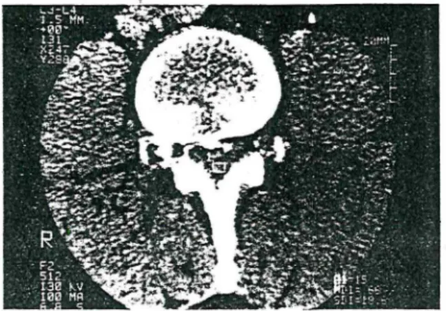
Spinal CT done on admission to hospital (Fig. 1) showed that the spinal canal was wide enough. but L3-4. L4-5. L5-S I disc protrusions filled the epidural
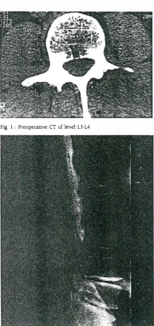
Fig. I : Preoperative CT of level L3-L4
Fig. 2 : MyelograP.hY showed a complete block at L3 l"vd and narrowing of L4·5. L5-S l int.,rverrebral space.
75
space. Intrathecal metrizamid myelopraphic CT revealed a soft tissue mass which was pressing and displacing the dural sac anteriorly (Fig. 3).
Fig. 3 , Post myelographic CT revealed a soft tissue mass.
The patient was taken to urgent operation. There was no epidural fat when partial L3 hemilaminec tomy was done. The posterior longitudinal ligament of the L3-L4 level was intact. Dural sac was found to be pressed and displaced by hard yellowish-white cartilagenous extruded disc material. This se questrated material was taken out the dural sac ex panded and bilaterally compressed L3. L4 nerve roots were decompressed. Histopathology of the material revealed intervertebral disc.
The patients's pain disappeared in the early postoperative period and started to walk on the se cond postoperative day. He was discharged on the 7th day without any neurogicai deficit. Follow-up ex amination 2 months after the operation also show ed no neurological deficit. Control CT was normal.
DISCUSSION
There are two reported case of posterior epidural migration of extruded lumbar disc. Lichtor in his case related the posterior migration partly to lumbar
....
-
...--
...�.
--
-
--
... ___ . ..__..._...____ ...--"---fusion (4). Our patient had no trauma or history of an operation related with .the speci&c region, In con trast to the case of (Lutz at al) who had sensorial deficit. our case had prominent motor deficit and had intact sacral elements (5). For investigation of the spinal region today. myelography. CT. MRI and postrnyelogram CT are widely used.
Although it is generally accepted that MRI is superior to other methods because sagittal sections can be elicited for the extruded disc material this is not so. Postrnyelogram CT proved to be most useful both on the reported cases and our patient (4.5). In the precontrast study we were not able to determine the cause of ��e posterior compression.
On postrnyelogram CT the subarachnoid space and dural sac the were found to be displaced anterior ly and recording of the density of the posteriorly plac ed mass. the patient's complaints of scia�ica and the width of the spinal canal made us think it was a posteriorly migrated extruded lumbar disc.
MRI was not performed. The operative findings showed full accordance with the postmyelogram CT.
Correspondence , Saffet Tiizgen. M.D ..
REFENCES
i.O. Cerrahpa�a Tip Fakiiltesi N6ro�iriirji Anabilim Dali Cerrahpa�a · Aksaray. istanbul
I. Fries JW. Abodeely DA. Vijungco JG. et al, Computed tomog raphy of herniated and extruded nucleus pulposus. J Comput Assist Tomogr 6,874-87. 1982
2. Manabe S. Tateishi A. Epidural migration of extruded cervical disc and its surgical treatment. Spine 11,873-8. 1988 J. Masaryk TJ. Ross JS. Modic MT. et al, High-resolution MR ima
ging of sequestered lumbar intervertebral disks. Am J Roent· gen 150,1155-62. 1988
4. Lichtor T. Posterior epidural migration of extruded lumbar disc. Surg Neural 32,311-2. 1989
5. Lutz JD. CT myelography of a fragrnant of a lumbar disc sequ estered posterior to the the cal sac. A JNR 1 L6 I 0-1. 1990