Review Article
Special Topic: Two-Dimensional Biomaterials in Regenerative Medicine
Recent advances in bioactive 1D and 2D carbon nanomaterials
for biomedical applications
Ozlem Erol
a, Idil Uyan
a, c, Meryem Hatip
a, b, Canelif Yilmaz
a, c,
Ayse B. Tekinay
a, b, c, Mustafa O. Guler
a, b, d,⁎
a
National Nanotechnology Research Center (UNAM), Bilkent University, Ankara, Turkey
b
Institute of Materials Science and Nanotechnology, Bilkent University, Ankara, Turkey
c
Neuroscience Program, Bilkent University, Ankara, Turkey
d
Institute for Molecular Engineering, University of Chicago, Chicago, IL, USA 60637 Received 22 October 2016; accepted 6 March 2017
Abstract
One-dimensional (1D) carbon nanotubes (CNTs) and the two-dimensional (2D) graphene represent the most widely studied allotropes of carbon. Due to their unique structural, electrical, mechanical and optical properties, 1D and 2D carbon nanostructures are considered to be leading candidates for numerous applications in biomedical fields, including tissue engineering, drug delivery, bioimaging and biosensors. The biocompatibility and toxicity issues associated with these nanostructures have been a critical impediment for their use in biomedical applications. In this review, we present an overview of the various materials types, properties, functionalization strategies and characterization methods of 1D and 2D carbon nanomaterials and their derivatives in terms of their biomedical applications. In addition, we discuss various factors and mechanisms affecting their toxicity and biocompatibility.
© 2017 Elsevier Inc. All rights reserved.
Key words: Carbon nanotube; Graphene; Graphene oxide; Biomedical applications; Biocompatibility
After the discovery of zero-dimensional fullerenes,1 one dimensional (1D) carbon nanotubes (CNT)2 and two-dimensional (2D) graphene sheets3 were developed and found to possess excellent materials characteristics for a variety of applications. Increasing attention has been given to these “next-generation” materials during the past decade, and much effort has been expended to explore their unique properties and utilize them in fields ranging from energy storage to biomed-icine. These materials have been widely used in biomedical applications and there is a growing and relevant concern for their toxicity and environmental effects, which remain modestly characterized.
The CNTs are hollow cylindrical sheets of hexagonal networks of carbon atoms. They exhibit very high aspect ratio, measuring a few nanometers in diameter and up to several microns in length. They can be metallic or semiconductive, depending on their chirality and add-atoms, and their electronic,
mechanical and optical properties are ideal for a broad range of purposes. In addition, CNTs have large surface area, small diameter and high curvature, which allow them to effectively interact with biomolecules through van der Waals,π-π stacking and hydrophobic interactions. These properties can also be used to facilitate the surface modification of CNTs in order to increase their solubility is aqueous media or modulate the covalent attachment of functional groups for biomedical applications.4 The biological utility of CNTs is further improved by their optical absorption in the NIR-IR window and fluorescence emission in the NIR-II window, which makes them well-suited for photothermal therapy, photoacoustic imaging and deep-tissue fluorescence imaging.5Due to their fibrous structure and strong mechanical integrity, CNTs can also be used as a reinforcing material in tissue engineering applications and provide electrical conductivity in regenerative scaffolds to direct cellular growth and differentiation.6
Nanomedicine: Nanotechnology, Biology, and Medicine 14 (2018) 2433–2454
nanomedjournal.com
Conflict of interest: The authors declare no conflicts of interest.
⁎Corresponding author at: Institute for Molecular Engineering, University of Chicago, Chicago, IL, USA 60637. E-mail address:[email protected](M.O. Guler).
http://dx.doi.org/10.1016/j.nano.2017.03.021
1549-9634/© 2017 Elsevier Inc. All rights reserved.
Please cite this article as: Erol O, et al, Recent advances in bioactive 1D and 2D carbon nanomaterials for biomedical applications. Nanomedicine: NBM 2018;14:2433-2454,http://dx.doi.org/10.1016/j.nano.2017.03.021
Graphene is a single layer of honeycomb carbon lattice and the basic building block of all graphitic forms. Similar to CNTs, graphene and its derivatives demonstrate unique mechanical, electrochemical and optical properties and interact with biomolecules throughπ-π stacking and/or electrostatic interac-tions, which are of great value for drug loading and biosensor design applications. In addition, the rich oxygen-containing groups that are attached to graphene oxide (GO) can be directly functionalized by biological ligands to facilitate targeted imaging and drug delivery. The high and intrinsic near-infrared (NIR) absorbance of GO allows it to be used as photo-thermal agents for cancer treatment with strong therapeutic outcomes.7
In this review, we present biomedical applications of CNT, graphene and GO based materials as representatives of 1D and 2D carbon nanostructures. We describe the structure, type, distinguishing properties, synthesis and purification methods, characterization techniques, and functionalization of CNTs, graphene and their derivatives. Then, their biocompatibility is discussed in terms of their cytotoxicity, genotoxicity and inflammatory responses elicited by these nanostructures. Lastly, their biomedical applications, including their use in tissue engineering, drug delivery, bioimaging and biosensors, are presented with examples from the recent works.
1D and 2D carbon nanomaterials Carbon nanotubes
CNTs are one-dimensional, hollow, tubular, nanostructured allotropes of carbon. They are composed of one or more layers of graphene sheet(s) that are rolled into a seamless cylinder, with at least one end typically capped with hemisphere of buckyball. CNTs have attracted great attention in various applications; including energy storage,8 polymer reinforcement,9 sensors,10 photonics,11catalysis12and recently biomedical engineering13 because of their unique and unusual structural, electrical, mechanical, thermal, and optical properties.
Structure and properties of carbon nanotubes
The discovery of CNTs dates back to the 1960s, with the identification of graphite whiskers in the form of rolled-up sheets of graphite layers by Bacon.14 This was followed by the discovery of fullerenes, which are singular sheets of graphene that are curved spherically to form closed cages, in 1985.1Iijima subsequently demonstrated the production of multi-walled carbon nanotubes (MWCNTs) in 1991, using a method similar to that used for fullerene synthesis.2
The backbone of CNT is built from the hexagonal carbon bonds of sp2-hybridized carbon atoms, which are arranged into hollow, cylindrical nanostructures. Although CNTs have similar hexagons of sp2 hybridized C-C bonds like graphene, they possess distinctive properties because of their high aspect ratios, large surface areas, ultra-small diameters and non-planar nature. When a graphene layer bends to form CNTs,σ-bonds that lie in the sp2 plane are shifted out of plane and reside outside the curvature of the nanotube sidewall, which causesπ-orbitals to be more delocalized outside the nanotube, inducing electron cloud distortion and resulting in a richπ-electron conjugation outside
the tubular structure.15This structural difference imparts CNTs with unusual electrochemical and thermal properties as well as high mechanical strength.
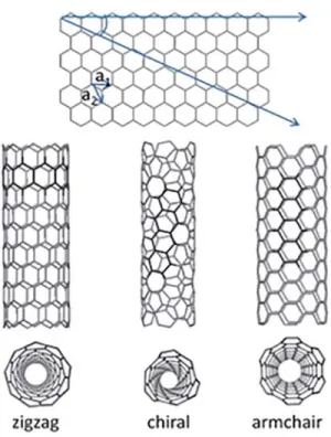
The CNTs can be classified as single or multi-walled. The outer diameter of single-walled CNTs (SWCNTs) varies between 0.4 nm to 2 nm, while that of multi-walled CNTs (MWCNTs) ranges from 2 to 100 nm, depending on the synthesis conditions.16Both SWCNTs and MWCNTs can reach lengths of 0.2 to several mm, resulting in very high (N10000) aspect ratio in many cases. Chirality (i.e., the angle between the C-C bonds and the nanotube axis) is another essential parameter for the structure and properties of CNTs. Representation of chiral, achiral zig-zag and armchair structures are shown in
Figure 1. This classification can be used to determine whether a particular SWCNT arrangement is semiconductive or metallic.17 On the other hand, the chirality of each wall is different in the case of MWCNTs, which exhibit metallic character. Electrical conductivity can reach to 106Scm-1for SWCNTs and 104Scm-1 for MWCNTs.
A one-dimensional tubular morphology and sp2 hybridized C-C bonds (which are stronger than sp and sp3 bonds) also provide CNTs with unique mechanical properties, such as Young’s moduli (~1 TPa) and tensile strengths (~50 GPa) that are much higher than those of steel.19In addition, CNTs have lower density compared to many high-strength materials. These good mechanical characteristics, in tandem with their light weight and flexibility, make CNTs effective candidates for reinforcing composites. CNTs also have excellent thermal conductivity and stability, which are important issues in the electronics industry to prevent structural damage.20
In general, large-scale production of MWCNTs is relatively easier compared to SWCNTs, which enhances the former’s popularity for biomedical applications. Although MWCNTs are more stable than SWCNTs, they are also more inert and less soluble in aqueous media. The CNTs tend to entangle and form bundles or crystalline ropes due to their high aspect ratio and strong van der Waals (more specifically π-π stacking) interactions between them.21 The CNTs also exhibit strong optical absorption in NIR-IR window (750–1000 nm), enabling their use in photothermal therapy and photoacoustic imaging. SWCNTs are particularly suitable in deep-tissue fluorescence imaging as fluorescence contrast agents due to their intrinsic fluorescence emission in the NIR-II window (1000–1700 nm).5 Synthesis of carbon nanotubes
The method used to synthesize CNTs is a very important part of CNT research, because the diameter, length, morphology, structure, chirality, quality and purity of the resulting structure depend strongly on the preparation method. Arc-discharge method is one of the most widely used techniques to obtain CNTs. In this method, an electric discharge is produced between two graphite electrodes in an inert atmosphere. This electric discharge produces a high temperature (around 3000 °C), resulting in the evaporation of a carbon electrode and its subsequent deposition on the other electrode. If the graphite electrode contains metal catalysts such as cobalt, nickel or iron, SWCNTs are formed; while a lack of metal catalysts produces MWCNTs.22The diameter of the CNTs can be controlled by
adjusting the pressure, current, and combination of catalysis.23 This method is amenable to large-scale production efforts, but the amount of byproducts can be up to 30%.24
An alternative to arc-discharge is known as laser ablation involving the use of a laser beam instead of an electric current as the energy source, was developed in 1995.25 In this method, intense laser pulses are used to vaporize a target containing graphite and transition metal catalysts at around 1200 °C in an inert environment. The produced CNTs are then collected on a cool collector by the inert gas flow.25The purity of CNTs is higher than those produced by the arc-discharge method, but laser ablation is less cost-effective.
Another method is chemical vapor deposition (CVD), which is used for the scaled up synthesis of CNTs, and it is also the most widely used technique for producing carbon filaments.26In the CVD process, hydrocarbon gases such as CH4, acetylene or
CO serve as the carbon source, which is decomposed over a silica catalyst or zeolite support in a tube reactor at a temperature range of 500–1200 °C. CVD is a fairly cost-effective method, but it results in relatively high density defects along the nanotube walls. The diameter, wall number, and length of CNTs can be controlled in the CVD method by varying the structure and composition of catalysts.27 Lastly, high-pressure carbon mon-oxide (HiPco) provides high quality, ease of purification and large scale commercial production capacity compared to the other methods. In this technique, CNTs are synthesized in a continuous flow of CO gas as a carbon source, while metal catalyst clusters are produced in situ by the decomposition of organometallic catalysis precursors introduced to the reactor at high temperatures.28In addition, several novel techniques have been developed to obtain CNTs with high quality and yields.27
Purification of carbon nanotubes
In all above-mentioned synthesis methods, CNTs contain impurities that are mostly metallic catalyst particles, amorphous carbon, and carbonaceous fragments which alter the electrical and mechanical properties of CNTs. These impurities can also be detrimental for their biomedical use. Differences in aspect ratios, sizes, oxidation rate and solubility between impurities and CNTs can be utilized to eliminate the former and improve the material properties of the latter. Various purification techniques, such as liquid and gas phase oxidations, filtration, and microwave heating, have been developed for this purpose.29It is essential to eliminate the impurities without destroying the tubular structure and intrinsic properties of CNTs. Oxidation is the most commonly used purification method, and includes liquid phase oxidation (such as acid treatment with HNO3, HCl, H2SO4etc. and/or refluxing in
water or H2O2) and gas phase oxidation (heating in air, oxygen, or
other gases).29,30Liquid phase oxidation typically removes metal catalysts, amorphous carbon and some fullerenes by refluxing CNTs with acids. The disadvantages of this method are that it causes defects on the surface of the CNTs and introduces oxygenated functional groups such as carboxylic acids. In contrast, gas phase oxidation selectively removes the carbonaceous impurities by heating the CNT at a controlled rate to temperatures around 330 °C. This method is unable to eliminate metal catalysts, which necessitate acid treatment such as HCl washing. In addition, as a chemical oxidative-based method, microwave heating treatment followed by treatment of HCl drastically reduces processing times to ~1h compared to conventional acid reflux methods (45h).31 Besides the chemical purification techniques, mass- or size-based purification methods such as microfiltration and chromatography are also used for the elimination of CNT impurities. These are relatively mild methods compared to their chemical counterparts.29
Graphene and graphene oxide
Graphene is the first two-dimensional (2D) lattice of carbon atoms to be discovered, and consists of a peculiar honeycomb structure and layers that are one atom thick each. Graphene and its oxidized derivatives (GO or reduced graphene oxide (rGO)) uniquely combine properties such as high electronic and thermal conductivities, mechanical strength and impermeability to gases.32 While graphene could be cytotoxic, pharmaceutical and biomed-ical applications can benefit greatly from non-toxic, biocompatible and water-dispersible graphene layers that are produced through chemical functionalization with various ligands.33
Structure and properties of graphene and graphene oxide Graphene family materials include few-layer graphene, gra-phene sheet, GO, and rGO sheet (Figure 2).34Graphene consists of a single-layer sheet of trigonally bonded sp2carbon atoms that is 0.35–1.6 nm in thickness33 and displays a compactly packed honeycomb crystal structure.35 GO, which is produced by the oxidation of graphite crystals, instead contains partly tetrahedrally bonded sp3 carbon atoms, which are located slightly above or below the graphene plane. Several observations confirm the presence of defective regions on GO surfaces, which interact readily with functional groups. According to Pandey et al.,36the random distribution of non-oxidized and oxidized areas with
Figure 1. Definition of the roll-up vector as linear combinations of the base vectors a1and a2for SWCNTs. Image reproduced with permission of The
oxygen-containing groups cause most of the carbon atoms to remain in sp2hybridization in the GO layers. Because of these differences in hybridization patterns, orientation and defects, layer surfaces are rougher in the GO sheets. These GO sheets can be also partially reduced to the so-called rGO sheets by removing oxygen-containing functional groups.
Graphene has a relatively high Young’s modulus, effective moisture barrier and fine electron mobility, which provides it with high electrical and thermal conductivity.37As a rolled-up graphene sheets, CNTs have similar strength and stiffness with graphene but the conduction properties are different (i.e., metallic or semi-conducting). The thermal conductivity of CNT is comparable to graphene and the mobility of graphene is higher than CNT.38
The GO can be produced with high yields by using inexpensive graphite. It is highly hydrophilic and provides pH dependent negative surface charge and colloidal stability. The GO has a higher surface area than other carbon-based nanomaterials and unique amphiphilic surface properties that allows the adsorption of proteins, dye molecules and water-insoluble drugs through noncovalent interactions. Thanks
to its fine-tuning ability, GO can be used in smart materials and systems that allow control over the release of small molecules.39 Production of graphene and graphene oxide
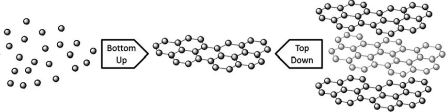
Numerous methods have already been established for producing various kinds of materials from the graphene family (Figure 2). These methods can be divided into two major approaches, top-down and bottom-up (Figure 3).40Top-down approaches include mechanical exfoliation,41 electrochemical exfoliation,42 solvent-based exfoliation43 and unzipping CNTs44; while epitaxial growth45 and CVD46 are among the bottom-up approaches. These different techniques not only yield graphenes with various sizes, shapes and compositions in various environments, but also provide different opportunities for the material’s subsequent functionalization.
In the micromechanical cleavage method, graphene is isolated from graphite by using adhesive tape47; and mono-, di-, and few-layer graphene sheets are produced through repeated cleavage. Due to its slow production process, this technique provides high quality sheets and it is usually preferred if the fundamental properties of graphene are only investigated.
Mechanical exfoliation is the initial step of the numerous discoveries of electronic and mechanical properties of graphene and the development of new production methods.48Exfoliation of graphite involves the weakening of van der Waals forces to separate the layers from each other. In electrochemical exfoliation, diluted H2SO4 or KOH are used as a reactive
sacrificial electrode solvent. Graphene is collected from this electrolyte solution that contains pyrene derivatives, H2SO4,
KOH, or surfactants such as sodium cholate, cetyltrimethylam-monium bromide, as bilayer or few-layer sheets.49 With this approach, a mixture of different thicknesses of graphite sheets can be isolated by centrifugation. However, it is difficult to remove surfactant molecules, which might influence the electrical and electrochemical features of graphene.50
Solvent-assisted or thermal exfoliation of graphite43is based on increasing the total area of graphite crystallites via immersing the graphene precursor into a solvent with sufficient surface tension.32 The Hummers method is commonly used for GO exfoliation.51This procedure involves the oxidation of graphite in H2SO4with KMnO4as an oxidant for the reaction at 45 °C.
Reduced GO is obtained following the exfoliation of GO. This method is highly popular and can yield graphene derivatives in addition to pristine graphene, but it also suffers from a high number of defects due to the harsh conditions of the production process. In addition, removing the expensive and hazardous components of the reaction is difficult, which may affect the final properties of graphene. Depending on the concentration of graphene, a decrease in flake size and increase in defect contamination might also occur.
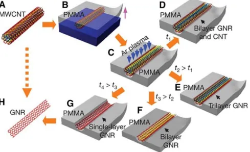
Another way of producing graphene and GO is longitudinally cutting (or“unzipping”) CNTs.44The unzipping method allows for better control and chemical functionalization capacity compared to other fabrication techniques. The graphene nanoribbons that are produced using the unzipping method were found to be conductive; however, they are electronically inferior compared to the large-scale graphene sheets because of the presence of oxygen defect sites.52Jiao et al. have showed that graphene nanoribbon formation can start from a dispersion of MWCNTs and proposed an unzipping opening mechanism that involves the exposure of a poly(methyl methacrylate) (PMMA)-MWCNTs film to an Ar plasma for varying time periods (Figure 4).44
The four aforementioned techniques constitute the major top-down synthesis methods of graphene and its derivatives. As
a bottom-up fabrication technique, the epitaxial growth tech-nique entails the sublimation of Si atoms and layer-by-layer reorganization of carbon atoms.46In this method, thin layers of graphene form on the entire surface of silicon carbide (SiC) wafers at precisely defined time and temperature intervals.53 This provides relatively high quality, but graphene sheets with more than two layers can rarely be obtained as side products. Nevertheless, some studies of these structures have revealed the presence of numerous holes and cavities on the graphene surface due to very weak bonding and rotation of the individual layers, which cause high surface roughness.54New methods have been developed to obtain more stable and homogeneous monolayers, which generally involve the formation of graphene on SiC at high temperatures (N1000 °C) and under ultrahigh vacuum conditions.50 SiC substrates designed for the production of graphene layers are commercially available; however, they are too expensive for commercial applications.
CVD55 is another useful bottom-up method for the production of graphene materials. Graphene that is produced by this technique has better crystallinity compared to those produced through other methods.35 Uniform polycrystalline graphene films with large surface areas are grown on Cu foils and films of metal such as Ni, Cu, TiOx, and so on, by CVD.46Even
though the complete process usually requires transfer from the copper support to a dielectric surface or other substrate of interest, square meters of graphene production has been achieved using this technique.56 CVD can also be used to fabricate monolayer graphene with high quality. The major drawback of this method is that the synthesis requires long period of time due to the difficulty in separation of the layers. A general comparison of advantages and disadvantages of the methods used to produce graphene are shown inTable 1.
Functionalization strategies
Functionalization of carbon nanotubes
Pristine CNTs have limited solubility and tend to aggregate in most types of solvents and biological media due to strongπ-π stacking and van der Waals interactions. Their limited water solubility makes them difficult to process and restricts their use for biomedical applications. For this reason, it is necessary to functionalize CNTs to improve their water dispersibility and biocompatibility. It should also be mentioned that the surface
chemistry of CNTs highly affects their behavior in vitro (e.g. for cellular uptake) and in vivo (e.g. for blood circulation time and biodistribution).57
CNTs exhibit chemical reactivity to many reactants depend-ing on both their aromaticity and diameter and might be more reactive at their ends than in areas along the sidewalls due to increased curvature at the terminal caps.58In addition, reactive locations mainly exist at or close to structural defects, including topological defects, points of rehybridization, vacancies in the CNT lattice and substitutional dopant impurities, which are formed during the fabrication process or occur during post-processing treatments such as purification and separation.59 These defects are a possible starting point for functionalization, and the high specific surface area of CNTs enables the adsorption or covalent bonding of various structural motifs at a high density. In summary, considerable effort has been spent to develop novel functionalization methods. Main approaches for the functionalization of CNTs can be generally classified in three categories: covalent, noncovalent, and endohedral functionalization.60The present section details these strategies, with emphasis on the methods that are especially suitable for biomedical applications.
Covalent functionalization of carbon nanotubes
The covalent modification of CNTs provides stronger bonds between the CNT and desired functional groups and offers a greater measure of control compared to methods based on noncovalent functionalizations. However, this approach changes the conjugatedπ-electron framework of CNTs (by hybridization from sp2 to sp3) and introduces defects onto the sidewalls of nanotubes, resulting in the loss of some intrinsic properties such as NIR fluorescence and Raman scattering characteristics. Therefore, covalent functionalization of CNTs has been widely utilized for drug and gene delivery, but is usually not ideal for sensing and bioimaging applications.21 Various strategies are
used for the covalent functionalization of CNTs for biomedical purposes, including surface oxidation of CNTs, cycloaddition reactions, radical additions and their subsequent derivatizations with biologically relevant molecules.60
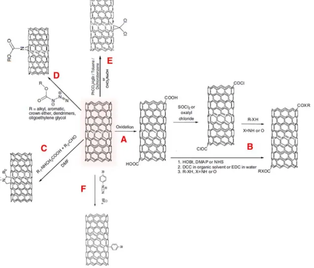
The most common method involves reactions with carboxylate groups generated on both the sidewall and nanotube ends of CNTs by oxidative treatment (Figure 5, A) under a wide variety of experimental conditions, such as sonication, refluxing in oxygen-containing acids (e.g., HNO3,61 HNO3/H2SO4,62 and
H2SO4/KMnO463) and applying ozone or H2O2.63However, the
oxidation treatment can shorten the length of CNTs and open up the end caps as a direct consequence of functionalization by oxygen-containing groups. For further derivatizations, these groups are used as anchor sites for esterification or amidation reactions (Figure 5, B), which are widely used for the conjugation of water-soluble organic molecules, hydrophilic polymers like polyethylene glycol (PEG), nucleic acids (DNA or RNA), or peptides, resulting in the production of multifunctional CNTs.60
Another widely used method involves direct additional reactions such as 1,3-dipolar, nitrene and carbene cycloaddition reactions. The cycloaddition reactions occur uniformly and densely at the CNT sidewalls, rather than the ends or defect sites (as is the trend for the oxidation functionalization strategy), and provide remarkable solubility in water, many organic solvents and physiological conditions.64 1,3-dipolar cycloaddition of azomethine ylides results in the formation of pyrolidine rings on the CNT surface (Figure 5, C), which can be substituted with many functional groups like amino acids, peptides,65therapeutic agents66 and fluorescent molecules67 for diverse biomedical applications.
The nitrene addition on the sidewall of CNTs is achieved by reactive alkyloxycarbonyl nitrenes obtained from alkoxycarbo-nyl azides through thermal decomposition or photolysis of organic azides (Figure 6, D).68 A broad range of nitrene precursors are used for the covalent binding of a variety of
Figure 4. Graphene nanoribbon formation from MWCNTs that are embedded in PMMA and then treated with an Ar plasma. Reprinted by permission from Macmillan Publishers Ltd: [Nature],44Copyright (2009).
different groups. The dichlorocarbene moiety has also been conjugated onto the CNTs using a chloroform/NaOH or a phenyl(bromodi-chloromethyl) mercury reagent (Figure 5, E), but X-ray photoelectron spectroscopy (XPS) results suggest that the degree of functionalization is low.69
Aryl diazonium coupling is frequently used to covalently functionalize CNT surfaces via radical addition reactions because of its simplicity and high yield. In this method, aryl diazonium salts are prepared ex situ from tetrafluoborate salts (Figure 5, F) or formed in situ via the reaction of aromatic amines with isoamyl nitrite and/or with NaNO2in ionic liquids
(or even without a solvent).18The reaction rate depends on the metallic character of CNTs. On the other hand, this technique has been only rarely employed for biological applications. Further details about covalent functionalization can be obtained from the relevant reviews.70
Noncovalent functionalization of carbon nanotubes
Noncovalent functionalization of CNTs is a useful and convenient technique to obtain highly dispersible and process-able CNTs without disturbing the carbon lattice, typically through the attachment of amphiphilic molecules ranging from small molecules to polymers. The principal advantage of this method is that it preserves the structure and optical properties of CNTs. Ideally, noncovalent functionalization should provide higher water solubility, biocompatibility, stability in various biological solutions and functional groups that are available for further bioconjugation.57Hydrophobic interactions,π-stacking and/or van der Waals forces between CNTs and guest molecules are the driving forces for this method.
The high specific surface area of CNTs provides high loading capacity for adhering molecules.60Many amphiphilic molecules, including pyrene, naphthalene derivatives, proteins, RNA, DNA, peptides, polymers and surfactants have been successfully adsorbed or wrapped onto the CNTs through noncovalent interactions.70The dispersion of the CNTs usually depends on the chemical characteristics of nanotube surfaces, the type and concentration of the amphiphilic molecule and the solvent, and dispersing conditions such as ultra-sonication and temperature. Steric stabilization by adsorbed nonionic surfactants or polymer
layers is dominant if nonionic surfactants or polymers are used. In contrast, ionic amphiphilic molecules facilitate the dispersal of CNTs through electrostatic repulsion between similarly charged groups that are oriented towards (or away from) the solution.72 Paloniemi et al. reported that specific interactions, like charge transfer and ion-π interactions, between CNTs and surface elements are important for the attachment of small aromatic molecules. The morphology of the aromatic moiety also affects the strength ofπ-π interactions by altering the curvature of the nanotube surface.73In particular, molecules containing polyaro-matic components generally demonstrate a stronger affinity towards the basal plane of CNT surfaces compared to single aromatic moieties. This effect is readily observed in pyrene-containing molecules, which have attracted considerable attention as noncovalent modification agents in recent studies.74 Cationic, anionic or nonionic surfactants have been success-fully used to produce stable aqueous dispersions of CNTs. The acquisition of stable CNT dispersions depends strongly on the length of the hydrophobic regions and the structure of the hydrophilic head group of the surfactant. However, surfactants face several problems in biological environments, including high critical micellar concentrations, lower stability and limited interaction with cellular proteins. Many of these problems can be circumvented by the use of PEG-modified phospholipids, which are non-toxic, biocompatible and amphiphilic polymers that possess various functional groups that can be further functionalized with therapeutic and targeting molecules.75
The potential of peptides as noncovalent modifiers for CNTs has been explored through the development of novel bioactive nanomaterials and may lead to new advances in biosensor and tissue engineering applications.76Peptides containing histidine and tryptophan residues at specific locations were identified by phage display to exhibit specific affinity to CNTs.77In addition, successful CNT dispersions have been prepared with the aid of amphiphilic peptide sequences that contain phenylalanine at specific locations78 and fold intoα-helixes on CNT surfaces. Self-assembling peptide amphiphile molecules, containing charged amino acid sequences covalently coupled to either a hydrophobic alkyl tail79or phenylalanine,76have also been used to disperse MWCNTs in aqueous solutions, with stable
Table 1
Advantages and disadvantages of techniques currently used to produce graphene.40,50
Advantages Disadvantages
Micromechanical cleavage
-High quality graphene sheets are produced -Slow method Electrochemical
exfoliation
-Produces a mixture of different thicknesses of graphite flakes -Difficult to remove surfactant molecules
-Few-layer graphene stacks can occur in the mixture
-Defects influence the electrical and electrochemical properties of graphene Solvent-based
exfoliation
-Common and easy production
-Increase in the total area of graphite crystallites -Suitable for mass production
-Expensive and hazardous solvents or surfactant molecules -Difficult to remove byproducts that affect properties of graphene -Harsh production conditions cause defects
Unzipping CNTs -Few-layers of graphene can be synthesized -Allows better control and chemical functionalization -Nanoribbons with different widths can be produced
-Electronically inferior due to oxygen defect sites
Epitaxial growth -Most even films (of any method) -Large-scale area
-Difficult to control the morphology and adsorption energy -High-temperature process
Chemical vapor deposition
-No need to prepare substrate
-Possible to prepare monolayer graphene
dispersions being obtained when the noncovalently functional-ized CNTs were highly charged. As amphiphilic polypeptides, proteins have also been widely used to disperse CNTs, and their performance depends on diverse parameters including primary structures and pH. But direct noncovalent modification of CNTs with proteins may partially unfold them and results in the loss of their biological functions. Nevertheless, indirect adsorption of proteins is possible with the aid of linker molecules such as 1-pyrenebutanoic acid succinimide ester, which immobilize proteins onto the CNT surface while maintaining the native protein structure.80
Endohedral functionalization of carbon nanotubes
In addition to the functionalization of the outer surface of CNTs, molecules can also be encapsulated within the hollow cavity formed by the nanotubes. Depending on the physico-chemical properties and stability of the filler molecules, several endohedral encapsulation strategies are used for this purpose. Some of these strategies include high-temperature processes such as the CVD of filler metals, melting and capillary encapsulation of metals, gas phase filling-polymerization of
polycyclic aromatic molecules, and in situ thermal decompo-sition of organometallic precursors. Metal and magnetic compound-filled CNTs obtained by these methods are useful for some biomedical (i.e., theranostics) applications; however, milder reaction conditions are required to insert biomolecules inside the cavities of CNTs. Supercritical CO2 extraction,
nano-extraction and nano-condensation are among the methods used for the encapsulation of heat-sensitive biomolecules. Detailed information about this approach can be found in recent review.81
Functionalization of graphene and graphene oxide
Functionalization of graphene and GO can be either covalent or noncovalent. Covalent functionalization of GO can be performed through functional oxygen groups on the surface, carboxylic acid groups on the edges or epoxy/hydroxyl groups on the basal plane. Covalent functionalization can be achieved by several chemical reactions such as nucleophilic substitution, electrophilic substitution, condensation, and addition reactions. Nucleophilic substitution reactions target the epoxy groups of
Figure 5. Covalent functionalization of CNTs; oxidation (A) and further esterification and amidation reactions (B), 1,3 dipolar cycloaddition (C), nitrene cycloaddition (D), carbine cycloaddition (E) and radical addition of aryl diazonium salt (F). Images reprinted and adapted with permission from The Royal Society of Chemistry.18,71
GO. A variety of groups, such as alkyl amines, amino acids,82 dopamine83 and polyglycerol,84 can be substituted in this manner. Regarding the hydrophilic nature of GO, it is able to conjugate amino acids through the epoxy groups of GO and obtain a flat orientation of the functional groups on the surface of the GO as a result of a nucleophilic reaction in alkaline solution.82In another example, chemically converted graphene she ets were produce d fr om the re act ion b et ween 3-aminopropyltriethoxysilane (APTS) and GO.85 These sheets can be homogeneously dispersed in common solvents.
Functionalization through noncovalent interactions is achieved by several approaches such as polymer wrapping, adsorption of small molecules, and interaction with host molecules like porphyrins or biomolecules like deoxyribonucleic acid (DNA) and peptides.33The functionalization of GO with these molecules is relatively common and well-characterized in the literature, though noncovalent modifications with various agents such as poly(sodium 4-styrenesulfonate),86pyrenebutyric acid87 and amine terminated polymers88have also been demonstrated. In addition, Liu et al. have reported a noncovalent functionalization technique for graphene, based onπ-π interactions between the π orbitals of graphene and poly(N-isopropylacrylamide).89 Yang et al. have reported the supramolecular functionalization of rGO by the conjugation of poly(2,5-bis(3-sulfonatopropoxy)-1,4-ethynylphenylene-alt-1, 4-ethynylphenylene) polyelectrolyte through electrostatic interactions90 which, imparted long dispersion stability and high electrical conductivity to the material.
Characterization
The characteristic properties of carbon based nanostructures require characterization after synthesis or functionalization processes to determine their structural, morphological, thermal and electrical properties. For biomedical applications, conven-tional in vitro studies are usually performed in 2D plate cultures as opposed to the deposit forms used in in vivo studies. Classical techniques used for characterization of biomaterials are per-formed in 2D forms and are a representative of 3D assembly formed by these nanomaterials.
Scanning electron microscopy (SEM) and transmission electron microscopy (TEM) are widely used for the preliminary evaluation and determination of the morphology of CNTs and their impurities. TEM provides more accurate information on the diameter, number of walls, structural integrity and defects of the nanotube architecture. Energy-dispersive spectroscopy can also be used in conjunction with SEM and TEM to determine not only the metallic impurity content but also non-metallic and heteroatom identity and quantity after functionalization of carbon-based nanomaterials. After functionalization, if the attached groups are large enough to be resolved and/or have enough contrast against the CNT background, they can also be identified morphologically by SEM and TEM.27 Grazing incidence small-angle X-ray scattering allows the real time in situ study of growing nanotubes on substrates and provides information about the three-dimensional structure and orienta-tion of CNTs, as well as morphological and structural data. It is also a very useful method if the nanostructures prove difficult to visualize by conventional means.91
Microscopic corrugations of graphene and its derivatives with a lateral dimension of 8 to 10 nm and height displacements of 0.7 to 1 nm can be estimated by TEM.92Although some scanning tunneling microscopy imaging studies indicated a limited correlation between b0.5 nm height corrugations and local electrical properties,93bigger ripples (2–3 nm in height) appear to be indicative of strain-induced local conductance modulations.94 Computational models, such as molecular dynamics simulations, can be employed to understand the mechanical properties of graphene monolayers.95 Experimen-tally, the Young’s modulus of a few layers of graphene was also investigated by force-volume measurements in AFM. In a recent study, for defect-free graphene, a Young's modulus of 1.0 TPa and a fracture strength of 130 GPa were determined by nanoindentation using AFM.96 In another study, the average elastic modulus and the highest fracture strength of GO platelets were reported as ~32 GPa and ~120 MPa, respectively.97
Fourier transform infrared (FTIR) spectroscopy allows the qualitative identification of functional groups on carbon nanostructures through the interpretation of characteristic adsorption bands. However, FTIR spectra often have insufficient
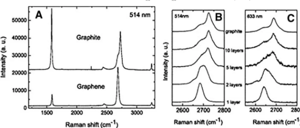
Figure 6. Raman spectra at 514 nm for bulk graphite and graphene (A), change of the spectra at 514 nm with the number of layers (B), change of the Raman spectra at 633 nm with the number of layers (C). Reprinted and adopted with permission from the American Physical Society,99Copyright (2006).
quality due to the strong electronic adsorption and light scattering properties of carbon nanostructures. To overcome this undesired behavior, the sample should have the smallest possible particle size. Attenuated total reflectance and diffuse reflectance methods can also be used to obtain higher quality spectra, and nuclear magnetic resonance spectroscopy can be used to confirm the presence of specific functional groups on nanostructures. XPS provides further insight into the chemical composition and environment of nanoparticle surfaces, but is limited to a depth of less than 10 nm into the structure.
Raman spectroscopy is a powerful tool to distinguish between the two distinct sp2carbon nanostructures that characterize CNTs and graphene. Useful information about crystallite size, clustering of the sp2 phase, the presence of sp2-sp3 hybridization and chemical impurities, the magnitude of the mass density, the optical energy gap, elastic constants, doping, defects and other crystal disorders, edge structure, strain, number of graphene layers, nanotube diameter, chirality, curvature, and metallic/semiconducting behavior can be obtained utilizing Raman spectroscopy.98The Raman spectrum of carbon materials typically yields three main characteristic bands; namely D, D’ and G modes in the 1000 to 2000 cm-1range and the overtone and combination bands G’=2D in the 2400 to 3400 cm-1
range. The G mode is centered around 1590 cm-1and attributed to the graphitic structure. D and D’ modes arise from amorphous disorder and defects in the carbon lattice. The crystallite and amorphous region densities of the structure are calculated from the intensity ratio of D and G modes (ID/IG). The radial breathing mode
(RBM) includes various vibrational transitions of radial expansion and contraction of the CNTs, and is usually observed in the region between 100 and 350 cm-1. The frequency of RBM is inversely proportional with the diameter of nanotubes.27With this tool, the quality of the graphene sheets can also be determined by evaluating the extent of disorder and number of defects on the graphene surface. In addition, the number of the layers can be determined by the shape, width, and position of the 2D peak.99Graphene and its derivatives exhibit prominent graphite peaks that are known as the“G band” (at 1580 cm-1) and the“2D peak” (at around 2700 cm-1).92Ferrari et al. demonstrated that increasing the number of graphene layers causes the 2D peak to shift to higher wavelength ranges (Figure 6).99
Thermogravimetric analysis (TGA) measures the mass change of a sample, which is examined as a function of temperature under a pre-defined atmosphere. It is also possible to identify the gas phase composition of the sample with the aid of TGA in conjunction with FTIR. TGA is frequently utilized to evaluate the thermal stability of carbon nanostructures, and also provide information about the proportion of carbon and other impurities in the CNTs. Additionally, TGA can measure the degree of functionalization due to the loss of organic moieties that occurs when functionalized carbon nanostructures are heated in an inert atmosphere.100
Biocompatibility Cytotoxicity
Carbon-based nanomaterials have been widely researched in biological systems in recent years due to their diverse potential and unique physicochemical and morphological structures.
Unlike graphite, which is a naturally occurring stable form of carbon; graphene, GOs and CNTs are artificially synthesized materials with uncertain effects on biological systems. Conse-quently, the toxicity of carbon-based nanomaterials has become a topic of great interest, and considerable effort has been spent to characterize their cytotoxic effects and behavior in microorgan-isms, cells and organmicroorgan-isms, both to evaluate the risks associated with their spread to the environment and to determine their potential utility in biotechnological and biomedical applications. In general, there are several factors that affect the toxicity of nanoscale biomaterials such as graphene, GO and CNTs; including size, number of layers, shape, structure, purity, exposure route, dose, charge, structural defects and hydrophobicity.101Surface hydrophobicity of a material directly affects its dispersion characteristics under physiological condi-tions. As such, surface functionalization of carbon nanomaterials can greatly alter their bioaccumulation, toxicity and cellular and systemic responses. Both pristine graphene and CNTs are highly hydrophobic, and functional groups are often used to increase the water solubility of these materials, which also enhances their absorption in biological context.
The main cellular uptake mechanisms for graphene are receptor mediated endocytosis, phagocytosis and membrane adsorption, whereas CNTs are internalized via micropinocytosis, receptor-free endocytosis and piercing (rupturing and self-healing of cellular membrane) in addition to the same internalization pathways as graphene.102 The GO is also known to be internalized via non-phagocytotic pathways as well as phagocytotic pathways.103 Pristine graphene has been shown to display lower dispersibility, inducing oxidative stress at 10μg/mL and leading to apoptosis in neural-like cells (PC-12) in a dose and time dependent manner. Graphene exposure was also associated with an increase in the level of reactive oxygen species (ROS) production and cell death through the induction of MAP kinases and the transforming growth factor beta pathway in macrophages. Graphene was initially observed to cause stronger metabolic activity compared to SWCNTs at lower concentrations; however, this trend was reversed at higher doses.104In another study, pristine graphene was reported to cause toxicity by accumulating on the cell membrane and eliciting an apoptotic response due to oxidative stress, whereas carboxyl-functionalized hydrophilic graphene was internalized normally and exhibited no cytotoxicity.105Both GO and carboxylated graphene was observed to be toxic to nonphagocytotic epithelial lung carcinoma cells by penetrating through the membrane, concentrating as vesicles in the cytosol, and increasing ROS; though lower concentrations of both materials (b4 μg/mL) did not exert any cytotoxic effect.106 However, incubation of graphene with 10% fetal bovine serum effectively decreased its toxicity, possibly because of the coating of the graphene surface with a protein layer.107The GO was also reported to induce necrotic death in macrophages by causing the activation of Toll-like receptor-4 signaling and production of tumor necrosis factor alpha via autocrine signaling in vitro.108 The in vivo biocompatibility of the GO is not well understood. It was shown to be effectively and rapidly cleared from zebrafish without lasting effects,109whereas lung administration of the GO to mice caused severe and persistent lung damage while pluronic dispersion of graphene minimized the damage.110 PEGylation is a common
approach in order to mask the material against immune system and reduces the oxidation of graphene. No toxicity was observed in mice treated with PEGylated graphene (20 mg/kg), as observed in histological and hematological analyses.111
Pristine CNTs were also shown to cause cytotoxicity through inflammation, activation of the immune system, and production of ROS. However, surface functionalization can be used to eliminate some of these problems. Functionalization is usually performed to increase the hydrophilicity and water dispersion of CNTs. and plays a vital role in facilitating non-destructive interactions between nanomaterials and cellular interfaces.112 PEGylation of SWCNTs, for example, was reported to reduce their toxicity and downregulate ROS production-related path-ways compared to uncoated SWCNTs,113 leading to lower cellular ROS levels. A number of studies hypothesize that surface functionalization improves the biocompatibility of 1D and 2D carbon nanomaterials111,114;however, some studies also suggest that the alteration of surface properties promotes and facilitates cellular uptake of the materials and increases their toxic potential. The assessed effect of 1D and 2D carbon nanomaterials having different functionalization type and dosage are summarized in Table 2. More comprehensive studies are necessary in order to understand the toxicity mechanisms of 1D and 2D carbon-nanostructures and to rationally design surface functionalization types that are able to mitigate the risks associated with these materials.
Genotoxicity
Genotoxicity is defined as the property of the material to directly or indirectly cause damage to the DNA. ROS produced in response to graphene-related materials can conceivably create an increase in cellular acidity, leading to DNA damage and possibly cancer. There are relatively few studies regarding the genotoxicity of graphene-based materials compared to cytotox-icity studies. Previously, the genotoxic potential of various nanoparticles and nanomaterials, including graphene, GO and
CNT were shown; and graphene exhibited genotoxicity at the lowest concentrations (N1 μg/mL). The CNTs, in contrast, produced adverse effects on the cellular genome atN100 μg/mL concentration.115 Wang et al. showed that GO interacted with DNA in a concentration-dependent manner, completely frag-menting DNA at 600μg/mL.116GO also inhibits the S phase of the cell cycle and leads to cell cycle arrest at the G0/G1phase.
Intravenously injected GO at a dose of 4 mg/kg for 5 days induced micronucleated polychromic erythrocytes in mice.117 Akhavan et al. reported that rGOs elicited size- and concentration-dependent increases in DNA fragmentation and produced chromosomal aberrations in human mesenchymal stem cells (MSCs).118 Pristine CNTs have been shown to cause genotoxicity mainly due to their capability to cause increased ROS production, which further causes DNA breaks through oxidation.119 Poulsen et al. suggested that CNTs with smaller surface areas (and therefore larger sizes) are associated with increased genotoxicity, as observed in mice studies.120However, a recent study by Girardi et al. suggests that embryonically applied PEGylated SWCNTs did not cause any genotoxicity in zebrafish between the concentration values of 0.1 and 1 ppm.121 Inflammatory response
It is not completely clear what happens to the 1D and 2D carbon nanomaterials once they are administered into the body; however, the immune reactions that occur in the body strongly depend on the route of administration. Graphene sheets with PEG and radioactive iodine has been administered intravenously to mice and graphene sheets were found to accumulate in reticuloendothelial system, liver and spleen, followed by a gradual clearance after 315 days.122Graphene and GO are more damaging when they are directly administered to the blood-stream or inhaled. Macrophages, as the first step of the immune defense machinery, respond immediately to foreign materials that are administered to blood. In addition, other immune cells such as phagocytes and dendritic cells are more sensitive to
Table 2
The effects of 1D and 2D carbon nanomaterials and their functionalization types and dosage.
Material Functionalization type Assessed effect Dose Ref.
Graphene Pristine Cytotoxic effect 10μg/mL 105
Graphene Carboxyl functionalization Cytotoxic effect N4 g/mL 106
Graphene Incubation of graphene with FBS Reduced cytotoxic effect 107
Graphene Pluronic dispersion Reduced cytotoxic effect 110
Graphene PEGylation No cytotoxic effect 20 mg/kg 111
CNT PEGylation Reduced cytotoxic effect 113
Graphene Pristine Genotoxic effect N10 μg/mL 115
CNT Pristine Genotoxic effect N100 μg/mL 115
GO Pristine Complete DNA fragmentation N600 μg/mL 116
GO Pristine Micronucleated polychromic erythrocytes 1 mg/kg 117
CNT PEGylated SWCNT PEGylated SWCNTs, no cytotoxic effect in zebrafish 0.1 and 1 ppm 121
Graphene Pristine Inflammatory response, inhalation N10 mg/m3 124
CNT MWCNT Inflammatory response, inhalation N0.5 mg/m3 124
GO Radiolabeled GO Inflammatory response N10 mg/kg 125
GO Pristine Inflammatory response in A549 lung epithelial cell N50 mg/mL 126
rGO PEGylation Inflammatory response in A549 lung epithelial cell N25 mg/mL 126
CNT Pristine Inhaled, mouse and rat, dose dependent fibrosis 5–40 μg 128
graphene than stem cells, osteocytes and chondrocytes. Wang et al. showed that the Th2 immune response was activated in the lungs of mice following the intravenous administration of graphene nanosheets, which was reflected by a strong neutro-philic influx and the secretion of interleukins IL-5, IL-13 and IL-33 in the bronchoalveolar lavage fluid.123 In a comparative study, mice were exposed to graphene-related materials by inhalation, and the inflammation process was initiated at concentrations of 10 mg/m3 for graphene and 0.5 mg/m3 for MWCNTs.124On the other hand, the GO was observed to cause no immune response or systemically pathological changes following IV injection in mice below the concentrations of 10 mg/kg, however, significant inflammatory and immune response was triggered by higher concentrations.125 In another recent study, non-functionalized rGOs were found to adhere to cell membranes, potentially binding Toll-like receptors and activating inflammatory responses mediated by NF-κB; while PEGylated rGOs were directly internalized by the cells but nonetheless created a NF-κB mediated inflammatory response.126
The SWCNT and MWCNT both caused immune activation, immune cell proliferation, secretion of inflammatory cytokines, chemokines, tumor necrosis factor-α, IL1-ß, monocyte chemo-tactic protein-1 and transforming growth factor-1 in RAW264.7 macrophages.127Furthermore, the CNTs were observed to cause lung inflammation and fibrosis in mice and rats, as they accumulated in inflammatory cells and triggered cytokine and growth factor secretions in the lungs, resulting in an increase in bronchoalveolar lavage fluid and the formation of epithelioid granulomas in the lung parenchyma.128In addition, intraperito-neal and intrapleural injections of CNTs were observed to cause inflammation and fibrosis in the regions in which they were administered.129 Although it is clear that inflammation is a frequent result of CNT exposure, further molecular, cellular and animal studies are nevertheless necessary to better determine the pathways in which 1D and 2D carbon materials are able to elicit immune responses.
Bioactivity and applications
As discussed above, 1D and 2D carbon nanomaterials have a number of unique properties that enable them to be used in biomedical applications. We provide a brief summary of the mentioned properties, and how they are exploited for biomedical applications inTable 3.
Tissue engineering
Bone regeneration
Major bone defects are one of the main challenges in bone tissue engineering and require implantation of either autologous bone grafts or scaffolds produced from various natural or artificial materials. To date, a variety of synthetic or natural materials have been used for promoting osteogenesis, however, further applicability and regenerative success of these materials depend on whether they meet some criteria. An ideal bone implant material should facilitate the attachment, proliferation and differentiation of the bone cells, as well as stimulating and sustaining a bioactive signal that promotes bone regeneration. Stem cell based-therapies are still being investigated for this purpose; however, these cells require in situ scaffolds that promote viability, attachment, spreading and differentiation in order to effectively replace lost tissue.
The 1D and 2D carbon nanomaterials serve as promising materials as bone scaffolds, coating materials and drug delivery agents to support bone regeneration due to their strong mechanical properties, excellent flexibility and nanotopography. Both graphene and CNTs exhibit high conductivity profiles, which have positive effects on the healing process. It has been suggested that osteoblast activity, adhesion and proliferation are modified by external electrical stimulation.130Graphene, with its great conductivity, charge carrier mobility and tensile strength, is considered to have a great potential as a material for bone regeneration.96The CVD graphene has been shown to increase the proliferation of mesenchymal stem cells and osteoblast-like
Table 3
Applications of 1D and 2D carbon nanomaterials and their utilized properties.
Property Usage Graphene GO CNT
π-π interactions Used for hydrophobic/inorganic drug loading, pH dependent release, RNA delivery
177,185 172,174,184,186 171,173,175,176,181,182,183 Photothermal effect Utilized in increasing effectivity
of drug delivery systems, often coupled with targeting moieties or drugs
179,180 178
Vehicle/Backbone
Conjugated with fluorophores, or filled
with magnetic particles for bioimaging 193,195,196 190,191,192,194 Fluorescence
quenching
Utilized in FRET-like fluorescent biosensors, where analyte binding alters proximity of fluorophore to carbon nanomaterial
197,198,199
Topography Patterning helps promote tissue regeneration 131,149,151,164,166 135,136,139,140,153,163 141,143,155,156,159,167 Conductivity Regeneration: Conductive materials mimic
action potentials
131,149,150,152,166 147,148,153 142,143,154,155,156,157, 158,159,160,161,165,167,168 Biosensors: Used as electrodes, or electrode
functionalization of biosensors
200 Mechanical properties:
stiffness and elasticity
Carbon nanomaterials provide mechanical strength comparable to that of natural bone
131,133,164 135,136,137,138,139,140 143,154 Protein adsorption Provides stability and increases local
concentration of growth factors and chemokines
SAOS-2 cells.131 Pristine graphene itself did not improve proliferation, however, the osteoblastic differentiation of stem cells was enhanced by the material.132The 2D graphene layers also enhanced osteogenic commitment compared to GO and PDMS,133and MSCs cultured on CVD graphene-coated glass and Si/SiO2surfaces were shown to express higher amounts of
osteocalcin, a late osteogenic marker.133
The MSCs cultured on GO nanoribbons also exhibited a 3.7 fold increase in mineralization compared to the those cultured on PDMS and glass.134These effects could be attributed to the high modulus stiffness and elasticity of graphene, as well as the lateral cytoskeletal tension that causes the induction of cytoskeletal organization in a way that promotes osteogenic differentiation.133 Combination of other molecules with GO has also been one of the common approaches. Chitosan-GO scaffolds were observed to significantly enhance osteoblast attachment, proliferation and extracellular matrix formation,135 and another, similar study reported higher cellular attachment, proliferation, growth and mineralization rates following the attachment of GO carboxyls to the amine groups of chitosan.136 Calcium phosphate-mineralized GO/chitosan scaffolds could also adsorb bone morphogenic protein-2 (BMP-2) encapsulated bovine serum protein nanoparticles and silver nanoparticles for bioactivity and anti-bacterial purposes, respectively. In vitro and in vivo studies showed that bone marrow stromal cell differentiation and proliferation were enhanced in the presence of the former system, contributing to improved bone regeneration.137 GO was also incorporated into a gelatin hydroxyapatite matrix in order to increase its mechanical strength, and the composite material was found to be biocompatible, biodegradable and capable of enhancing the osteogenic differentiation of human adipose derived MSCs.138 Osteoinduction of MSCs was also observed on another polymer, poly(lactic-co-glycolic) acid (PLGA), following enrichment with GO.139 The mechanism for the enhancement of osteogenic differentiation and related pathways is hypothesized to lie in the combination of physiochemical properties of GO, including its stiffness, nanoscale roughness, presence of functional groups such as reactive oxygen, water retention capability and hydrophilic nature, as well as the direct adsorption of biomolecules onto the GO surface.140 The CNTs have also been used in the field of osteogenic regeneration, as their incorporation adds great strength to composite materials. Studies have shown that alkaline phosphatase activity, calcium deposition, and osteoblast cell adhesion were enhanced on CNT/polycarbonate urethane composite scaffolds.141The conduc-tive properties of CNTs are also attracconduc-tive for the field of bone regeneration. Poly(lactic acid) (PLA)/MWCNT composite scaffolds were reported to show increased cell proliferation and extracellular matrix calcium deposition of osteoblasts,142while electrospun PLA/ MWCNT composites induced cell alignment and proliferation in the presence of electrical stimulation.143In addition, CNTs were found to be effective in stimulating osteocyte proliferation in a time dependent manner, despite some initial cell death. Macrophages and neutrophils have been suggested to remove the toxic potential of CNT substrates, allowing the subsequent proliferation of the osteoblasts.144
Neural regeneration
The central nervous system has a very low regenerative capacity due to the inhibitory environment, low abundance of
neural stem cells (NSCs) and their low potential to compensate the loss of neurons. Current clinical treatments for nerve injuries have not advanced beyond minimizing secondary damage following initial neuronal loss. The NSC transplants are used to compensate for the lost neural tissue; however, NSCs require constant stimulation in order to survive and selectively differentiate into astrocytes when transplanted, highlighting the need for developing biomaterials that promote NSC survival and neuronal differentiation. Therefore, it is essential to investigate the mechanisms of NSC differentiation and develop biomaterials for this purpose in order to effectively facilitate nerve regeneration. Although regeneration is more favorable in the peripheral nervous system, nerve gaps longer than 2 cm still require the artificial connection of distal and proximal nerve stumps in humans. As such, effective neural treatment strategies require the elimination of the inhibitory signals that prevent regeneration, and scaffold biomaterials that restore the intercon-nections between nerve stumps.145
The 1D and 2D carbon nanomaterials have attracted tremendous interest in nerve regeneration due to their high conductivity, nanotopography and their ability to be functionalized for specific purposes. For instance, carbon nanotubes provide topographical cues for neurons, resembling the extracellular matrix fibers in the native environment of the nervous system. While certain polymers are similarly conductive, the rigid structure of polymers is not optimal for culturing neurons, since neural substrates must be designed to have lower Young’s moduli compared to other tissues.146The effect of graphene-based materials on neural differentiation has been widely studied. Lie et al. showed that culturing NSCs on 3D porous graphene foams promoted their differentiation towards neurons rather than astrocytes. In another in vitro study, human NSCs grown on rGO sheets exhibited accelerated neural differentiation following pulsed laser stimulation.147 More specifically, Yang et al. proved that embryonic stem cells showed enhanced dopaminergic neural differentiation on GO sheets in a dose dependent manner, whereas graphene and CNTs did not have such an effect.148In another study, graphene-poly(ε-caprolactone) (PCL) nanofiber hybrid systems were developed for guiding stem cell differentiation into oligodendrocytes in order to promote myelination. Without introducing differentiation inducers, NSCs were guided to differentiate into mature oligoden-drocytes due to the presence of permissive surfaces for the adhesion of cells and proteins, as well as the conductivity of the scaffold system, which could potentially be used for the treatment of myelination disorders.149Polypyrrole-functionalized graphene nano-fibers were also developed for improving the optic nerve regeneration, and retinal ganglion cells showed enhanced viability and neurite outgrowth in the presence of electrical stimulation on these substrates.150In an in vivo study, Zhou et al. showed that the implantation of colloidal graphene-coated electrospun microfiber networks into the subventricular zone prevented glial scars, decreased microglia and astrocyte activation, and supported neuroblast migration from the subventricular zone.151 A recent work further demonstrated that of the neurogenic differentiation of MSCs was promoted by electrical stimulation on rGO- poly(3,4-ethylenediox-ythiophene) (PEDOT) hybrid microfibers.152GO was also used for coating aligned PLLA nanofibrous scaffolds, and PC-12 proliferation and differentiation were found to be significantly improved on these scaffolds.153
Because of their conductive, fibrous and hollow structure, as well as their high surface to volume ratios, CNTs have been an attractive materials for neural regeneration studies. Materials such as polymers and biomolecules have been used to modify carbon nanotubes to effectively guide the outgrowth of neurons. Silk-CNT composites, for example, were able to enhance the neural differentiation and axonal lengths of human embryonic stem cells154; while Si2O cell culture surfaces patterned with
double-walled CNTs and CNT layers were observed to promote neural differentiation and adhesion of Neuro-2a cells to a greater extent than individual groups of CNT and Si2O surfaces.155In
another study, immature spinal cord neurons isolated from neonatal rat spinal cords showed rapid growth on pyrolidine-functionalized MWCNT coated surfaces.156In an in vivo study, electrospun collagen-PCL-MWCNT fibrous scaffolds were shown to assist in the recovery of rat sciatic nerve defect models and prevented muscle atrophy. In the same study, the scaffold also supported Schwann cell adhesion and elongation.157 Freeze-dried silk/SWCNT/fibronectin-based nerve guidance con-duits also showed higher nerve conduction velocities and more myelinated axons in a rat sciatic nerve deficient model.158 Furthermore, collagen isolated from rat tails was incorporated into composite hydrogels with CNTs and the composite system provided significant stimulation of MSC differentiation into the neural fate.159Koppes et al. showed that SWCNTs incorporated into Matrigel™ hydrogels showed enhanced neurite outgrowth of dorsal root ganglia compared to SWCNT-free Matrigel™ group, while external electrical stimulation promoted outgrowth within SWCNT-free control groups.160In a recent study by Usmani et al., 3D conductive MWCNT meshes were used to culture organotypic spinal cord slices that were allowed to spontaneously grow. 3D artificial scaffolds substantially boosted regrowth of nerve bundles into a random net (Figure 7).161
Muscle regeneration
Skeletal tissue is composed of bundles of muscle fibers, which in turn are clusters of fused myoblasts. Significant skeletal muscle loss may occur due to damage, trauma or surgery, and deficiencies in its regeneration may lead to permanent functional disabilities.162Although minor muscle injuries are often repaired rapidly with no medical intervention, the regeneration of bulk muscle tissue still remains a challenge in tissue engineering. It is known that muscle tissue is electrically responsive, and electrical cues have been utilized to promote myotube formation and muscle contraction. The 1D and 2D nanomaterials, with their outstanding flexibility, high conductivity, great mechanical properties and ultralow density, bear great potential as substrates for muscle regeneration.
The GO was reported to accelerate the myogenic differentiation of C2C12 cells due to better adsorption of serum proteins and nano-topographical cues.163Electrospun PLGA-collagen impreg-nated GO fibers were also observed to promote adhesion and proliferation of C2C12 cells.164 In another recent study, conductive PCL-graphene composite scaffolds promoted the adhesion and proliferation of C2C12 cells, as well as inducing the differentiation of these cells into multinucleated myotubes in a concentration-dependent manner.165 Stem cell based-treatments remain as promising strategies for cardiac regeneration, but despite
the ability of MSCs to differentiate into cardiomyocytes in vitro, their clinical efficacy is quite low. Park et al. demonstrated that MSC proliferation and commitment towards the cardiomyogenic lineage was promoted by culturing the cells on graphene.166 Moreover, carbon nanotubes also show great potential in cardiac regeneration, thanks to their electroactive nature. Cardiomyogenic differentiation of MSCs was enhanced when cells were seeded on carboxyl-modified, SWCNT-based PLA scaffolds, electrically stimulated, and treated with carboxyl modified SWCNTs in growth medium.167 In an in vivo study, electrospun PCL/ MWCNT/poly(acrylic acid)/poly(vinyl alcohol)(PAA/PVA) hydrogels promoted myogenic cell growth.168In another study, PCL-thiophene conjugated CNTs were shown to support cardio-myocyte proliferation,169and in a recent study, nanostructured CNT carpets combined with microscale aligned fibrous architec-ture effectively promoted multiple myocyte fusion into multinu-cleated myotubes.170
Drug delivery
Due to their high surface areas and loading efficiency, 1D and 2D carbon nanomaterials can be used as highly versatile backbones for drug delivery. Most chemotherapeutic agents are antimetabolites (i.e., nucleotide analogues), cell cycle inhibitors, alkylating reagents or cytotoxic molecules, all of which are small molecules with hydrophobic properties. This enables them to interact with carbon nanomaterials through strong π-π interactions, which are only reversed through an external stimulus such as pH change.
There is substantial research on the drug loading and release behaviour of carbon nanomaterials. Panczyk et al. has compu-tationally shown that the chemotherapeutic drug doxorubicin (DOX) and several other hydrophobic dye molecules are readily adsorbed onto SWCNTs, and due to the protonation of the molecules at lower pH values, their release becomes favourable in low-pH environments that characterize tumor niches.171 Exploiting this behaviour, Depan et al. has shown that both DOX and folate-chitosan (a targeting molecule) can be loaded on GO sheets simply by mixing the two components with GO dispersed in aqueous solution.172DOX was released slowly in pH 5.3, but was retained in physiological pH 7.4. More recently, Singh et al. devised a Paclitaxel (PTX) loaded system where they conjugated riboflavin and thiamine on amine-functionalized MWCNTs.173 This enabled selective targeting of MCF-7 cells in conjunction with the pH-dependent release of PTX. Gao et al. proposed a simple crosslinking-based method to functionalize GO sheets with PEGMA, which didn’t interfere with the drug adsorption capacity viaπ-π interactions and still permitted drug release in low-pH environments.174 Fedeli et al. produced MWCNTs functionalized with azido groups to make them compatible with click reactions. They then clicked biotin for the selective internalization of CNTs, BODIPY for imaging, and adsorbed DOX for cytotoxicity.175Cao et al. used oxidized MWCNTs and decorated them with Fe3O4nanoparticles to obtain magnetized
MWCNTs, and loaded them with chemotherapeutic chelerythr-ine for targeted drug delivery.176 Liu et al. used simple starch both as a reducing agent and as a functionalization molecule to obtain water-soluble graphene sheets in a facile one-pot
synthesis method.177 They then used this material to load hydroxycamptothectin, a chemotherapeutic that can be released in acidic pH to cause cytotoxicity in the tumor microenvironment.
Another important property of the carbon nanomaterials is the photothermal effect. When excited at near-IR wavelengths, they exhibit hyperthermia, experiencing a rapid increase of temper-ature (up to 40 °C)178 to induce targeted and controlled cytotoxicity. This property allows carbon nanomaterials to be utilized for photodynamic therapy as well. Han et al. used SWCNTs functionalized with phenoxylated dextran to this purpose. The phenoxy-group enabled π-π interactions with SWCNTs, while dextran facilitated the selective targeting of macrophages. Temperature increases from 20 °C to 60 °C were recorded following near IR irradiation, allowing the material to eliminate specific cells without damaging the surrounding tissue. Similarly, Sharker et al. utilized the pH responsive polymer poly(2-dimethyl amino ethyl methacrylate) (PDMEAEMA) to fabricate GO nanosheets that exert photothermal activity only at acidic pH.179 Battogtokh et al. also rendered GO sheets pH-sensitive by adding an aconityl-linked folate-pheophorbide A complex, folate (for tumor targeting) and pheophorbide A (as a photosensitizer), further improving GO’s innate photothermal capabilities.180
There are many other strategies and applications that utilize carbon nanomaterials as drug delivery platforms. For example, de Souza et al. showed that the adsorption of cisplatin into
SWCNTs is kinetically favorable, and that carbon nanomaterials can be used to deliver inorganic drugs to specific sites as well.181 Anderson et al. fabricated PEGylated SWCNTs, functionalized them with poly(allylamine hydrochloride), and adsorbed siRNAs on them to produce a CNT-based transfection agent.182 Similarly, Siu et al. has modified SWCNTs with lipid-modified poly(ethyleneimine) for successful siRNA de-livery both in vitro and in vivo.183 Weaver et al. proposed a novel polypyrrole-GO-dexamethasone complex that can release the anti-inflammatory drug dexamethasone upon electrical stimulation.184Mo et al. described an aptamer-based assembly, where ATP binding to the aptamers would disrupt the assembly and release the adsorbed DOX.185de Sousa et al. thiolated GO sheets in order to increase their mucoadhesive properties and potentially use the hydrophobic drug adsorbed to the sheets as an oral delivery agent.186 Last but not least, Singh et al. conjugated functional superoxide dismutase enzymes onto MWCNTs to alleviate oxidative stress, as a model for enzyme delivery with carbon nanomaterials.187
Bioimaging
Carbon nanomaterials, especially when modified through the above-mentioned methods, show desirable biocompatible char-acteristics, such as being soluble in aqueous media, low cytotoxicity, and biostability. In addition, they provide a means to functionalize the designed system with various other moieties,
Figure 7. (A) SEM micrographs (left) and confocal 3D reconstruction (right) of MWCNT meshes. (B) Spinal organotypic slices cultured in control and 3D CNF (carbon nanotube frame) at day 14 (βIII tubulin: red, neurofilament H: green, Nuclei (DAPI): blue). Reprinted with permission from AAAS.161Copyright (2016).