x263
ANALYTICAL SCIENCES 2008, VOL. 24 x263
2008 © The Japan Society for Analytical Chemistry
Much recent interest in the coordination chemistry of manganese with N and/or O containing ligands has been driven by the important roles in metalloenzymes and highly valuable catalysts in olefin expoxitation,1 optical and magnetic materials.2,3 In particular, manganese derivatives from tetradentate Schiff-base ligands have a clear tendency to form infinite linear or helical chains, due to the predisposition of these ligands to occupy a planar configuration in octahedral coordination geometry, leaving the axial positions free to develop polymerization through bidentate bridges.4 Recently, part of our research program has focused on Schiff-base MnIII complexes.2,3 As an extension of the research on the structural characterization of MnIII complexes, here the crystal structure of the title compound, which is a hydrogen-bonded linear chain of pseudodimers Schiff-base MnIII complex having elongated axial Mn–O bonds to two aqua ligands, is reported.
The ligand was prepared by the reaction of 1,2-diaminopropane (1 mmol) with 3-methoxysalicylaldehyde (2 mmol) in hot ethanol (100 mL). A yellow compound was precipitated from the solution upon cooling. The title compound was prepared by the addition of manganese(III) nitrate dihydrate (1 mmol) in 70 mL of hot ethanol to the ligand (1 mmol) in 100 mL of hot methanol. The resulting solution was stirred for 10 min. A deep-green solution was obtained, which was allowed to stand at room temperature. Several weeks of standing led to the
growth of deep-green crystals of the title compound, suitable for X-ray analysis. Anal. Calcd for C19H25MnN3O10: C, 44.71; H, 4.94; N, 8.23%. Found: C, 44.50; H, 5.02; N, 8.35%.
Diffraction measurements were made on three-circle CCD diffractometers using graphite-monochromated Mo-Ka radiation at 100 K. The intensity data were integrated using the SAINT program. The structure was solved by direct methods, and refined using full-matrix least squares against F2 using SHELXTL. All non-hydrogen atoms were refined anisotropically. The 1,2-diaminopropane portion of the ligand is disordered over two positions, which manifests itself as a terminal methyl group (atoms C19A or C19B) being attached to either C17 or C18, with 67% and 33% occupancy, respectively. The water molecule O10 is disordered over two positions (O10a/ O10b) with occupancies of 0.4/0.6. The positions of the water H atoms were located from difference Fourier maps. The O–H distances were restrained to 0.80(2)Å, with Uiso(H) = 1.5Ueq(O).
In complex (I), the molecule comprises a manganese(III) centre coordinated by the nearly planar Schiff-base ligand [the
X-ray Structure Analysis Online
Crystal Structure of Diaqua[N,N¢-bis(3-methoxysalicylidene)propane-1,2-
diaminato]manganese(III) nitrate monohydrate
Hulya K
ARABalikesir University, Faculty of Arts and Sciences, Department of Physics, TR-10145 Campus, Balikesir, Turkey
The title compound, a hydrogen-bonded linear chain of pseudodimers, Mn(C19H19N2O4)(H2O)2]NO3·H2O, (I), has been determined. The complex affords an elongated octahedral MnN2O4 coordination environment, geometry with the four donor atoms of the tetradentate Schiff base in the equatorial plane and with two aqua ligands in axial positions with Mn–O = 2.238(4) and 2.256(3)Å.
(Received June 6, 2008; Accepted September 16, 2008; Published on web October 31, 2008)
† To whom correspondence should be addressed.
E-mail: [email protected]
Fig. 1 Schematic diagram of the title compound.
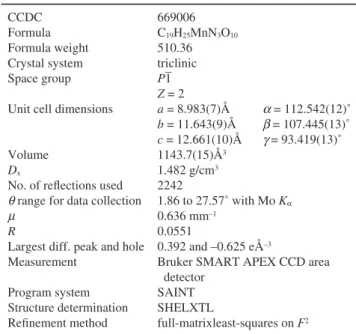
Table 1 Crystal and experimental data CCDC
Formula Formula weight Crystal system Space group Unit cell dimensions Volume
Dx
No. of reflections used
q range for data collection m
R
Largest diff. peak and hole Measurement Program system Structure determination Refinement method 669006 C19H25MnN3O10 510.36 triclinic P1 Z = 2 a = 8.983(7)Å a = 112.542(12)˚ b = 11.643(9)Å b = 107.445(13)˚ c = 12.661(10)Å g = 93.419(13)˚ 1143.7(15)Å3 1.482 g/cm3 2242 1.86 to 27.57˚ with Mo Ka 0.636 mm–1 0.0551 0.392 and –0.625 eÅ–3
Bruker SMART APEX CCD area detector
SAINT SHELXTL
x264 ANALYTICAL SCIENCES 2008, VOL. 24
angle between the least-squares planes of the aromatic rings of the ligands is 11.53˚] with Mn–Ophenol bond lengths of 1.885(3)Å and 1.890(3)Å, together with Mn–Nimin, bond lengths of 1.967(4)Å and 1.981(4)Å. The coordination sphere of the manganese centre is completed by two water molecules [Mn– Owater = 2.238(4)Å, 2.256(3)Å]. The central MnIII ion adopts an elongated octahedral coordination geometry, with the displacement of the Mn1 ion from the O1/N1/N2/O2 least-squares plane being 0.002(2)Å. In the crystal structure of (I), adjacent molecules are linked by hydrogen bonds [O5·O1i = 2.875 Å, O5·O2i = 2.941 Å, O5·O3i = 2.934 Å and O5·O4i = 3.003 Å; symmetry code: (i) –x+1, –y, –z], to form hydrogen-bonded pseudo-dimers, with additional face-to-face p-p stacking
interactions between the benzene groups (C7·C15 = 3.681 Å
and C6·C10 = 3.825 Å); symmetry code: (i) [–x+1, –y, –z]. Moreover, intermolecular hydrogen bonds [O6·O8 = 3.126 Å and O6·O9 = 2.893 Å; symmetry code: (ii) x+1, y, z, and O6·O10B = 2.670 Å (iii) –x+1, –y+1, –z] between these pseudo-dimers and nitrate counterions result in hydrogen-bonded linear chains.
Acknowledgements
This work was supported by Balikesir University under grant number 2007/16. The author would like to thank TUBITAK for a NATO-B1 fellowship for financial support and Prof. Guy Orpen (School of Chemistry, University of Bristol, UK) for his hospitality.
References
1. M. L. Ludwig, K. A. Pattridge, and W. C. Stallings, “Manganese in Metabolism and Enzyme Function”, 1986, Academic Press, New York, chap. 21, 405.
2. A. Karakas, A. Elmali, Y. Yahsi, and H. Kara, J. Nonlinear
Optical Phys. Mater., 2007, 16, 505.
3. H. Kara, Anal. Sci., 2008, 24, x79; Z. Naturforsch., 2008,
63b, 6; Z. Naturforsch., 2007, 62b, 691.
4. Q. Shi, R. Cao, X. Li, J. Luo, M. Hong, and Z. Chen, New.
J. Chem., 2002, 26, 1397.
5. Bruker (2002). SMART, SAINT & SHELXTL. Bruker AXS Inc., Madison, Wisconsin, USA.
Table 2 Selected geometric parameters [Å, ˚]
Fig. 2 (a) ORTEP drawing of the title compound with atom labeling; (b) Stick representation of the hydrogen-bonded (dashed lines) linear chain of pseudodimers formed in (I).
Table 3 Hydrogen bonding geometry [Å, ˚]
D-H·A D-H H·A D·A D-H·A
Symmetry codes: (i) –x+1, –y, –z; (ii) x+1, y, z; (iii) –x+1, –y+1, –z.
Fig. 3 Spacefill representation of the hydrogen-bonded linear chain of pseudodimers formed in (I).
![Table 2 Selected geometric parameters [Å, ˚]](https://thumb-eu.123doks.com/thumbv2/9libnet/5964425.124712/2.892.454.815.144.357/table-selected-geometric-parameters-å.webp)
