urtscher retinopathy is a traumatic angiopathy first described by Otmar Purtscher evaluating a head trauma patient in 1910.1 The
ethology is not known clearly but believed to be associated with oc-clusion of the arterioles that supplies the peripapillary capillaries.2Although
the first case described in literature was a trauma case, Purtscher like retinopathy has been defined in different conditions such as acute pancre-atitis, childbirth, chronic renal failure, barotrauma, steroid injections in and around the orbit and nasal passages, retrobulbar anesthesia, retinal vasculi-tis associated with autoantibodies, preeclampsia, lupus and other non-trauma related cases.2-4The diagnostic clinical findings of Purtscher retinopathy is
seen bilaterally in 60% of cases.3They are localized to the peripapillary area
and posterior pole of the retina. It is a clinical diagnosis with opthalmoscopic Turkiye Klinikleri J Ophthalmol 2018;27(1):65-7
65
A Case of Unilateral Purtscher Like Retinopathy
After Spinal Surgery
AABBSS TTRRAACCTT Purtscher like retinopathy has the same presentation like Purtscher retinopathy but without any trauma history. A 48-year-old female presented to our clinic with a one month his-tory of severe visual loss in her left eye. She had no systemic illness or trauma hishis-tory. She had undergone disc herniation surgery one month before when applied to as. Her laboratory findings were normal but a slightly elevated total cholesterol level. Her vision was 20/20 in the right eye and counting fingers at 50 centimeter in the left eye. Fundus examination showed patched pat-tern of retinal whitening and hemorrhages. The clinical presentation of the patient might be re-lated to increased orbital pressure due extended interval of prone positioning that led to chest compression.
KKeeyywwoorrddss:: Purtscher like retinopathy; spinal surgery; trauma Ö
ÖZZEETT Purtscher benzeri retinopati Purtscher retinopati ile aynı klinik bulgulara sahiptir ancak hastada travma öyküsü bulunmaz. Kırk sekiz yaşında kadın hasta bir aydır var olan sol gözde görme kaybı şikayeti ile başvurdu. Hastanın travma ya da sistemik hastalık hikayesi yoktu. Hasta kli-niğimize başvurusundan bir ay önce disk herni ameliyatı geçirmişti. Laboratuvar incelemesinde ha-fifçe artmış total kan kolesterol düzeyi dışında bir anormallik yoktu. Görme keskinliği sağ gözde tam, sol gözde 50 santimetreden parmak sayma düzeyinde idi. Fundus muayenesinde yama şek-linde retinal beyazlaşma ve kanamalar mevcuttu. Hastanın klinik görünümünün, uzun süre yüz-üstü pozisyonun yol açtığı göğüs kompresyonunun orbita basıncını arttırması ile ilişkili olabileceği düşünüldü.
AAnnaahh ttaarr KKee llii mmee lleerr:: Purtscher benzeri retinopati; spinal cerrahi; travma
Esin SÖĞÜTLÜ SARI,a
Işıl KUTLUTÜRK,b
Alper YAZICI,a
Nesime TISKAOĞLUa aDepartment of Ophthalmology, Balıkesir University Faculty of Medicine, Balıkesir
bClinic of Ophthalmology, Kocaeli State Hospital, Kocaeli
Re ce i ved: 02.03.2016
Received in revised form: 12.06.2016 Ac cep ted: 13.06.2016
Available online: 22.02.2018 Cor res pon den ce:
Esin SÖĞÜTLÜ SARI
Balıkesir University Faculty of Medicine, Department of Ophthalmology, Balıkesir, TÜRKİYE/TURKEY
Cop yright © 2018 by Tür ki ye Kli nik le ri
DOI: 10.5336/ophthal.2016-51185
features consisting of cotton woolspots, Purtscher flecken, retinal hemorrhages and a pale oedematous optic disc.5
CASE REPORT
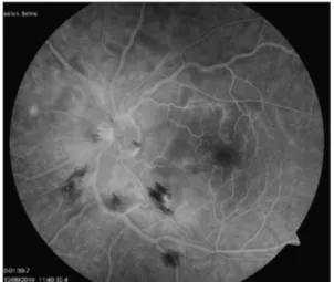
A 48-year-old female presented to our clinic with severe visual loss in her left eye for one month. She had undergone surgery for a disc herniation one month before. The surgery was a two-hour proce-dure with no complication. Her laboratory findings were normal but a slightly elevated total cholesterol level. Her vision was 20/20 in the right eye and counting fingers at 50 centimeter in the left eye. On fundus examination, the right eye was normal but the left eye revealed cotton wool spots surrounding the optic disc in a concentric pattern, pre and in-traretinal hemorrhages and Purtscher flecken with pale edematous optic nerve head (Figure 1). A fun-dus fluorescein angiogram (FFA) revealed blockage by intraretinal hemorrhages and cotton wool spots (Figure 2). Macular optical coherence tomography showed retinal nerve fiber swelling (Figure 3). In-formed consent was obtained from the patient.
DISCUSSION
Purtscher retinopathy is a rare condition seen mostly in male patients who have suffered trau-mas.5While the exact pathogenesis of the disease is
still unknown, many theories have been put for- ward such as lymph extravasation due to an in-crease in intracranial pressure, venous dilatation due to an increased intrathoracic pressure, vasculi-tis secondary to lipase and microemboli in the ar-terioles potentially arising from air, fat, fibrin clots and leucocyte aggregates. The most recent mecha-nism is the vascular endothelial dysregulation re-sulting from a rheological event at inner retinal layer of posterior pole.2,6-8 In Purtscher like
retinopathy, when there is non-traumatic ethol-ogy, an excessive activation of the complement sys-tem inrespiratory distress, acute pancreatitis, connective tissue disorders, amniotic fluid and renal failure can lead to leukocyte aggregation. Pa-tients generally present within 24-48 hours after the causing event with loss of visual acuity and ac-companying visual field loss but in some cases vi-sion loss of variable severity is realized in hours to days after the initial pathology.5,6 In the present
Esin SÖĞÜTLÜ SARI et al. Turkiye Klinikleri J Ophthalmol 2018;27(1):65-7
66
FIGURE 1: Color fundus photo reveals cotton wool spots and retinal
hemor-rhages at the posterior pole which are seen as hypo-fluorescent in angio-graphic images.
FIGURE 2: Late fundus fluorescein angiography image demonstrated
fluo-rescein blockage areas related to the cotton wool spots and retinal hemor-rhage.
case although there was no trauma or sudden tho-racic compression or any other reasons of Purtscher like retinopathy ethology history, it was present to clinical findings of Purtscher retinopathy. Be-cause the patient had a long time prone position after spinal surgery. This event supports the theory of venous reflux occurring after sudden chest trauma or interventions of prolonged prone posi-tion.9
Although, no current consensus on the treat-ment of this disorder; the steroids have been re-ported to be ameliorative effect in some cases. Agarwal et al reported a spontaneous visual recov-ery in 50% of the cases.6Spontaneous recovery of
visual acuity and field is generally seen within 3-6 weeks. Our patient did not have any apparent rea-son when applied. We believe the loss of vision is associated with the surgery she had before. Purtscher flecken, cotton wool spots and retinal hemorrhages have been reported to might persist for a month. Atrophy of the retinal pigment ep-ithelium and optic disc pallor are associated find-ings of Purtscher retinopathy.5,7
The ethology is not still clear because of lim-ited reports of the different type of Purtscher retinopathy cases. To our knowledge, this is the first case of Purtscher like retinopathy following
spinal surgery. Our case is also unique due to late admission and the lack of improvement in symp-toms.
S
Soouurrccee ooff FFiinnaannccee
During this study, no financial or spiritual support was received neither from any pharmaceutical company that has a direct connection with the research subject, nor from a company that provides or produces medical instruments and materials which may negatively affect the evaluation process of this study.
C
Coonnfflliicctt ooff IInntteerreesstt
No conflicts of interest between the authors and / or family members of the scientific and medical committee members or members of the potential conflicts of interest, counseling, ex-pertise, working conditions, share holding and similar situa-tions in any firm.
A
Auutthhoorrsshhiipp CCoonnttrriibbuuttiioonnss
I
Iddeeaa//CCoonncceepptt:: Esin Söğütlü Sarı; DDeessiiggnn:: Esin Söğütlü Sarı, Işıl Kutlutürk; CCoonnttrrooll//SSuuppeerrvviissiioonn:: Esin Söğütlü Sarı; DDaattaa CCooll--l
leeccttiioonn aanndd//oorr PPrroocceessssiinngg:: Esin Söğütlü Sarı, Nesime Tıskaoğlu; A
Annaallyyssiiss aanndd//oorr IInntteerrpprreettaattiioonn:: Esin Söğütlü Sarı, Nesime Tıskaoğlu, Alper Yazıcı; LLiitteerraattuurree RReevviieeww:: Alper Yazıcı, Işıl Kutlutürk, Esin Söğütlü Sarı, Nesime Tıskaoğlu; WWrriittiinngg tthhee A
Arrttiiccllee:: Alper Yazıcı, Işıl Kutlutürk, Esin Söğütlü Sarı, Nesime Tıskaoğlu; CCrriittiiccaall RReevviieeww:: Alper Yazıcı, Işıl Kutlutürk, Esin Söğütlü Sarı, Nesime Tıskaoğlu; RReeffeerreenncceess aanndd FFuunnddiinnggss:: Esin Söğütlü Sarı; MMaatteerriiaallss:: Alper Yazıcı, Işıl Kutlutürk, Esin Söğütlü Sarı, Nesime Tıskaoğlu.
1. Yalçınbayır Ö, Toka F. [Purtscher’s retinopa-thy and Terson’s syndrome]. Ret-Vit 2012;20 Özel Sayı:71-5.
2. Buckley SA, James S. Purtscher’s retinopa-thy. Postgrad Med J 1996;72(849):409-12.
3. Jeon SY, Jung E, Seol HJ, Hur YJ. Develop-ment of Purtscher-like retinopathy after pre-eclampsia combined with acute pancreatitis. Obstet Gynecol Sci 2013;56(4):261-4. 4. Mayer C, Khoramnia R. Purtscher-like
retinopathy caused by acute pancreatitis. Lancet 2011;378(9803):1653.
5. Agrawal A, McKibbin M. Purtscher’s retinopa-thy: epidemiology, clinical features and out-come. Br J Opthalmol 2007;91(11):1456-9. 6. Agrawal A, McKibbin MA. Purtscher’s and
Purtscher-like retinopathies: a review. Surv Opthalmol 2006;51(2):129-36.
7. Günenç Ü, Önal A, Erkin E, Saatçi AO, Maden A, Ergin M. [A case of bilateral Purtscher retinopathy]. Turkiye Klinikleri J Ophthalmol
1995;4(2):142-5.
8. Miguel AI, Henriques F, Azevedo LF, Loureiro AJ, Maberley DA. Systematic review of Purtscher's and Purtscher-like retinopathies. Eye (Lond) 2013;27(1):1-13.
9. Niggemann P, Seifert M, Förg A, Schild HH, Urbach H, Krings T. Positional venous MR an-giography: an operator-independent tool to evaluate cerebral venous outflow hemody-namics. AJNR Am J Neuroradiol 2012;33(2): 246-51.
REFERENCES
Esin SÖĞÜTLÜ SARI et al. Turkiye Klinikleri J Ophthalmol 2018;27(1):65-7