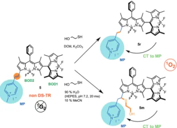
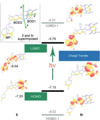
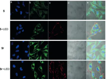
Intracellular modulation of excited-state dynamics in a chromophore dyad: differential enhancement of photocytotoxicity targeting cancer cells
Tam metin
Şekil
Benzer Belgeler
敝人於住院期間,感受到護理站的護士及人員,總是熱心地向病人解說病情,且態度親切、愛心與耐心兼
臺北醫學大學今日北醫: 雙和開幕滿月喜 溫馨好事一連連
Serebral bi- lateral anterior “watershed” enfarktları, sistemik hipoperfüzyon veya hipovolemik şok, pontin ve ekstrapontin miyelinolizis, serebral ve servikal travma
Table Page 1 Brief information about included school types………...25 2 Summary of six proficiency levels with minimum scores for mathematics, reading and
But, the number of P cells of islets of Langerhans and insulin immunoreactivity of P cells (Table 1) in the diabetic group given glurenorm increased in
Örgütsel Sadakat ve Örgütsel Bağlılık: Siyasi Partiler Açısından Bir Analiz Akyay UYGUR Gazi Üniversitesi [email protected] Hakan KOÇ Gazi Üniversitesi
Objective: The aim of this study was to compare the clinical and pathological properties of cases of hepatitis C virus (HCV) infection with respect to the presence or absence
In this study, we measured the expression levels of P53, BAX, BCL-2, CAS3, CAS9, BIRC, and PPIA (housekeeping) genes related to apoptosis on A549 human lung carcinoma
