24
ISSN:1307-9972 e-ISSN:1308-0679
Three-Dimensional Analysis with Computed Tomography of
Kidneys in the Kangal Dogs
Ömer ATALAR
1, Mustafa KOÇ
2, Asuman ARKAŞ ALKLAY
3, Hasan Hüseyin
ARI
41Fırat University Faculty of Veterinary Medicine Department of Anatomy, 23119, Elazığ, Turkey 2Fırat University Faculty of Medicine, Department of Radiology, 23119, Elazığ, Turkey 3Dicle University Faculty of Veterinary Medicine Department of Anatomy, 21280, Diyarbakır, Turkey 4Cumhuriyet University Faculty of Veterinary Medicine Department of Anatomy, 58140, Sivas, Turkey
Introduction
Kangal dog is a Turkish shepherd and guard dog is racial. It is also known as Anatolian Shepherd Dog or Sivas Kangal Dog. The average length of this breed 72-77 cm in females, 77-86 cm in males, and the weights are 41-54 kg in females and 50-66 kg in males (1). The computerized tomography method is regarded as the most accurate and fastest way to detect the anatomical differences of these animals
from other dog breeds without damage to the animal. Three-dimensional computerized tomography (3D CT) images of the kidneys are important in determining the pathological conditions in this region, in the correct detection of anatomical features, and also in the training of anatomy (2,3,4). It is known that computed
Dicle Üniversitesi Veteriner Fakültesi Dergisi
http://www.dicle.edu.tr/veteriner-fakultesi-dergisi
Araştırma Makalesi
Summary
It has been found that there is no study done with two and three dimensional imaging related to kidneys of the Kangal dogs in the literature review. The aim of this investigation is to seen that for the first time in Kangal dogs; to obtain the measurements and localizations of the kidneys using the multidetector computed tomography (MDCT) image. In the study, 5 males and 5 females of a total of 10 adult Kangal dogs were used. The images obtained from MDCT were stacked and overlaid to reconstruct the 3D model of the kidneys using 3D modeling software (VITAL, Vitrea 2, HP XW6400 Workstation, USA). The locations of kidneys, their width, length and thickness, and their distance from columna vertebralise of the centers were measured and analyzed statically. In all animals except one material, right kidney measurements were higher than in the left kidney. In a female dog, rotation anomaly was detected in the right kidney. Whereas, no significant statistical difference was observed in the statistical analysis results. It has been concluded that the results of the research will be a source for both anatomical investigations and similar studies in the future.
Keywords: Computed Tomography, Kangal Dog, Kidney, Three-Dimensional Image
Kangal Köpeklerinde Böbreklerin Bilgisayarlı Tomografi ile Üç Boyutlu İncelenmesi Özet
Literatür taramaları sonuçlarına göre; Kangal köpeklerinde böbreklerin, iki ve üç boyutlu incelenmesi üzerinde; herhangi bir çalışma bulunmadığı tespit edilmiştir. Bu araştırmanın amacı, Kangal köpeklerinde ilk kez; çok detektörlü bilgisayarlı tomografik görüntüler kullanılarak, böbreklerin lokalizasyonunu ve ölçümlerini incelemektir. Çalışmada, 5 erkek ve 5 dişi olmak üzere toplam 10 erişkin Kangal köpeğinden faydalanılmıştır. Multidetektör bilgisayarlı tomografi (MDBT) cihazından elde edilen görüntüler, üç boyutlu (3D) modelleme yazılımı (VITAL, Vitrea 2, HP XW6400 Workstation, ABD) ile, böbreklerin 3D modelinin oluşturulması için kullanılmıştır. Böbreklerin yerleşim alanları, en, boy ve kalınlıkları ile merkezlerinin columna vertebralis’e olan uzaklıkları ölçülmüş ve istatistiksel olarak analiz edilmiştir. Bir materyal hariç bütün hayvanlarda, sağ böbrek ölçümleri sol böbrekten daha yüksek olarak tespit edilmiştir. Bir dişi köpekte, sağ böbrekte rotasyon anomalisi olduğu saptanmıştır. İstatistiksel analiz sonuçlarında ise anlamlı bir istatistiksel fark gözlenmemiştir. Araştırma sonuçlarının, hem anatomi alanındaki incelemelere, hem de ileride yapılacak benzer alanlardaki çalışmalara kaynak teşkil edeceği kanaatine varılmıştır.
25 tomography (CT) images are also frequently used
in Veterinary Medicine (5,6).
Although many information is available on the anatomy of the kidneys and the excretory system in dogs (2,7,8,9); in the case of Kangal species, no literature data on the three- dimensional computerized tomography of the kidneys were found.
In this study; it is thought that it will provide important contributions to literature information both in terms of both material and methods as well as presenting the first findings in this subject.
Material and Methods
A total of 10 adult Kangal dogs, 5 females and 5 males, were used for this study. General anesthesia was obtained with the combination of Xylazine (1.1 mg / kg, i.m., Rompun ®, Bayer) and Ketamine (22 mg / kg, i.m., Ketalar ®, Eczacıbaşı).
Multi-detector computerized tomography (MDCT) images of dogs admitted to the Toshiba Aquilion 64 section CT device were taken in a prone position. These images were between kVp 120, mAs 150-200 and 0.5 mm parallel cross-sectional thickness, 0.5 mm reconstruction interval, diameter FOV (30 cm) and range value 1-1.5. In this study, dosage parameters and screening were done within standard protocols (10, 11, 12).
High-resolution MDCT images were obtained from the kidneys of materials. The axial images were stored in DICOM format and the results were forwarded to the study center (VITAL, Vitrea 2, HP XW6400 Workstation, USA). After the images were evaluated within the research area, the findings were recorded. First, relevant parts were selected from two- dimensional (2D) images. Then three dimensional (3D) modeling was done.
After three-dimensional (3D) modeling of the kidneys was established, statistical analysis of biometric measurements was performed at 5% confidence interval (P <0.05). Statistical analysis was performed with SPSS 21.0 Windows Computer Packaged Software. The avarage and standard deviation values of the materials were determined by the Mann Whitney U test.
As a terminology was based on Nomina Anatomica Veterinaria (13).
Results
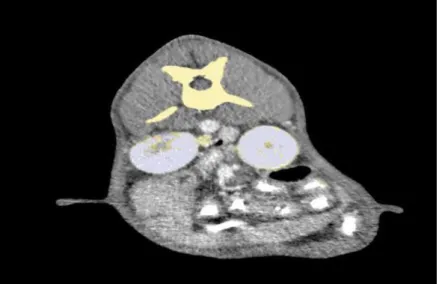
In the transversal sections of computed tomography images, the kidneys were initially identified as two-dimensional (2D) (Figure 1). Then dimensional imaging (Figure 2) and three-dimensional modeling (Figures 3 and 4B) were performed.
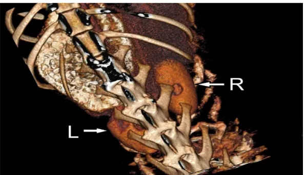
26 Figure 2. Dorsal plan, 3D image of the right (R) and left (L) kidneys.
Figure 3. Anterior plan image with 3D Reconstructive. W: Width, between the margo lateralis and margo medialis. T: Thickness, between the facies dorsalis and facies ventralis.
The mean measurement values of the kidneys in male and female Kangal dogs were reported as the difference between the standard deviation values and the mean values by sex. All
statistical results; the table 1 is presented with the details (Table 1). (Kidney measurements: P <0.05 and SD (± mm).
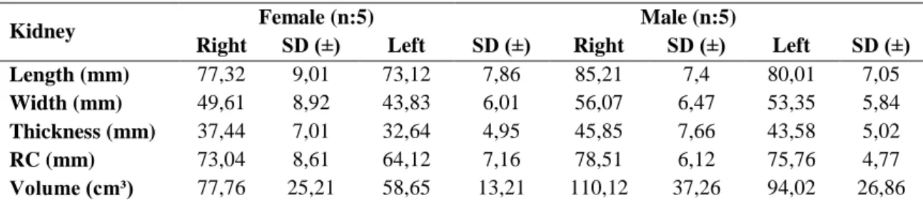
27 Table 1. Measurement values of kidneys with computed tomography.
Kidney Female (n:5) Male (n:5)
Right SD (±) Left SD (±) Right SD (±) Left SD (±)
Length (mm) 77,32 9,01 73,12 7,86 85,21 7,4 80,01 7,05
Width (mm) 49,61 8,92 43,83 6,01 56,07 6,47 53,35 5,84
Thickness (mm) 37,44 7,01 32,64 4,95 45,85 7,66 43,58 5,02
RC (mm) 73,04 8,61 64,12 7,16 78,51 6,12 75,76 4,77
Volume (cm³) 77,76 25,21 58,65 13,21 110,12 37,26 94,02 26,86
Length: Between the extremitas cranialis and extremitas caudalis. Width: Between the margo lateralis and margo
medialis. Thickness: Between the facies dorsalis and facies ventralis. RC: Between the hilus renalis and the columna vertebralis. Volume: Volumes of the kidney.
According to the results of our study; the right kidney has always been found to be higher than the left kidney (Table 1). In all subjects except one male, right kidney measurements were higher than in the left kidney. In a male material, left kidney length, width and thickness ratios (83,07 X 55,68 X
46,21); right kidney length, width and thickness (81,72 x 54,03 x 43,79) were measured. In parallel with this, the results of the volume measurements of the kidneys; in males and in the right kidneys of volume was found to be higher. In a male dog; the left kidney (108,92 cm³), which has a higher measurement value, was found to be larger than the
right kidney (99,15 cm³) of the same animal. In the nine animals, the localization of the kidneys was found to be normal. In a female dog, it was determined that rotation anomaly of the right kidney was formed (Figures 4A and 4B). In the biometric measurements, it was determined that the left kidney is closer to the columna vertebralis in
both male and female. The distances of the hilus renalis from the columna vertebralis are expressed together with the mean values and standard deviations in the table. The standard deviation values for all measured parameters show a consistent variably between sexes. No statistically significant difference was found in the data.
Figure 4. Rotation anomaly of right kidney. A: ventral plan image with 3D. B: dorsal plan image with 3D reconstructive. R: right kidney. L: Left kidney. Le: Lenght, between the extremitas cranialis and extremitas caudalis.
28 Discussion
Researchers report that the right kidney is always ahead of the left kidney, that there is no complete symmetry between the kidneys and that the left kidney is closer to the columna vertebralis (2, 3, 7, 8). In the morphological research of Evans (8) related with in dogs; it was determined that the size of the kidney and the localization of the right kidney are higher than that of the left kidney. Eken et al. (14) indicate that the measurements and percentage differences from the right kidney, regardless of gender, are higher in all rabbits studied than in the left kidney. Some researchers emphasize that in humans, rarely the left kidney can carry higher measured values than the right kidney, which is not a pathological condition (15). In this study, we have assessed both gender and direction (right and left kidney) distinction measures on Kangal Dogs. According to our results, higher measurements were obtained in males and right kidneys compared to females and left kidneys. There is also a male subject; it is seen that the left kidney data are superior to the right kidney data as is the case in humans.
It has been stated that kidney rotation anomalies rarely seen in humans can be in both male and female, both in the right and left kidney (15). Such anomalies have also been reported in cats and dogs (16). In this research we made on Kangal dogs; in a female animal, rotation anomaly of the right kidney was detected. With this research, rotation anomaly in Kangal dogs is reported for the first time.
The confidence interval of the measurements made with two and three-dimensional images obtained with a multidetector computerized tomography device is considered to be very high (14, 17, 18). In the results obtained from our research results and standard deviation values; the
units in mm and cm³ provide two decimal digits after the comma, (with the same precision ratings in this respect) so they have the same precision ratings as this rule supports.
Eken et al. (14) reports that the three-dimensional images obtained from rabbits are closest to reality. The same researchers say that artificial kidneys can be made almost as close as possible to the measurements they make. These researchers support the work we have done; thanks to the data we obtained from Kangal dogs; we are convinced that we have obtained findings for this species that can obtain artificial kidneys very close to the truth.
Kidneys are the organs that often have problems with diseases related to the urinary system. The first step for the correct resolution of these organs in diagnosis, treatment and pathological cases; The anatomy of the organ will be known most accurately in every direction (19). This valuable dog breed, which cannot always be studied in our research, can be used in every direction of the kidney anatomy; it is meaningful that both two- and three-dimensional images, as well as their measurements, are presented for the first time and the confidence level is high.
Therefore, it is thought that this research is important not only because it is the first report in its field, but also in terms of contributing to future studies and training of Veterinary Medicine. References
1. Yılmaz O. Turkish Kangal (Karabash) Shepherd Dog (in English)
https://www.researchgate.net/publication/263468806_Tur kish_Kangal_Karabash_Shepherd_Dog_in_English; 20.04.2017
2. Smallwood JE, George TF. (1993). Anatomic Atlas for Computed Tomography in the Mesaticephalic Dog: Thorax and Cranial Abdomen. Vet Radiol Ultrasound. 34: 65-169 84.
29 3. Regedon S, Franco A, Garin JM, Robina A Lignereux
Y. (1991). Computerized Tomographic Determination of the Cranial Volume of the Dog Applied to Racial and Sexual Differentiation. Acta Anat.142: 347-50.
4. Eken E, Gezici M. (2002). The Influence of Stomach Volume on the Liver Topography in Cats. Anat Histol Embryol. 31: 99-104.
5. Kara M, Turan E, Dabanoglu I, Ocal MK. (2004). Computed Tomographic Assessment of the Trachea in the German Shepherd Dog. Ann Anat.186: 317-21. 6. Onar V, Kahvecioglu O, Cebi V. (2002). Computed Tomographic Analysis of the Cranial Cavity and Neurocranium in the German Shepherd Dog (Alsatian) Puppies. Vet Arhiv. 72: 57-66.
7. Dursun N. Veteriner Anatomi I. (2006). 11th ed, Medisan, Ankara.
8. Evans HE. (1993). Skeleton. In: Miller’s Anatomy of the Dog. H.E. Evans (ed). 3rd ed.197-204. W.B. Saunder Company. Philadelphia.
9. Özdemir D, Özüdoğru Z, Malkoç İ. (2009). Internal Segmentation of the Renal Arteries in the Kangal Dog. Journal of the Faculty of Veterinary Medicine, Kafkas University. 15 (1): 41-44.
10. Hu H, He HD, Foley WD, Fox SH. (2000). Four Multidetector-Row Helical CT: Image Quality and Volume Coverage Speed. Radiology. 215: 55-62. 11. Prokop M. (2003). General Principles of MDCT. Eur J Radiol. 45: 4-10.
12. Kalra MK, Maher MM, Toth TL, Hamberg LM, Blake MA, Shepard JA, Saini S. (2004). Strategies for CT Radiation Dose Optimization. Radiology. 230: 619-28.
13. Nomina Anatomica Veterinaria. (2012). Prepared by the International Committes on Veterinary Gross Anatomical Nomenclature and Authorized by the General Assembly of the World Association of Veterinary Anatomists, The Editorial Committee Hannover, Sapporo, Japan.
14. Eken E, Çorumluoğlu Ö, Paksoy Y, Beşoluk K, Kalaycı İ. (2009). A Study on Evaluation of 3D Virtual Rabbit Kidney Models by Multidetector Computed Tomography Images. International Journal of Experimental and Clinical Anatomy. 40-44.
15. Glodny B, Petersen J, Hofmann KJ, Schenk C, Herwig R, Trieb T, Koppelstaetter C, Steingruber I, Rehder P. (2009). Kidney Fusion Anomalies Revisited: Clinical and Radiological Analysis of 209 Cases of Crossed Fused Ectopia and Horseshoe Kidney. BJUI. 103: 224-235.
16. Kaufmann ML, Osborne CA, Johnston GR. (1987). Renal Ectopia in a Dog and a Cat. J Am Vet Med Assoc. 190: 73–77.
17. Aldur MM. (2005). Creating Computer Aided 3D Model of Spleen and Kidney Based on Visible Human Project. Saudi Med J. 26: 51-6.
18. Turkvatan A, Ozdemir M, Cumhur T, Olcer T. (2009). Multidetector CT Angiography of Renal Vasculature: Normal Anatomy and Variants. Eur Radiol.19: 236-44.
19. Janoff DM, Davol P, Hazzard J, et al. (2004). Computerized Tomography with 3-Dimensional Reconstruction for the Evaluation of Renal Size and Arterial Anatomy in the Living Kidney Donor. J Urology. 171: 27-30.
Yazışma Adresi:
Prof. Dr. Ömer ATALAR
Fırat Üniversitesi Veteriner Fakültesi Anatomi Anabilim Dalı, 23119, Elazığ, Türkiye